Fig. 69.1
(a) Foot skeleton. Dorsal view.(1) Calcaneus, (2) talus, (3) trochlea tali, (4) navicular bone, (5) cuboid bone, (6) cuneiform bone, (7) metatarsal bones, (8) phalanges (Reproduced with permission from Danilo Jankovic). (b) Foot skeleton. Lateral view. (1) calcaneus, (2) tuber calcaneus, (3) talus, (4) sinus tarsi, (5) navicular bone, (6) cuneiform bones, (7) cuboid bone, (8) subtalar joint, (9) talocalcaneonavicular joint (Reproduced with permission from Danilo Jankovic)

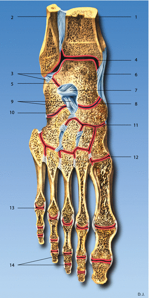
Fig. 69.2
(1) Tibia, (2) fibula, (3) posterior talofibular ligament, (4) talocrural joint, (5) subtalar joint, (6) deltoid ligament, (7) talocalcaneus ligament, (8) talonavicular ligament, (9) bifurcated ligament, (10) calcaneocuboid ligament, (11) cuneonavicular ligament, (12) tarsometatarsal ligament, (13) metatarsophalangeal ligament, (14) articulationes digiti (Reproduced with permission from Danilo Jankovic)
The joint surfaces of the talocrural joint are formed by the malleolar mortise (mortise joint) at the trochlea of the talus, with its superior facet and medial and lateral malleolar facets. The ligaments of the upper ankle joint are the medial collateral ligament (deltoid ligament), anterior talofibular ligament, posterior talofibular ligament, and calcaneofibular ligament (Figs. 69.3 and 69.4a, b).


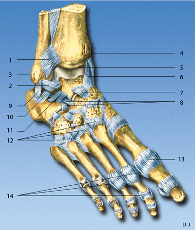
Fig. 69.3
Foot joints. Lateral view. (1) Anterior tibiofibular joint, (2) anterior talofibular ligament, (3) lateral malleolus, (4) medial malleolus, (5) talocrural joint, (6) deltoid ligament (medial collateral ligament), (7) talonavicular ligament, (8) bifurcated ligament, (9) calcaneofibular ligament, (10) talocalcanean ligament, (11) interosseous talocalcaneal ligament, (12) dorsal tarsometatarsal ligaments, (13) articular capsule, (14) collateral ligaments (Reproduced with permission from Danilo Jankovic)

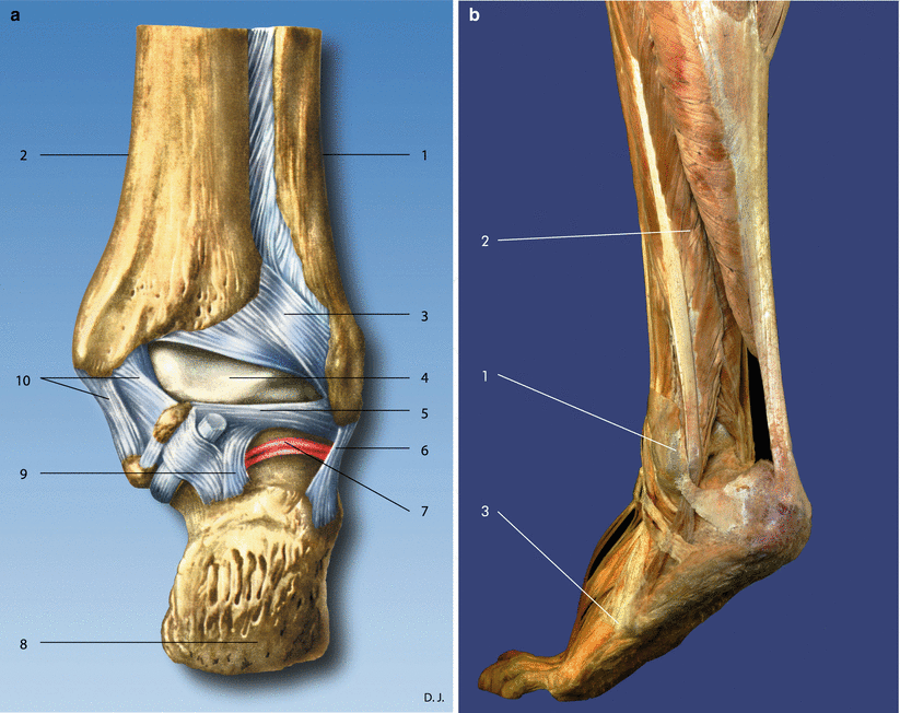
Fig. 69.4
(a) A Foot joints. Dorsal view. (1) Fibula, (2) tibia, (3) posterior tibiofibular ligament, (4) talus, (5) posterior talofibular ligament, (6) calcaneofibular ligament, (7) subtalar joint, (8) calcaneus, (9) posterior talocalcaneal joint, (10) deltoid ligament (Reproduced with permission from Danilo Jankovic). (b) Ankle joint. Lateral view. (1) Lateral malleolus, (2) peroneal muscles, (3) tuberosity of the metatarsal bone 5. With tendon of the peroneus brevis and tertius muscle (Reproduced with permission from Danilo Jankovic)
The lower ankle joint consists of two separate joints – the subtalar joint, which forms the posterior part of the lower ankle joint, and the talocalcaneonavicular joint, which forms the anterior part of it. These two separate joints act together. The joint surfaces in the subtalar joint are formed by the talus and calcaneus (Figs. 69.3 and 69.4a, b).
The vascular supply to the ankle is from branches of the posterior tibial artery (the medial and lateral plantar arteries) and from the anterior tibial artery (the dorsalis pedis artery). The ankle is innervated from the tibial nerve (lateral and medial plantar nerves and calcaneal branches) and from the deep fibular (peroneal) nerve (Fig. 69.5a–c).


Fig. 69.5


(a) Foot. Vascular supply and innervation. (1) Tibial nerve, (2) medial plantar nerve, (3) lateral plantar nerve, (4) calcaneal branches, (5) posterior tibial artery, (6) posterior tibial vein (Reproduced with permission from Danilo Jankovic). (b) Cutaneous innervation areas of the back of the foot. Lateral view. (1) Superficial peroneal(fibular) nerve, (2) deep peroneal(fibular) nerve, (3) anterior tibial artery, (4) sural nerve, (5) lateral malleolus (Reproduced with permission from Danilo Jankovic). (c) Cutaneous innervation areas in the region of the back of the foot (from the front). (1) Superficial peroneal (fibular) nerve, (2) deep peroneal (fibular) nerve, (3) saphenous nerve, (4) sural nerve, (5) lateral malleolus, (6) medial malleolus (Reproduced with permission from Danilo Jankovic)
Stay updated, free articles. Join our Telegram channel

Full access? Get Clinical Tree


