Fig. 65.1
Anterior and posterior divisions of the obturator nerve between the adductor muscles (ALM adductor longus muscle, ABM adductor brevis muscle, AMM adductor magnus muscle) (With permission from Jens Kessler)
Indications for an obturator nerve block are the suppression of the adductor reflex for transurethral lateral bladder wall resections [4], adjunct to femoral nerve blocks for postoperative pain management of the medial knee joint [7] and the treatment of adductor spasm.
The block is performed in supine position with slight abduction of the hip and external rotation of the thigh. After skin disinfection and appropriate covering of the transducer (Fig. 65.2) to maintain sterility, the transducer is placed in the medial thigh to visualize the obturator divisions and the adductor brevis muscle in a short-axis view. It is also possible to start with the femoral nerve, artery, and—by compression—vein, moving medially and slightly caudal to the pectineus and the adductor muscles.
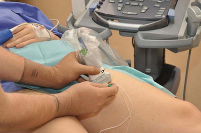
Fig. 65.2
Out-of-plane approach to obturator nerve block in the medial thigh (With permission from Jens Kessler)
Color Doppler can help detecting the adjacent obturator arteries to avoid hemorrhage due to unintentional vessel puncture. Both divisions should be blocked separately, beginning with the posterior division. In case of inadequate visualization of both divisions, the local anesthetic can be injected between the three adductor muscles [3, 5].
A linear transducer with a higher frequency would be appropriate for this kind of nerve block. Both out-of-plane [2] and in-plane approaches are possible to inject the local anesthetic around the divisions by using only one needle insertion point.
References


