Fig. 24.1
A schematic diagram showing the arrangement of the four rotator cuff muscles: subscapularis, supraspinatus, infraspinatus, and teres minor (Reproduced with permission from Philip Peng Educational Series)
Fig. 24.2
Anterior view of the shoulder showing the subscapularis and supraspinatus muscles. The anterior portion of the deltoid muscle was reflected to show the underlying rotator cuff muscle (Reproduced with permission from Philip Peng Educational Series)
Fig. 24.3
Posterior view of the shoulder to show the infraspinatus and teres minor muscle. The posterior portion of the deltoid muscle was partially removed to show the underlying muscle (Reproduced with permission from Philip Peng Educational Series)
Sonoanatomy
The patient is examined in sitting position or semi-inclined position on an examination bed with the shoulder over the edge of the bed. The hand is put in modified Crass position (similar to the posture “putting the money back in the back pocket”). This maneuver allows a longer segment of supraspinatus tendon to be scanned. A high-frequency (6–13 MHz) linear ultrasound probe is placed over the SS tendon. A good reference point is the long head of biceps (LHB) tendon. The long-axis view of the SS tendon is almost parallel to the LHB tendon, and the SS tendon should be within 1.5 cm from the LHB tendon. Further away from the LHB tendon is the intercalated zone between the supraspinatus and infraspinatus tendon. To examine the presence of pathology of SS tendon, both the long- and short-axis scans are required. The SASDB appears as a hypoechoic space between the deltoid muscle and the SS tendon (Figs. 24.3, 24.4, and 24.5).
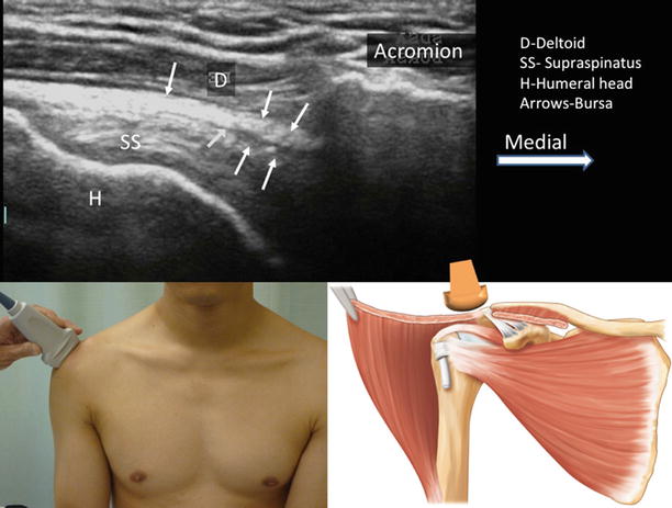
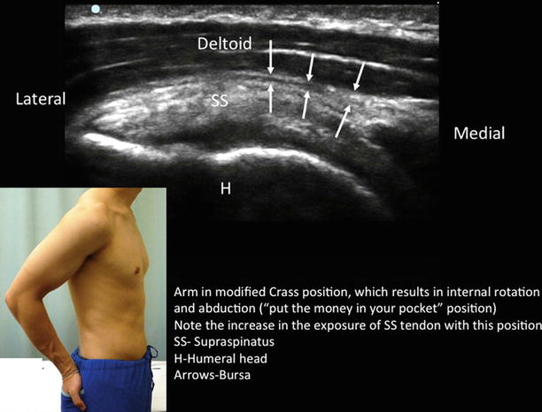
Fig. 24.4
Ultrasound image of the subacromial subdeltoid bursa (SASDB). The supraspinatus tendon (SS) was seen attached laterally onto the greater tuberosity of the humeral head (H). The insert on the left shows the position of the patient and the ultrasound probe and the one on the right shows the probe and the structures underneath. The deltoid muscle was reflected to show the underlying supraspinatus muscle (Reproduced with permission from Philip Peng Educational Series)
Fig. 24.5


Ultrasound image of the supraspinatus tendon when the arm is put in the modified Crass position. Note that the portion of the supraspinatus tendon lateral to the acromion process was significantly increased by this maneuver. The insert shows the position of the modified Crass position. SS supraspinatus tendon, H humeral head, D deltoid muscle. Line arrows outline the peribursal fat of the SASDB (Reproduced with permission from Philip Peng Educational Series)
Related posts:


Stay updated, free articles. Join our Telegram channel
Full access? Get Clinical Tree
