Fig. 22.1
Glenohumeral joint showing various ligaments and the joint capsule. The anterior capsule is reinforced by the superior, middle, and inferior glenohumeral ligament (Reproduced with permission from Philip Peng Educational Series)
The ACJ is stabilized by both the static and dynamic stabilizers. The static stabilizers include the acromioclavicular ligaments (superior, inferior, anterior, and posterior), the coracoclavicular ligaments (trapezoid and conoid), and the coracoacromial ligament. The dynamic stabilizers include the deltoid and trapezius muscles (Fig. 22.2).
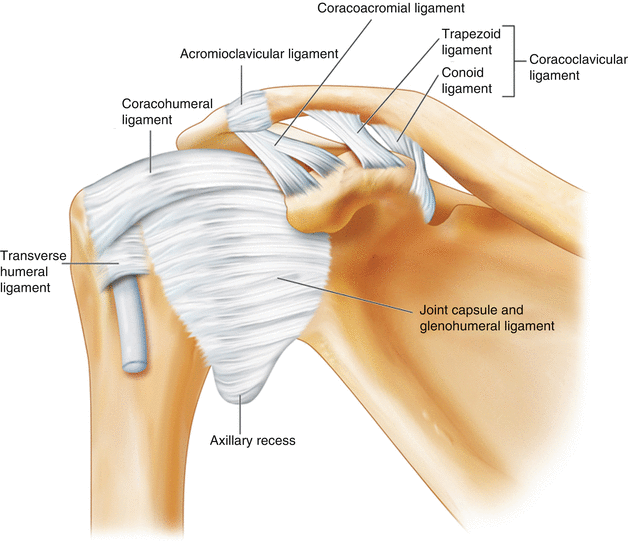
Fig. 22.2
The acromioclavicular joint is a synovial joint with the articular surfaces separated by a wedge-shaped fibrocartilaginous disc (asterisk). The inferior surface of the joint is in direct contact with the subacromial bursa and supraspinatus muscle and may play a role in the development of the impingement syndrome (Reproduced with permission from Philip Peng Educational Series)
Sonoanatomy
The patient is placed in sitting with arm in neutral position, in which the deep joint space of ACJ is the widest. Because of the superficial location of the joint, linear high-frequency (6–13 MHz) ultrasound probe is appropriate. The probe is initially placed over the joint in the coronal plane (medial part of probe in line with clavicle initially). The lateral end of the probe is then rotated in an anterior and posterior direction to obtain the best view of ACJ (Fig. 22.3). The sonogram should show the hyperechoic ends of acromion and distal clavicle, fibrocartilaginous disc, and superior joint capsule.
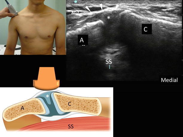
Fig. 22.3
Ultrasound image of the acromioclavicular joint. The upper insert shows the position of the probe and the patient, and the lower insert shows the position of the probe and the structures underneath. A, acromion process; C, clavicle; *, the wedge-shaped fibrocartilaginous disc; arrowheads, superior joint capsule; SS, supraspinatus muscle (Reproduced with permission from Philip Peng Educational Series)
Patient Selection
The ACJ injection can be used as a diagnostic or therapeutic block. It is commonly used as a diagnostic test to localize the source of pain when ACJ injury is suspected. The main therapeutic indication for ACJ injection is arthritis (post-traumatic and osteoarthritis) of this joint. Patient usually complains of anterior shoulder pain brought about by daily activities such as reaching the back pocket or unhooking a brassiere. Physical examination reveals point tenderness over ACJ and positive cross-arm adduction test.
Related posts:


Stay updated, free articles. Join our Telegram channel

Full access? Get Clinical Tree


