Fig. 21.1
Glenohumeral joint showing various ligaments and the joint capsule. The anterior capsule is reinforced by the superior, middle, and inferior glenohumeral ligament. The insert shows the articular surface, the glenoid process, and the labrum (Reproduced with permission from Philip Peng Educational Series)
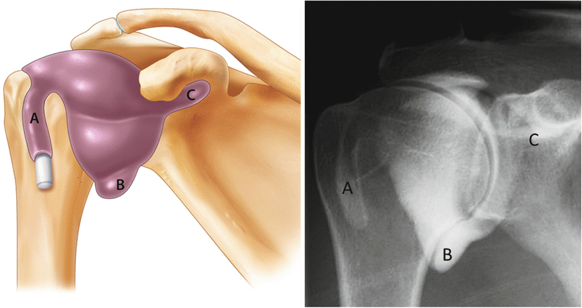
Inside the joint capsule, the synovial membrane overlies the long head of biceps (LHB) tendon and forms three synovial recesses: the biceps tendon recess anteriorly, the subscapularis recess medially, and the axillary pouch inferiorly (Fig. 21.2). The biceps tendon recess allows a portal of entry to the GHJ through the rotator cuff interval [1–2].


Fig. 21.2
The drawing of three main recesses of the joint (left)—A, the biceps tendon sheath; B, the axillary pouch; C, the subscapular recess—and the corresponding radiographic (arthrogram) appearance (right) (Reproduced with permission from Philip Peng Educational Series)
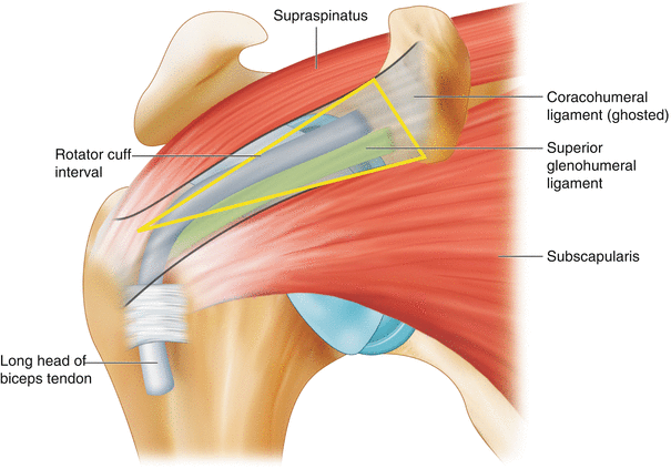
The rotator cuff interval (RCI) is a triangular space defined by the subscapularis (SSC) tendon, supraspinatus (SS) tendon, and the base of the coracoid process. It is a space where the GHJ synovial lining extends around the biceps tendon and where the surgeon inserts the arthroscope into the GHJ to avoid damaging the cuff tendons. Thus, this allows access for GHJ injection by the interventionist (Fig. 21.3). The contents of RCI are the LHB tendon and the superior glenohumeral ligament.


Fig. 21.3
The anterosuperior view showing the rotator cuff interval, which is a triangular space between the tendons of subscapularis (anterior) and supraspinatus (posterior) muscles, and the base of the coracoid process. The roof is the coracohumeral ligament (ghosted), and the contents are the long head of biceps tendon (blue) and superior glenohumeral ligament (green) (Reproduced with permission from Philip Peng Educational Series)
Sonoanatomy
In general, there are three approaches to access the GHJ: anterior, posterior, and rotator cuff interval [1–2]. Only the posterior approach and rotator cuff interval approach are described here.
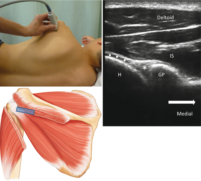
For the posterior approach, the patient is placed in either sitting position or lateral decubitus position with the injection side as the nondependent side. In both cases, the patient’s hand is placed on the contralateral shoulder to open up the GHJ space. A high-frequency linear probe (6–13 MHz) is used unless the patient is of very high BMI or very muscular. The lateral end of the probe is placed just caudal to the acromion over the infraspinatus tendon. This scan should reveal the glenoid process, labrum, capsule, humeral head, and infraspinatus muscle and tendon (Figs. 21.4 and 21.5).


Fig. 21.4


Ultrasound image of the posterior glenohumeral joint. The glenoid process and humeral head both appear as hyperechoic structures with anechoic shadow. The insert on the top shows the position of the patient and the ultrasound probe while one below shows the probe position and the structures underneath. IS infraspinatus muscle, H humeral head, GP glenoid process, * glenoid labrum, ♦ the articular cartilage of the humeral head (Reproduced with permission from Philip Peng Educational Series)
Stay updated, free articles. Join our Telegram channel

Full access? Get Clinical Tree


