, Corinna Eleni Psomadakis2 and Bobby Buka3
(1)
Department of Family Medicine, Mount Sinai School of Medicine Attending Mount Sinai Doctors/Beth Israel Medical Group-Williamsburg, Brooklyn, NY, USA
(2)
School of Medicine Imperial College London, London, UK
(3)
Department of Dermatology, Mount Sinai School of Medicine, New York, NY, USA
Keywords
LacerationSutureStitchesScarScarringWoundHealingPreventionKeloidHypertrophic scarHemostasisInflammationGranulationTissueRemodelingSunscreenHydrogel dressingIntralesional steroidChemical peelLaser resurfacing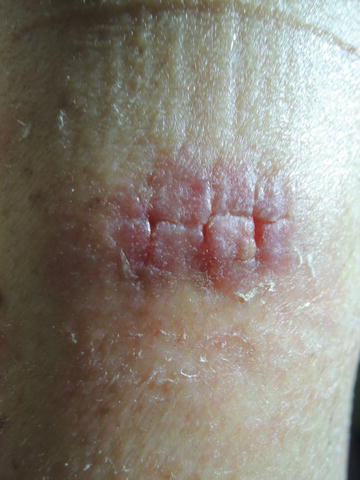
Fig. 39.1
Post suture removal with good approximation of edges in this arm wound
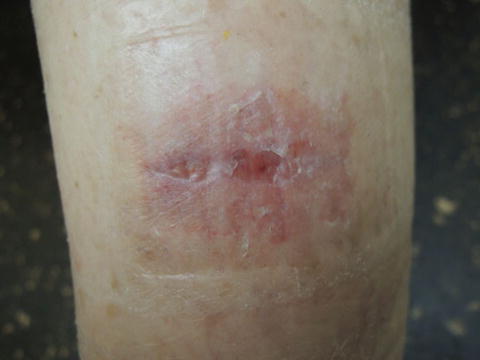
Fig. 39.2
Mild wound gaping noted centrally, poor approximation of edges, and higher risk of cosmetically less desirable scarring in this leg wound
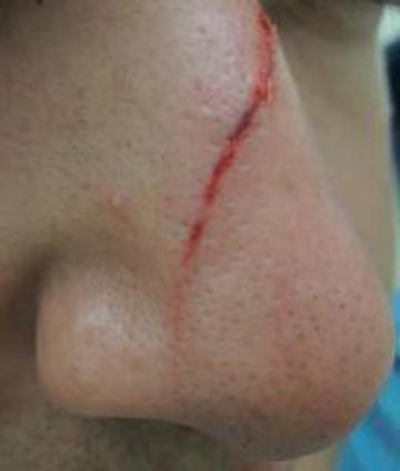
Fig. 39.3
Linear abrasion, partial thickness
Primary Care Visit Report
The first patient, a 56-year-old female with newly diagnosed invasive squamous cell carcinoma on her lower leg, presented for removal of stitches from a recently biopsied lesion on her left upper arm. The sutures had been placed 14 days prior to this visit.
Vitals were normal. On exam, her left upper arm had a 2 cm × 2 cm erythematous raised lesion with three sutures, knots buried, and some erythematous papular irritation surrounding the lesion from the bandage. Her left lower leg featured a well-healing lesion from prior SCC removal.
The stitches were removed from the left arm lesion.
The second patient was a 28-year-old male with no past medical history who presented with a laceration on his nose. About an hour prior, a fluorescent light box had fallen from the ceiling and cut his nose.
Vitals were normal. On exam, there was a 3 cm linear laceration extending from the left side of his nose superiorly to the right side of his nose inferiorly (crossing the bridge of his nose) with minimal bleeding.
A plastic surgeon was consulted, and advised that, even though this laceration appeared to be superficial, stitching would help reduce scarring. The patient was referred to this plastic surgeon for suturing.
In both cases, the patients were concerned with scarring from these skin lesions and wanted to know how to minimize or prevent scarring. They were advised to ensure the lesions were covered with sunblock and to avoid direct sun exposure.
Discussion from Dermatology Clinic
Differential Dx
Scar
Hypertrophic scar
Keloid
Favored Dx
Both patients have actively healing partial thickness wounds that may result in scarring.
Overview
Scars are formed by fibrous tissue, which replaces normal tissue after an insult or injury to the skin. Following damage to tissue, wounds undergo a dynamic healing process that is characterized by four stages: hemostasis , inflammation , proliferation or granulation , and maturation or remodeling [1]. This healing process can last between months and years [2]. Depending on the depth and nature of the wound, the healing process can result in formation of a scar.
Hemostasis is the first stage of tissue healing. After the continuity of skin is disrupted, platelet activation and the coagulation cascade act to stop hemorrhage. The inflammatory phase is marked by initiation of the inflammatory cascade, with neutrophils, macrophages, and other mediators infiltrating the wound site, and contributing to removal of damaged tissue. Tissue loss triggers vascularization and formation of new tissue, termed granulation tissue, and over the course of up to 2 years, fibroblasts and collagen are produced and deposited, and the skin is remodeled [3]. Rather than occurring sequentially, these events are dynamic and represent an interplay between several healing mediators.
A number of different characteristics of wounds affect the prognosis of healing and scar formation. Wounds can be classified according to the depth of injury, with superficial wounds limited to the epidermis, partial thickness wounds involving some of the dermis, and full-thickness wounds extending fully through the dermis, and into subcutaneous tissue in some cases. Superficial wounds, caused by friction, mild burns, or abrasions, often resolve without scarring, as the healing process typically only requires re-epithelialization of the epidermis of otherwise continuous, intact skin [4]. Partial thickness wounds typically heal in a similar fashion to superficial wounds; however, their size and shape would influence the healing outcome. Wounds that cover a large surface area, or involve jagged edges, are more likely to result in a scar. Full thickness wounds often require suturing and resolve with a scar.
Surgical incisions are the most common cause of scarring, with an estimated incidence of 100 million new scars per year following elective procedures in the developed world [2]. Acne scars are estimated to affect up to 14 % of people suffering from acne [5]. Other common causes of scarring are discoid lupus erythematosus, scarring alopecia, and varicella. Exaggerated healing responses can lead to hypertrophic and keloid scarring. Between 5 and 15 % of wounds are thought to result in keloid scars [6]. Keloid scars are more common in people under the age of 30, are 15 times more likely to occur in African Americans and 5 times more like to occur in Asians than Caucasians [5, 7]. Scars can cause anxiety and depression, and more than half of people with scars report dissatisfaction with their scars’ appearance [2].
Related posts:


Stay updated, free articles. Join our Telegram channel

Full access? Get Clinical Tree


