Fig. 48.1
Lateral view of the sacroiliac joint with the synovial surface (blue) and the ligamentous (lig) area. In the ligamentous area, the joint surfaces are connected with an intricate set of ligamentous connection (Reproduced with permission of Philip Peng Educational Series)

Fig. 48.2
Schematic and CT scan pictures of the upper and lower sacroiliac joint (SIJ). (a) In the upper half of the SIJ, the ilium is seen as a prominent bony structure from the posterior SIJ surface. (b) Corresponding CT scan. (c) In the lower half of the SIJ, the ilium is seen flat from the posterior joint surface. The synovial portion is indicated with black arrow heads. (d) Corresponding CT scan (Reproduced with permission of Philip Peng Educational Series)

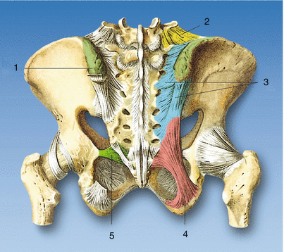
Fig. 48.3
Anatomy. (1) Posterior superior iliac spine, (2) iliolumbar ligament, (3) dorsal sacroiliac ligament, (4) sacrotuberous ligament, (5) sacrospinous ligament (Reproduced with permission from Dr. Danilo Jankovic)
The innervation of the SIJ is complex. The posterior joint is better understood and more relevant for treatment purposes. A recent detailed anatomical study revealed that the SIJ was innervated by the posterior sacral network, which is 100 % contributed by S1–S2, 88 % by S3, 8 % by L5, and 4 % by S4 [6].
Clinical Features
Fortin et al. performed contrast injection and provocative test in volunteers and patients in an attempt to identify an SI pain pattern. He found that patients with SIJ pain present with buttock pain extending into the posterolateral thigh [7, 8]. Compared with other source of back pain (e.g., facetogenic and discogenic), patients with SIJ pain are more likely to report lateral pain rather than central pain [9, 10], pain radiation into the groin [11], unilateral pain, pain arising from sitting, and absence of lumbar pain [12].
Controversy exists whether medical history or physical examination maneuvers were reliable in the diagnosis of SIJ pain (Slipman, Dreyfuss, Laslett) [13–15]. Szadek et al. performed a systematic review and concluded that three positive provocation tests had significant discriminative power (diagnostic odds ratio: 17.16) for diagnosing SIJ pain using the reference standard of two positive blocks [16].
Thus, the presence of three or more positive provocative tests appears to have reasonable sensitivity and specificity in identifying those individuals who will positively respond to diagnostic SIJ injections [4].
Intervention
Interventional procedures to SIJ include injection to SIJ (intra-articular or extra-articular) and radiofrequency lesioning of the lateral branches of sacral nerves. The last one will be out of the scope of this chapter.
In determining the types of articular block (intra-articular or extra-articular), several factors need to be considered: the clinical evidence supporting a putative diagnosis, the evidence supporting the treatment and any anatomical considerations that may affect the decision-making process (e.g., spondyloarthropathy or multiple previously failed interventions) [17]. In elderly, the source of pain is usually related to arthritis (intra-articular pathology), while in young, active people, the SIJ pain is more related to the soft tissue structures (i.e., ligaments and muscles) that comprise the SI articulation (extra-articular pathology). As discussed previously, immunohistological studies suggest that the source of nociception can come from the SIJ capsule, surrounding ligaments and subchondral bone. Depending on the patient, both intra-articular and extra-articular injections may provide benefit.
Literature on periarticular injection with local anesthetic and steroid includes two controlled trials [18, 19], one on patients with seronegative spondyloarthropathy and the other on patients with nonspondyloarthropathic SIJ pain. Both supported the analgesic efficacy of extra-articular injection up to 2 months. For intra-articular injections, there is only one controlled trial supporting analgesic efficacy for spondyloarthropathy and anecdotal evidence for a beneficial effect in nonspondyloarthropathy SIJ pain [4].
Technique
With landmark guidance, the success rate ranged from 12 to 22 % [20, 21]. Fluoroscopic (FL) guidance is commonly used to improve accuracy of this procedure, and more recently, computed tomography (CT), magnetic resonance imaging (MRI), and ultrasound have also been utilized. This chapter will discuss the technique with fluoroscopy and ultrasound guidance.
Fluoroscopy-Guided Technique
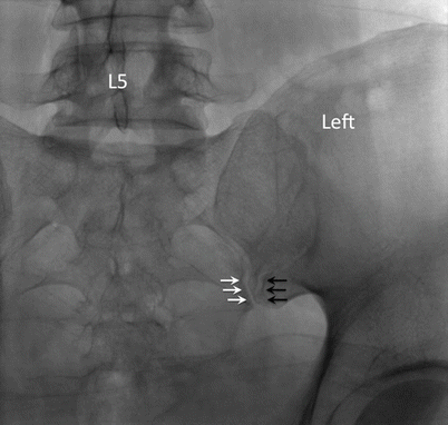
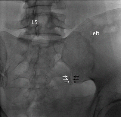
The patient is placed in prone position. The C-arm is placed initially in neutral position. The target point is the inferior, posterior aspect of the joint, approximately 1–2 cm cephalad of the most inferior end [22]. In this view, both anterior and posterior joint lines are seen and the posterior joint line is usually the more medial one (Fig. 48.4). The C-arm is rotated and adjusted until the medial cortical line of the medial silhouette is maximally crisp, which coincides with the beam directed into the posterior opening of the inferior joint space (Fig. 48.5). Usually, this view is obtained with 5–20° of contralateral rotation. A 22- or 25-gauge spinal needle is used and directed toward initially to the sacrum first to appreciate the depth required. Once the bone is contacted, the needle is redirected toward the joint line. The operator should note the depth of the initial contact so that the needle should not penetrate a few more millimeters deeper than this depth (Fig. 48.6). Once the needle has entered the joint space, 0.3–0.5 mL of contrast is injected. In posteroanterior view, the contrast material should be seen traveling rostral along the joint (Fig. 48.7). A lateral X-ray should be used to confirm the needle position (Fig. 48.8). The joint volume in asymptomatic individual is approximately 1.6 mL but is about 1.08 mL in patients requiring SIJ injection. Thus, the injectate is usually 1 mL.


Fig. 48.4
Straight posteroanterior X-ray of the sacroiliac joint. In this view, both anterior (black arrows) and posterior joint (white arrows) lines are seen. The posterior joint line (white arrows) is usually the more medial one (Reproduced with permission of Philip Peng Educational Series)

Fig. 48.5


Contralateral oblique X-ray of the sacroiliac joint. The anterior and posterior joint lines aligned to form a crisp silhouette of the joint (Reproduced with permission of Philip Peng Educational Series)
Stay updated, free articles. Join our Telegram channel

Full access? Get Clinical Tree


