Fig. 49.1
Anatomic specimen. Lateral view of the lumbosacral plexus. (1) Lumbosacral plexus from segments L5 to S3, (2) sacrospinal ligament, (3) sacrum (Reproduced with permission from D. Jankovic)
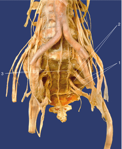
Fig. 49.2
Anatomic specimen. Sacrum, ventral view. (1) Sacral plexus, (2) anterior sacral foramina, (3) sympathetic trunk (Reproduced with permission from D. Jankovic)
The sacral canal contains the five paired sacral nerves. They course caudally and exit through the sacral foramina.
The superior gluteal nerve (L4–S1) supplies the gluteus medius and gluteus minimus muscles.
The inferior gluteal nerve (L5–S2) supplies the gluteus maximus.
The posterior cutaneous nerve of the thigh (S1–S3) is a purely sensory nerve and gives off branches to the lower edge of the buttocks (the inferior clunial nerves) and to the perineal region (perineal branches).
The pudendal nerve (S2–S5) leaves the pelvis through the infrapiriform foramen, courses dorsally round the ischial spine, and passes through the lesser sciatic foramen into the ischiorectal fossa. In the fossa, it courses along the lateral wall in the pudendal canal to below the symphysis and with its terminal branch to the dorsal side of the penis or clitoris. Numerous branches are given off in the pudendal canal (cf. Chap. 56):
The inferior rectal nerves, which may also arise directly from sacral nerves S2–S4, provide the motor supply to the external anal sphincter and sensory supply to the perianal skin and the lower two thirds of the anal canal.
The perineal nerves are involved in the innervation of the external anal sphincter muscles and bulbospongiosus, ischiocavernosus, and superficial transverse perineal muscles.
The muscular branches (S3, S4) supply the levator ani and coccygeal muscles.
The urinary bladder, urethra, and external genitalia are mainly innervated by nerves from S2 to S4.
The external genitalia, bladder, and rectal orifice are border areas between the autonomic smooth muscles and voluntary striated muscles. Autonomic and somatomotor fibers are therefore intertwined in them.
Indications
Surgical
None.
Diagnostic
1.
For differentiation between pain states in the lower extremities (localization of the affected segment in cases of lumbosacral radiculopathy), suprapubic region, and around the perineum.
2.
Bladder dysfunction (bladder sphincter spasm in spinal cord injuries—provided that cystometry has confirmed there is good tone in the bladder muscles).
3.
The S1 nerve root block is commonly performed for diagnostic block in patients with radicular pain of uncertain contribution from lumbosacral nerve root (selective spinal nerve root block).
Therapeutic
1.
Treatment of bladder sphincter spasm (see above).
2.
Sciatic pain with transforaminal S1 nerve root injection.
3.
Neurolytic blockade: to support pain treatment in malignant processes (pelvis, perineum, or suprapubic pain) as an alternative to intrathecal neurolysis (with less severe incontinence effects or motor dysfunction).
4.
Bladder dysfunction. The musculature of the inferior urinary tract has parasympathetic, sympathetic, and somatic innervation. The motor innervation of the detrusor muscle is via parasympathetic efferents, the motor neurons of which are closely adjacent to the (somatic) anterior horn cells of the pudendal nerve and originate from the second to fourth sacral segments. The preganglionic parasympathetic neurons course as the pelvic nerve to the pelvic plexus, from which they reach the bladder wall. Preganglionic efferents arise from segments T11–L2, pass through the sympathetic trunk, and then form the hypogastric nerve. From the latter, branches course to the pelvic nerve and together with it form the pelvic plexus. Postganglionic sympathetic fibers mainly innervate the bladder neck region. The striated external urethral sphincter and the pelvic floor muscles are supplied by the somatic pudendal nerve (S2–S4). Sensory impulses are derived from stretching and pain stimuli in the bladder and are conducted both via the pelvic nerve and also the hypogastric nerve. The pudendal nerve also has afferent functions and conducts signals from the urethra that convey the sensation of urinary flow, as well as proprioceptive impulses from the pelvic floor muscles. The sacral micturition center ensures urination without residual urine (muscular urination control, S2–S4).
5.
Bladder emptying after paraplegia. When the spinal cord is severed above the sacral medulla, reflex emptying is no longer observed after bladder filling in either animals or humans (spinal shock), and the urinary bladder becomes flaccid and atonic for a period of days to weeks. In the chronic state, this phase gradually shifts to the reflex bladder phase, in which minor bladder filling leads to reflex contraction of the bladder detrusor and frequent urination. The reflex arc has a spinal course. The motor neurons to the external urethral sphincter no longer have reflex inhibition, but instead reflex stimulation. This leads to detrusor–sphincter dyssynergia. Paraplegic patients can learn bladder emptying control. If cystometric examinations show good bladder tone in paraplegic patients, a transsacral blockade of the second and third sacral nerves bilaterally can trigger sphincter spasm.
Contraindications
Absolute
1.
When declined by the patient
2.
Coagulation disturbances and anticoagulant treatment
3.
Local infections (skin diseases) at the injection site
Relative
Obesity.
Procedure
Preparations
Check that the emergency equipment is complete and in working order; sterile precautions; and intravenous access, ECG monitoring, pulse oximetry, intubation kit, ventilation facilities, and emergency drugs.
Related posts:


Stay updated, free articles. Join our Telegram channel

Full access? Get Clinical Tree


