Fig. 6.1
Eyelids and lacrimal apparatus. (1) Cornea, (2) conjunctiva, (3) medial angle of the eye, (4) lacrimal caruncle, (5) lacrimal papilla, (6) inferior eyelid, (7) pupil, (8) lateral angle of the eye, (9) superior eyelid (With permission from D. Jankovic)
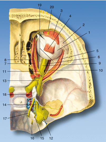
Fig. 6.2
Anatomy of the eye. (1) Eyeball, (2) lacrimal gland, (3) levator palpebrae superioris muscle, (4) superior rectus muscle, (5) lateral rectus muscle, (6) inferior rectus muscle, (7) medial rectus muscle, (8) superior oblique muscle, (9) optic nerve, (10) ciliary ganglion, (11) nasociliary nerve, (12) trigeminal ganglion, (13) frontal nerve, (14) ophthalmic nerve, (15) trochlear nerve, (16) abducent nerve, (17) oculomotor nerve, (18) internal carotid artery, (19) retrobulbar fat, (20) supraorbital nerve (With permission from D. Jankovic)
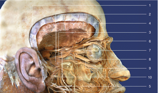
Fig. 6.3
Anatomy. (1) Spinal dura mater, (2) trigeminal ganglion, (3) internal carotid artery, (4) trochlear nerve, (5) oculomotor nerve, (6) supraorbital nerve, (7) superior rectus muscle, (8) ciliary ganglion, (9) inferior rectus muscle, (10) ophthalmic nerve (With permission from D. Jankovic)
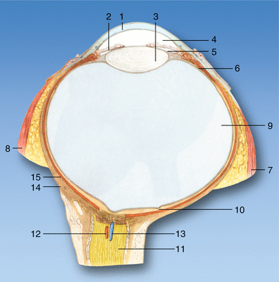
Fig. 6.4
Horizontal section through the eyeball. (1) Cornea, (2) iris, (3) lens, (4), anterior chamber of the eyeball, (5) posterior chamber of the eyeball, (6) ciliary body, (7) lateral rectus muscle, (8) medial rectus muscle, (9) vitreous body, (10) central retinal fovea, (11) optic nerve, (12) central retinal artery, (13) central retinal vein, (14) sclera, (15) choroid (With permission from D. Jankovic)
Underneath this lies the crystalline lens which is located in front of the iris, with its central opening, the pupil. The conjunctiva covers relatively the tough sclera. The optic nerve enters the globe on the hind surface of the globe slightly medial to the axis. The anterior chamber of the eye is bounded by the cornea, the iris, and the lens. The posterior chamber of the eye encircles the lens in a ringlike shape, and the posterior chamber of the eye contains the vitreous body.
The movements of the globe are made possible by four straight muscles, inferior rectus (oculomotor nerve), lateral rectus (abducent nerve), medial rectus (oculomotor nerve), and superior rectus (oculomotor nerve), and two oblique, superior oblique muscle (trochlear nerve) and inferior oblique muscle (oculomotor nerve) (Figs. 6.2, 6.3, and 6.5). The rectus muscles arise from the annulus of Zinn near the apex of the orbit and insert anterior to the equator of the globe thus forming an incomplete cone. Thus, the orbit is divided into two compartments although not completely separate, an extraconal compartment and an intraconal compartment. Injected local anesthetics are easily able to cross the barrier between two compartments by diffusion. Within the annulus and the muscle cone lie the optic nerve (II), the oculomotor nerve (III containing both superior and inferior branches), the abducent nerve (VI nerve), the nasociliary nerve (a branch of V nerve), the ciliary ganglion, and vessels.
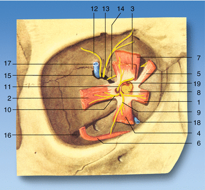

Fig. 6.5
Eye: muscles, nerves, and vessels. (1) Medial rectus muscle, (2) lateral rectus muscle, (3) superior rectus muscle, (4) inferior rectus muscle, (5) superior oblique muscle, (6) inferior oblique muscle, (7) levator palpebrae superioris muscle, (8) optic nerve, (9) oculomotor nerve, (10) abducent nerve, (11) nasociliary nerve, (12) lacrimal nerve, (13) frontal nerve, (14) trochlear nerve, (15) superior orbital fissure, (16) inferior orbital fissure, (17) superior ophthalmic vein, (18) inferior ophthalmic vein, (19) ophthalmic artery (With permission from D. Jankovic)
The sensory supply of the orbit is provided by lacrimal, frontal, and nasociliary branches of the ophthalmic division of the trigeminal nerve. (See Chap. 10 Trigeminal Nerve, pp. 135–7) Autonomic fibers run from the ciliary ganglion situated within the cone near to the orbital apex (Figs. 6.2 and 6.3). The ciliary ganglion, a tiny collection of nerve cells, lies in the posterior part of the orbit between the optic nerve and the lateral rectus muscle. The sensory and sympathetic roots of the ciliary ganglion are provided by the nasociliary nerve and the neural network around the internal carotid artery but do not always connect to the ciliary ganglion. Their fibers can reach the eye directly via the ciliary nerves. The sympathetic fibers which are already postganglionic after they have switched to the cervical sympathetic trunk ganglia can accompany the ophthalmic artery and its branches on the way to their destination. Stimuli from the cornea, iris, choroid, and intraocular muscles are conducted in the sensory fibers.
The eye and orbital contents receive their main arterial supply from the ophthalmic artery (Fig. 6.6). The ophthalmic artery is a branch of the internal carotid artery. In the orbital cavity, the artery runs forward for a short distance lateral to the optic nerve and medial to the lateral rectus muscle, the abducent and oculomotor nerves, and the ciliary ganglion. The artery then turns medially and crosses above the optic nerve, accompanied by the nasociliary nerve. The venous blood of the orbit is drained by the superior and inferior ophthalmic veins which in turn drain into the cavernous sinus. The superior ophthalmic vein crosses the optic nerve with the ophthalmic artery. The veins of the orbit are tortuous and freely anastomose with one another, and they have no valves. These vessels are known to be damaged when a long needle is inserted deep into the apex.
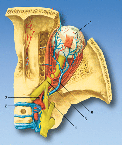

Fig. 6.6
Blood supply in the eye. (1) Eyeball, (2) internal carotid artery, (3) central retinal artery, (4) ophthalmic artery, (5) superior ophthalmic vein, (6) optic nerve (With permission from D. Jankovic)
Tenon capsule (fascial sheath) is a thin membrane that envelops the eyeball and separates it from the orbital fat. The inner surface is smooth and shiny and is separated from the outer surface of the sclera by a potential space the sub-Tenon space. Crossing the space and attaching the fascial sheath to the sclera are numerous delicate bands of connective tissue (Fig. 6.7). Anteriorly, the fascial sheath is firmly attached to the sclera about 3–5 mm posterior to the corneoscleral junction. Posteriorly, the sheath fuses with the meninges around the optic nerve and with the sclera around the exit of the optic nerve. The tendons of all six extrinsic muscles of the eye pierce the sheath as they pass to their insertion on the globe. At the site of perforation, the sheath is reflected back along the tendons of these muscles to form a tubular sleeve. The local anesthetic is injected beneath this part of the sub-Tenon space.
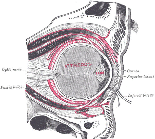

Fig. 6.7
Sub-Tenon space showing multiple connective tissue bands (With kind permission from www.bartleby.com)
Anatomy Relevant to Blocks
The terminology used for regional orbital blocks is controversial. An intraconal (retrobulbar) block involves the injection of a local anesthetic agent into the muscle cone behind the globe formed by four recti muscles and the superior and inferior oblique muscles. An extraconal (peribulbar) block involves the injection of local anesthetic outside the muscle cone. Many studies confirm that there are multiple communications between two compartments, and it is sometimes difficult to differentiate if the injection is intraconal or extraconal. A combination of intraconal and extraconal blocks is described as combined retroperibulbar block.
The average distance between the orbital margin and the ciliary ganglion is approximately 38 mm (range 32–44 mm). If a 38-mm needle is used, it may reach the optic nerve in a large number of patients leading to serious catastrophic events. A needle <31 mm must be used to protect the globe, optic nerve, and other major structures behind the globe during both extraconal and intraconal blocks. In cadaver studies, Karampatiakis et al. found that when a retrobulbar block was carried out with a needle 40 mm long, the needle tip would reach the posterior optic nerve in 100 % of cases; even with needles 35 mm long, the covering of the optic nerve would be touched [2].
Average axial length (anteroposterior distance of the globe) of the eye varies from 22 to 24 mm. Eye with axial lengths >26 mm are more prone to globe damage. The axial length measurement is available during cataract surgery. If a patient is scheduled for non-cataract surgery, spherical power of the spectacles may offer some clue. If a patient has very thick prescription glasses, it indicates high myopia. The risk of damage to the globe and optic nerve is also greater when the globe is rotated during injection. Therefore, it is recommended that the globe should be in the neutral gaze during the injection. It is safer to introduce a needle as far lateral as possible because if it is introduced at the junction of medial 2/3rd and lateral 1/3rd of the inferior orbital rim, the inferior rectus and inferior oblique muscles and their nerves may be damaged.
In sub-Tenon block, local anesthetic agent is injected under the Tenon capsule. This block is also known as parabulbar block, pinpoint anesthesia, and medial episcleral block.
Physiology
The physiological pressure (intraocular pressure) interior of the eye is between 10 and 20 mmHg. It is higher in patients with a large-diameter eyeball and in the recumbent position. It is higher in the morning than in the evening. It increases during coughing, physical exertions, and vomiting. An increase in the plasma concentration of carbon dioxide and decrease in oxygen concentration increase the intraocular pressure. Intraocular pressure increases after the needle technique, and this is due to increase in the volume behind the globe. There is little or no increase in intraocular pressure after sub-Tenon block. The anterior and posterior chambers of the eye contain 250 μl of an aqueous liquid (rate of synthesis ca. 2.5 μl/min).
Indications and Contraindications
Both intraocular procedures (cataract extraction, vitrectomy) and extraocular procedures (strabismus surgery, retinal detachment) can be carried out under local anesthesia in suitable patients. There are only a few absolute contraindications such as patient refusal to accept local anesthesia, allergy to local anesthetic agent, local infection, excessive abnormal body movements, severe breathlessness, psychiatric conditions, and in situations where it is difficult to establish a good communication between patients and health-care workers, but the list is shrinking and local anesthesia is increasingly used even in the above group.
Patient’s Assessment and Preparation
Most ophthalmic surgical procedures are carried out as outpatient basis. There is debate as to the degree of preoperative assessment and investigation required and they vary worldwide. In the UK, the Joint Colleges Working Party Report recommended that routine investigations are unnecessary and the patients are not fasted [3]. Routine investigation of patients undergoing cataract surgery under regional anesthesia is not essential because it neither improves the health nor improves the outcome of surgery, but tests can be done to improve the general health of the patient if required [4].
The preoperative assessment should always include a specific enquiry about bleeding disorders and related drugs. There is an increased risk of hemorrhage, and this requires that a clotting profile is available (and recorded) prior to injection (see later) [5, 6]. Patients receiving anticoagulants are advised to continue their medications. Clotting results should be within the recommended therapeutic range. Currently, there is no recommendation for patients receiving antiplatelet agents. Blood pressure should be controlled in hypertensive patients. Diabetic patients are allowed to take their usual medications, and active control of blood sugar is not necessary. There is no need for antibiotic prophylaxis in patients with valvular heart disease, and surgery is not performed if patient has suffered myocardial infarction during the last 3 months [3]. The presence of a long eye, staphyloma, or enophthalmos, the use of faulty technique, a lack of appreciation of risk factors, an uncooperative patient, and the use of unnecessarily long needles are some of the contributing causes. Knowledge of axial length measurement is essential. A precise axial length measurement is usually available for intraocular lens diopter power calculation before cataract surgery [7]. If the block is performed for other surgery and the axial length measurement is not known, close attention to the diopter power of patients’ spectacles or contact lenses may provide valuable clues to globe dimension (patients usually provide this history).
General Considerations and Preparation Before Block
The patient and the person performing the block must be involved in full discussion of the proposed technique. The anesthetic and surgical procedures are explained to the patients. Patient is placed in a comfortable position, and oxygen is administered via a suitable device. All monitoring and anesthetic equipments in the operating environments should be fully functional. Blood pressure, oxygen saturation, and ECG leads are connected, and baseline recordings are obtained. Insertion of an intravenous line is a prerequisite for needle block, but its use has been questioned during sub-Tenon injections [2]. It is not unusual to observe adverse medical events in elderly patient, and a working intravenous line could only be a good clinical practice.
Sedation and Analgesia During Ophthalmic Blocks
The use of sedation varies in different parts of the world and may or may not be used. Sedation is used during block procedure, surgical procedure, or during block and surgery. However, sedation is common during topical anesthesia. Selected patients, in whom explanation and reassurance have proved inadequate, may benefit from sedation. Short-acting benzodiazepines, opioids, and small doses of intravenous anesthetic induction agents are favored, but the dosage must be minimal. An increased incidence of adverse intraoperative events is anticipated with sedation in elderly [6].
It is recommended that one of the following drugs is given immediately before carrying out regional anesthesia:
Midazolam 1 mg (+ remifentanil 0.33 μg/kg)
Propofol 0.5 mg/kg
Deep sedation should not accompany regional anesthesia; the patient should be capable of cooperating.
Conduction and Infiltration Block
Needle Techniques
Atkinson’s classical retrobulbar block involves insertion of a 38-mm-long needle through the skin at the junction of the medial 2/3rd and lateral 1/3rd of the inferior orbital margin in a rotated eye toward the orbital apex. A facial nerve block (7th nerve block) is essential (see Chap. 7).
The classical retrobulbar block has now been superseded by a higher-volume modern retrobulbar and peribulbar blocks.
Modern Retrobulbar Block
The needle is deliberately directed toward the apex and within the muscle cone (see above) while the globe is in neutral gaze.
Indications
Both intraocular and extraocular surgery may include cataract surgery, vitreoretinal surgery, panretinal photocoagulation, trabeculectomy, optic nerve sheath fenestration, delivery of drugs (steroids, neurolytic agents), and sometimes strabismus surgery in willing patients. Retrobulbar block is also performed for the delivery of neurolytic agents for the treatment of chronic orbital pain.
Stay updated, free articles. Join our Telegram channel

Full access? Get Clinical Tree


