Positioning


Positioning


Robert C. Morell

Richard C. Prielipp


What Baseline Information Is Important?


▪ MECHANISMS OF NERVE INJURY

Mechanisms that may contribute to the development of peripheral neuropathies include excessive pressure (compression), stretch, ischemia, metabolic derangement, toxins, disease states such as hypertension or diabetes, smoking, direct trauma or laceration of a nerve, and other factors that remain unknown (see Tables 57.1, 57.2). Nerve compression may occur from either external or internal mechanisms. In the perioperative setting, placement of noncompliant external objects or improper positioning may create external pressure on a peripheral nerve. For example, allowing the elbow to rest on the steel frame of a surgical table may compress the ulnar nerve because it lies within the rigid, bony canal of the superficial condylar groove at the elbow. A nerve may also be entrapped internally by the patient’s own anatomy. Examples of such internal compression include carpal tunnel syndrome, whereby the median nerve is compressed by the transverse carpal ligament (see Fig. 57.1) or cubital tunnel syndrome where the ulnar nerve may be compressed within the fibrous bands of the cubital tunnel. If pressure, either internal or external, is applied to a peripheral nerve of sufficient magnitude and/or duration, it may ultimately produce nerve ischemia and injury.1,2,3

Most peripheral nerves are intolerant of a stretch beyond 10% of the nerve’s normal length. Combinations of stretch and pressure may also occur, for example, persistent extreme elbow flexion may create two mechanisms leading to possible nerve injury—direct internal compression and internal fixation within the cubital tunnel, which may render the remainder of the nerve more vulnerable to stretch along its course. Figure 57.2 illustrates the cubital tunnel retinaculum that is lax while the forearm is extended, but becomes taut as the elbow is flexed, producing internal compression of the ulnar nerve.4

An additional factor that may contribute to perioperative nerve dysfunction is the phenomenon of the double crush syndrome. Double crush syndrome describes the coexistence of two (or more) clinical or subclinical insults along the course of a nerve. Double crush syndrome was first described in 1973 by Upton and McComas and reflects the phenomenon whereby one compressive lesion occurring along a nerve renders the nerve less tolerant of compression at the same or a second locus.5 Therefore, nerves with a preexisting injury or compression are at much greater risk of a second,
possibly subclinical, insult; together, these may result in a permanent nerve injury.5,6 This phenomenon is schematically illustrated in Figure 57.3. Although the exact mechanism is not definitively understood, it may involve disturbances in axonal flow and/or disruption of the architecture of neurofilaments. Clinically, proximal upper extremity nerve root pathology has been shown to lessen the median nerve compression necessary to produce symptoms of carpal tunnel syndrome and worsen outcome after carpal tunnel decompression.6 In addition, some evidence highlights the increased susceptibility of the ulnar nerve to ischemia, compared to either the radial or median nerves.7,8,9 Finally, certain medical diseases and/or concomitant drug therapy may have physiologic and/or toxic effects on peripheral nerves, rendering them more vulnerable to injury in the perioperative period. Smoking, hypertension, and diabetes may all contribute to microvascular changes that can contribute to the development of peripheral neuropathies and may well predispose peripheral nerves to be more vulnerable to relatively minor insults. Tables 57.1 and 57.2 list many of the diseases and conditions as well as medications and toxins that can predispose patients to neuropathic injury.


TABLE 57.1 Diseases and Conditions Which Predispose to Neuropathies


Acromegaly


Amyloidosis


Carcinoma


Cryoglobulinemia


Diabetes mellitus


Diphtheria


Hereditary predisposition to pressure palsy


Hypoglycemia


Hypothyroidism


Liver disease


Lymphoma


Macroglobulinemia


Malabsorption and vitamin deficiencies


Monoclonal gammopathy


Multiple myeloma


Polycythemia vera


Porphyrias


Uremia


Reprinted from Prielipp RC, Morell RC, Butterworth J. Ulnar nerve injury and perioperative arm positioning. Anesthesiol Clin N Am. 2002;20:589-603, with permission.


TABLE 57.2 Drugs and Chemical Toxins Which Predispose to Neuropathies


Acrylamide


Amiodarone


Arsenic


Aurothioglucose


cis-Platinum


Dapsone


γ-Diketone hexacarbons


Dimethylamino propionitrile


Disulfiram


Hydralazine


Isoniazid


Lead


Metronidazole


Organophosphates


Perhexiline


Phenytoin


Pyridoxin


Thalidomide


Thallium


Vincristine


Reprinted from Prielipp RC, Morell RC, Butterworth J. Ulnar nerve injury and perioperative arm positioning. Anesthesiol Clin N Am. 2002;20:589-603, with permission.


FIGURE 57.1 Ultrasonography of the median nerve as it is compressed by the transverse carpal ligament. Note the flattened cross-sectional profile of the nerve, labeled N between the upper and lower arrows. (Courtesy of Francis O. Walker, MD, Wake Forest University School of Medicine, Winston-Salem, NC.)


ULNAR NERVE ANATOMY AND INJURY

The ulnar nerve is a peripheral nerve that originates from the ventral nerve roots of C8 and T1 (motor fibers) and from the C8 dorsal root ganglion (sensory fibers). These nerve roots contribute to the lower trunk of the brachial plexus (see Fig. 57.4). After dividing into anterior and posterior divisions, the bulk of the fibers of the medial cord continue as the ulnar nerve, which courses along the medial head of the triceps muscle to the posterior aspect of
the medial epicondyle of the elbow (see Fig. 57.5). At this point, anatomic variations can result in nerve entrapment. The cubital tunnel retinaculum, which holds the ulnar nerve in position, comprises the 0.4 mm fibrous roof of the cubital tunnel, extending from the medial epicondyle to the olecranon. Figure 57.6 is a magnetic resonance image taken in the axial plane of the ulnar nerve as it courses through the rigid, superficial condylar groove at the elbow. Variations of the cubital tunnel retinaculum may increase the likelihood of either static or dynamic compression of the ulnar nerve during flexion or extension of the elbow4 (Fig. 57.2). Cubital tunnel syndrome is a collective term for a defined subgroup of ulnar neuropathies arising at the elbow. In addition, other variations at the elbow of ulnar nerve structures also exist, such as accessory epitrochleoanconeus muscles or other dense fibrous bands directly bridging the medial epicondyle to the olecranon, which have been implicated in ulnar neuropathy.


FIGURE 57.2 Illustrates the cubital tunnel retinaculum (CTR) that runs from the medial epicondyle (ME) to the olecranon insertion (OI). A: The CTR is lax while the forearm is extended. B: The CTR becomes taut as the elbow is flexed. (Reprinted from O’Driscoll SW, Horii E, Carmichael SW, et al. The cubital tunnel and ulnar neuropathy. J Bone Joint Surg Br. 1991;73:613-617, with permission.)

Men are three times as likely as women to develop a perioperative ulnar neuropathy. This fact may be explained, in part, by the anatomic differences of the elbow between men and women.3,10,11 Although there are no gross anatomic gender differences of the ulnar nerve itself, women exhibit a strikingly greater (2- to 19-fold) fat content on the medial aspect of the elbow, presumably providing a greater degree of subcutaneous padding for the superficial ulnar nerve along its course beneath the elbow.10 In addition, the tubercle of the coronoid process of the ulna is significantly larger in men and may impede nutrient blood flow to the nerve. Figure 57.7 illustrates the microvasculature of a peripheral nerve. These vessels are delicate, and the nerve is particularly vulnerable to changes or disruptions in its vascular supply. Indeed, there is evidence that the ulnar nerve may be more sensitive to ischemia than either the median or radial nerve, as demonstrated by a greater ischemiainduced decrease in somatosensory evoked potential (SSEP) amplitude.9


FIGURE 57.3 The double crush phenomenon is illustrated by two subclinical insults along the course of a nerve. The left hammer represents a proximal injury and the right hammer represents a distal injury. While either injury alone may be subclinical, two separate injuries along the course of a nerve may result in clinical symptoms.


Are Perioperative Nerve Injuries Always Preventable?

Perioperative ulnar neuropathy may occur despite the use of extensive arm and/or elbow padding during surgery and even with “proper positioning of the arms.” There are no concrete data to support or recommend one type of padding over another (such as gel padding vs. foam padding). In addition, it is possible that a given placement or type of padding may increase direct pressure on a peripheral nerve. An analysis of perioperative nerve injuries detected by the American Society of Anesthesiologists (ASA) Closed Claims Database revealed that ulnar nerve injuries accounted for 34% of 227 perioperative nerve injuries.12 However, the mechanism of injury was clearly determined in only 10 (9%) of the 113 cases of ulnar nerve injuries. Of the 10 cases where a mechanism of injury was determined, 3 were associated with the performance of an axillary block, four were attributed to preoperative trauma, one was because of intraoperative trauma, one was caused by the use of crutches, and one resulted from the surgical procedure. Difficulty in determining specific mechanisms of injury is further amplified by the finding that ulnar nerve injury occurs with nearly equal frequency for both medical and surgical patients hospitalized for >2 days13,14 (see Table 57.3, for overlapping confidence intervals). Men are predisposed to ulnar nerve injury likely because of gender-based anatomic variations of the cubital tunnel. Prolonged periods of bed rest in the supine position—whether during or after surgery or for medical conditions—may contribute to the etiology of perioperative ulnar nerve injury, particularly in men.13,14,15


FIGURE 57.4 The brachial plexus originates at the C4-8 cervical nerve roots, with contributions from T1 as well. As the nerve roots join to become trunks and the trunks divide to become divisions and cords, the brachial plexus passes between the clavicle and the first rib. The median, ulnar, and radial nerves are shown as the plexus transitions to peripheral nerves. (Reprinted from Brown DL. Atlas of regional anesthesia, 2nd ed. Philadelphia: WB Saunders; 1999:15, with permission.)


FIGURE 57.5 This drawing illustrates the ulnar nerve as it runs along the ulnar groove adjacent to the medial epicondyle and against the tubercle of the coronoid process. (Redrawn from Contreras MG, Warner MA, Charboneau WJ, et al. Anatomy of the ulnar nerve at the elbow: Potential relationship of acute ulnar neuropathy to gender differences. Clin Anat. 1998;11:372-278, with permission.)

Most perioperative ulnar nerve injuries are not evident immediately after surgery. In fact, the ASA Closed Claims Database review demonstrated that only 21% of cases were evident in the immediate postoperative period, although 62% became evident between 1 and 28 days, with a median of 3 days.12 Along with the similar incidence of ulnar nerve injury in hospitalized nonsurgical patients, this delayed presentation raises the question of when a “perioperative” nerve injury actually occurs. Therefore, it is often difficult to determine if an injury occurred intraoperatively, in the PACU, or at some time after the patient returned to the ward or to home.


FIGURE 57.6 A magnetic resonance image taken in the axial plane of the ulnar nerve as it courses through the rigid, superficial condylar groove at the elbow. A and V refer to the artery and vein. N indicates the ulnar nerve. (Reprinted from Prielipp RC, Morell RC, Walker FO, et al. Ulnar nerve pressure: Influence of arm position and relationship to somatosensory evoked potentials. Anesthesiology. 1999;91:345-354, with permission.)

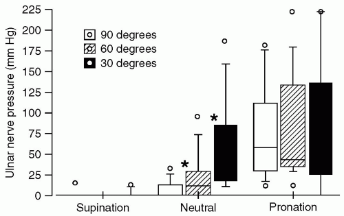
These confounding factors notwithstanding, it is important to minimize direct pressure on the ulnar nerve, particularly pressure exerted by noncompliant or rigid surfaces. When a supine individual has an arm abducted on an armboard, research data indicate that direct pressure against the ulnar groove is minimized by having the forearm supinated, with the palm up. Pronation results in the greatest pressure against the ulnar groove, although the neutral position was intermediate.
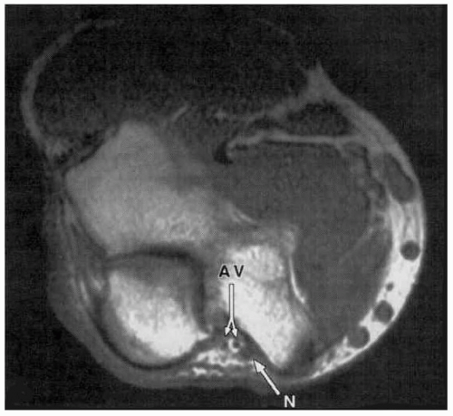
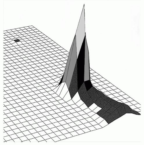
16 It is important to note that these data pertain to pressure only and do not address stretch. In addition, abduction to 90 degrees results in less direct pressure than abduction to 30 or 60 degrees. These data were obtained using a pressure-sensing pad that detected surface pressure distribution beneath the ulnar nerve with 1 cm2 resolution (see Fig. 57.8). Forearm supination minimized direct pressure exerted directly upon the ulnar nerve, because this position decreased contact with the weight-bearing surface (see Table 57.4 and Fig. 57.9). Conversely, pressure localized over the ulnar nerve was greatest with the forearm pronated. Indeed, with the forearm in supination, only 6 of 50 subjects manifest any pressure directly upon the ulnar nerve. With the forearm in neutral orientation, pressure over the ulnar nerve decreased as the arm was abducted from 30 to 90 degrees (see Fig. 57.10).


FIGURE 57.7 This drawing illustrates the microvasculature of a peripheral nerve with delicate vessels leaving the nerve particularly vulnerable to changes or disruptions in this vascular supply. end, endoneurium; p, perineurium; epi, epineurium; rv, regional feeding vessels; exv, extrinsic vessels. (Reprinted from Lundborg G. Nerve injury and repair. Edinburgh: Churchill Livingstone; 1988:43, with permission.)


TABLE 57.3 Incidence of Ulnar Neuropathy in All Hospitalized Patients


Primary
Diagnosis of
Hospitalized
Patients


Prospective
Incidence
of Ulnar
Neuropathy (after 48-72 h)


Percentage


Confidence
Interval


Medical


2/986


0.2


0.02-0.73


Surgical


7/1,502


0.47


0.2-1.0


Summed totals


9/2,488


0.36


n/a


Reprinted from Prielipp RC, Morell RC, Butterworth J. Ulnar nerve injury and perioperative arm positioning. Anesthesiol Clin N Am. 2002;20:589-603, with permission.


In cases where SSEP monitoring is being performed (such as for spinal cord surgery), the ulnar nerve may be monitored as an upper extremity control and may also provide information for positioning purposes. Interpretation of abnormalities generally requires good communication between the monitoring neuroelectrophysiologist, the anesthesia team, and the surgeon. Deepened levels of anesthesia, hypothermia, hypotension, and anemia may cause global increases in SSEP latency or decreases in SSEP amplitude. In cases where abnormalities are detected, assessment and decision making should consider global signals, surgical manipulation, mechanical factors, electric factors, and/or positioning. Figure 57.11 demonstrates the focal changes that occurred in a 62-year-old male patient undergoing an anterior cervical discectomy and fusion, during which the anesthesia level remained constant and unilateral decreases in ulnar SSEP amplitude were seen. Lower extremity SSEP signals were unchanged, making it unlikely that the SSEP changes were due to depth of anesthesia or global monitoring effects. The arm (which was previously tucked and padded with gel foam at the patient’s side) was therefore repositioned. The blood pressure cuff on that arm was also moved to the forearm, below the elbow. Within a few minutes, ulnar nerve SSEP amplitude began to recover. The patient was awakened at the conclusion of the operation and was neurologically intact with no ulnar nerve deficit throughout his postoperative course. While it is neither necessary nor recommended—nor practical—to use sophisticated neuroelectrophysiologic monitoring on all patients undergoing anesthesia and surgery, on those in whom this modality is utilized, unique information may be obtained regarding intraoperative nerve dysfunction, on a case-by-case basis.


TABLE 57.4 Pressure Recorded over the Ulnar Nerve in 50 Volunteers with the Forearm in Three Positions


Arm
Position


Total Arm
Pressure (mm Hg)


Total Arm Contact
Area (cm2)


Ulnar Nerve
Pressure (mm Hg)


Ulnar Nerve
Contact Area (cm2)


Number of
Subjects with
No Pressure on
the Ulnar Nerve


Mean


Median


Mean


Median


Mean


Median


Mean


Median


Supination


1,020


950


36


35


2


0


2.2


1


44


Neutral


1,000


890


42


41


69


22a


5.5


5a


14


Pronation


1,010


970


41


39


95


91a,b


5.8


6a


7


a p = 0.0001 by Mann-Whitney U-test (supine compared to pronated and neutral).

b p = 0.05 by Mann-Whitney U-test (pronated compared to neutral).


Reprinted from Prielipp RC, Morell RC, Walker FO, et al. Ulnar nerve pressure: Influence of arm position and relationship to somatosensory evoked potentials. Anesthesiology. 1999;91:345-354, with permission.


FIGURE 57.8 A three-dimensional graphic representation of the pressure beneath the ulnar nerve and olecranon obtained with the pressure-sensing mat (XSensor Technology Corporation, Calgary, Alberta, Canada). (Reprinted from Prielipp RC, Morell RC, Walker FO, et al. Ulnar nerve pressure: Influence of arm position and relationship to somatosensory evoked potentials. Anesthesiology. 1999;91:345-354, with permission.)


FIGURE 57.9 Box and whiskers plot of peak ulnar nerve pressure as measured by the pressure-sensing mat with the arm in supination, neutral position, and pronation and at 30, 60, and 90 degrees of abduction. (Reprinted from Prielipp RC, Morell RC, Walker FO, et al. Ulnar nerve pressure: Influence of arm position and relationship to somatosensory evoked potentials. Anesthesiology. 1999;91:345-354, with permission.)


FIGURE 57.10 Superimposed image demonstrating the three arm positions tested. (Reprinted from Prielipp RC, Morell RC, Walker FO, et al. Ulnar nerve pressure: Influence of arm position and relationship to somatosensory evoked potentials. Anesthesiology. 1999;91:345-354, with permission.)


FIGURE 57.11 A, B, and C demonstrate the ulnar SSEP changes that occurred in a 62-year-old male patient undergoing an anterior cervical discectomy and fusion, during which the anesthesia level remained constant and unilateral decreases in ulnar SSEP amplitude were seen. A: Shows the baseline cortical and subcortical ulnar SSEP tracing. B: Shows the decrease in amplitude occurring during the procedure. C: Demonstrates the recovery of SSEP amplitude after repositioning of the left arm. SSEP, somatosensory evoked potential. (SSEP tracings courtesy of Mr. Brian Conn, Certificate in Neurophysiologic Intraoperative Monitoring.)


RECENT SCIENTIFIC INVESTIGATIONS AND ANESTHETIC IMPLICATIONS

Relatively recent studies have tested assumptions regarding the etiology of ulnar neuropathy16,17,
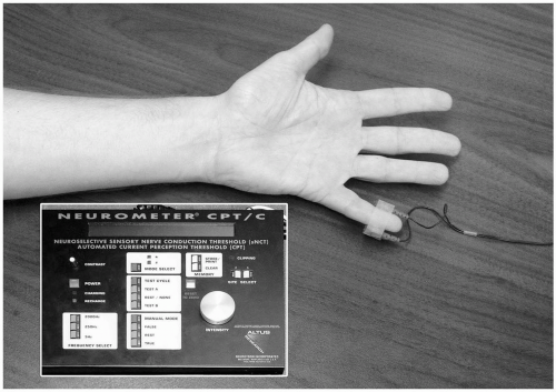
18 using quantitative and physiologic models of ulnar nerve stress. One such study characterized the ulnar nerve response to various experimental stressors (stretch, pressure, and ischemia), which might be encountered in the preoperative setting.18 Alterations in current perception threshold (CPT) were used as a surrogate marker of ulnar nerve dysfunction (see Fig. 57.12). CPT analysis also allowed the differentiation between nerve fibers subtypes. Nerve ischemia produced with an arm tourniquet inhibited all three fiber subtypes. Conversely, a model of ulnar nerve stretch (produced by arm flexion at the elbow to 110 degrees) failed to produce significant CPT increases at any of the three stimulating frequencies. However, direct pressure over the ulnar nerve produced significant CPT increases at 5 Hz and 250 Hz, indicating inhibition of both unmyelinated C fibers and myelinated Aδ fibers. In addition, C fibers demonstrated significant gender differences, with nerve pressure having a 1.7-fold (95% confidence interval, 1.2- to 2.4-fold) greater effect in men (see Table 57.5). This 70% increase in C-pain fiber susceptibility to direct pressure in men could be a partial explanation for the threefold greater frequency of perioperative ulnar neuropathies in men.


FIGURE 57.12 Alterations in ulnar current perception threshold (CPT) were measured using a Neurometer CPT machine as a surrogate marker of ulnar nerve dysfunction. (Reprinted from Morell RC, Prielipp RC, Harwood TN, et al. Men are more susceptible than women to direct pressure on unmyelinated ulnar nerve fibers. Anesth Analg. 2003;97:1183-1188, with permission.)

Lastly, in a comparison of the onset of clinical paresthesia to the onset and severity of SSEP electrophysiologic changes, intentional ulnar nerve compression was induced in 16 male volunteers by placing a wooden dowel snugly in the ulnar groove and allowing the full weight of the arm to rest directly on the wooden block for a maximum of 60 minutes, while recording maximal decreases in SSEP waveforms.16 Eight subjects complained of a progressive hand paresthesia 37 minutes after placement of the wooden block in the ulnar groove, and all eight of these subjects also manifested significant SSEP changes with a mean decrease in the N9-N9N amplitude of −44% (range of −20 to −71%). By contrast, eight volunteers reported no ulnar paresthesia during 60 minutes of a similar pressure from the wooden block in the ulnar groove. Nevertheless, these eight subjects demonstrated a mean SSEP decrease in the N9-N9N waveform amplitude of -44% (range of -19% to -72%) (see Table 57.6). These results suggest that up to one half of male patients who experience pressure on peripheral nerves sufficient to impair electrophysiologic function may be “at risk” because they do not perceive a concurrent paresthesia of that ulnar nerve. Therefore, significant ulnar nerve compression and dysfunction can occur in unsedated men in the absence of perceived symptoms.


TABLE 57.5 Current Perception Threshold (CPT) Data Demonstrating Gender Differences with Experimental Nerve Pressure


Stimulus


Time


Ratio (M/F)a


95% Confidence Interval of Ratio


p-Value


5 Hz


5 min direct pressure


1.7


(1.2-2.4)


0.0027b


10 min direct pressure


1.7


(1.2-2.4)


0.0036b


5-min recovery


1.1


(0.78-1.6)


0.5441


10-min recovery


1.3


(0.91-1.9)


0.1404


250 Hz


5 min direct pressure


1.1


(0.85-1.5)


0.4178


10 min direct pressure


1.1


(0.72-1.5)


0.7730


5-min recovery


0.82


(0.55-1.2)


0.3293


10-min recovery


1.0


(0.68-1.5)


0.9735


2,000 Hz


5 min direct pressure


1.1


(0.93-1.3)


0.2384


10 min direct pressure


1.1


(0.89-1.3)


0.5311


5-min recovery


1.0


(0.83-1.3)


0.9170


10-min recovery


1.0


(0.83-1.3)


0.7312


a Ratio M/F, the ratio of alteration in CPT measurements in men to the alteration of CPT measurements in women.

b Statistically significant gender differences.


Reprinted from Morell RC, Prielipp RC, Harwood TN, et al. Men are more susceptible than women to direct pressure on unmyelinated ulnar nerve fibers. Anesth Analg. 2003;97:1183-1188, with permission.


What Can I Do to Minimize the Likelihood of Nerve Injuries?


▪ PERIPHERAL NERVE

The ASA Practice Advisory for the Prevention of Perioperative Peripheral Neuropathies was published in 200019 and is a systematically developed report that is intended to assist our decision making, because clear scientific evidence is lacking. This advisory was a result of a process that included expert opinions, consensus surveys, open forums, as well as data analysis. The consensus recommendations of the task force are summarized in Table 57.7. Specific recommendations for the upper extremity included ascertaining that the patient can comfortably tolerate the anticipated surgical position, limiting arm abduction (in supine patients) to 90 degrees, attempting to decrease pressure directly on the ulnar groove, using a supinated or neutral position for arms that are abducted on arm boards, avoiding prolonged pressure on the radial nerve as it lies in the spiral groove of the humerus, and avoiding extension of the elbow beyond a comfortable range. It is important to note the advisory recommended that padded arm boards may decrease the risk of upper extremity neuropathy and that properly functioning automatic blood pressure cuffs, when used on the upper arm, do not increase the risk of a perioperative upper extremity neuropathy.


TABLE 57.6 Data from 16 Male Subjects with Somatosensory Evoked Potential (SSEP) Monitoring during Intentional Application of Pressure to Ulnar Nerve


Paresthesia
(Yes or No)


Number of Subjects


Parameters


Time to SSX (min)


% SSEP Change


Yes


8


Mean


37


-44


Median


33


-45


Range


20-59


-20 to -71


No


8


Mean


60


-44


Median


60


-45


Range


-19 to -72


p-value


0.0003


0.92


Subjects are grouped by those who reported (“Yes”) or denied (“No”) paresthesia during direct application of pressure to the ulnar nerve during the investigational protocol. The protocol was then terminated.


SSX, verbal confirmation of symptoms of ulnar nerve paresthesia by the subject; p-value, Mann-Whitney U-test comparing the group who reported ulnar nerve paresthesia (n = 8), to those who denied symptoms of ulnar nerve paresthesia (n = 8).


Reprinted from Prielipp RC, Morell RC, Butterworth J. Ulnar nerve injury and perioperative arm positioning. Anesthesiol Clin N Am. 2002;20:589-603, with permission.

Only gold members can continue reading. Log In or Register to continue

Stay updated, free articles. Join our Telegram channel

Jul 15, 2016 | Posted by in ANESTHESIA | Comments Off on Positioning

Full access? Get Clinical Tree

Get Clinical Tree app for offline access