Fig. 66.1
(a) Tibial nerve. (1) Tibial nerve, (2) medial plantar nerve, (3) lateral plantar nerve, (4) calcaneal branches (With permission from Danilo Jankovic). (b) Tibial nerve – sole of the foot. (1) Medial plantar nerve, (2) lateral plantar nerve, (3) medial calcaneal branches, (4) posterior tibial artery (With permission from Danilo Jankovic)
The tibial nerve in the ankle region is the direct continuation of the tibial nerve distal to the bifurcation of the sciatic nerve in the popliteal fossa. The nerve descends through the calf sandwiched between gastrocnemius/soleus and the deep flexors. It reaches the distal lower leg posterior to the medial malleolus running deep to the flexor retinaculum and is usually posterior to the artery. It gives off medial calcaneal branches to the heel and divides into its two end branches, the medial and lateral plantar nerves, which pass to the sole of the foot and provide it with its sensory supply.
Superficial Peroneal (Fibular) Nerve (Figs. 66.2 and 66.3)
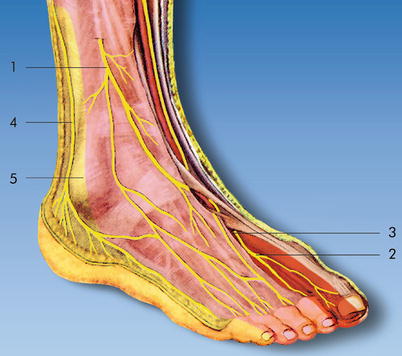
Fig. 66.2
Common, superficial, and deep peroneal (fibular) nerve and sural nerve. Cutaneous innervation areas of the back of the foot (lateral). (1) Superficial peroneal (fibular) nerve, (2) deep peroneal (fibular) nerve, (3) dorsalis pedis artery, (4) sural nerve, (5) lateral malleolus (With permission from Danilo Jankovic)
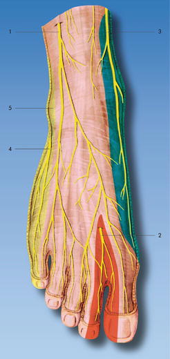
Fig. 66.3
Cutaneous innervation areas in the region of the back of the foot (from the front). (1) Superficial peroneal (fibular) nerve, (2) deep peroneal (fibular) nerve, (3) saphenous nerve, (4) lateral dorsal cutaneous nerve (sural nerve), (5) lateral malleolus (With permission from Danilo Jankovic)
This nerve arises from the common peroneal (fibular) nerve, runs through the peroneus longus muscle, and extends between the peroneus longus and brevis muscles. In the distal half of the leg, the nerve is sandwiched between peroneus brevis and extensor digitorum longus in the intermuscular septum that separates the anterior and lateral compartments of the leg and gradually ascends into a superficial location before eventually piercing the crural fascia. Subcutaneously, or still at the subfascial level, it divides into the thicker medial dorsal cutaneous nerve and the smaller intermediate dorsal cutaneous nerve, providing the sensory supply for the skin on the back of the foot and the toes.
Deep Peroneal (Fibular) Nerve (Figs. 66.2, 66.3, and 66.4)
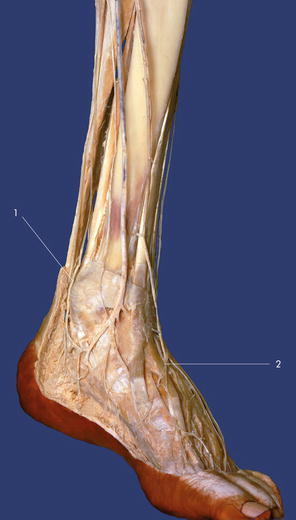
Fig. 66.4
(1) Tibial nerve, (2) deep peroneal nerve (With permission from Danilo Jankovic)
This nerve runs between the tibialis anterior muscle and the extensor hallucis longus muscle in the direction of the ankle, where it divides into a medial and a lateral end branch. The medial end branch continues in the same direction as the nerve trunk and passes with the dorsalis pedis artery to the first interosseous space, crossing under the tendon of the extensor hallucis brevis muscle to the distal end of the interosseous space. Here it joins with a strand of the superficial peroneal nerve and divides into the end branches for the facing sides of the dorsal surfaces of the first and second toes. The lateral end branch turns laterally and supplies the extensor digitorum brevis muscle, sending off three interosseous nerves.
Sural Nerve (Figs. 66.2 and 66.3)
The medial sural cutaneous nerve arises in the proximal part of the popliteal fossa, runs down between the two heads of the gastrocnemius muscle, and joins the peroneal communicating branch (lateral sural nerve) to form the sural nerve. Accompanied by the small saphenous vein, the sural nerve runs behind the lateral malleolus and is contained within the same superficial fascial sheath. It passes as the lateral dorsal cutaneous nerve along the lateral side of the foot, where it gives off a connecting branch to the intermediate dorsal cutaneous nerve and ends as the dorsalis digiti minimi nerve on the lateral edge of the dorsum of the small toe.
Behind the lateral malleolus, it sends off branches (the lateral calcaneal branches) to the skin there and at the heel. The branches for the lateral side of the ankle, for the anterior capsular wall, and for the tarsal sinus originate proximal to the malleolus.
Saphenous Nerve (Fig. 66.3)
Below the knee, it runs along the tibial surface close to the great saphenous vein. Its relationship to the saphenous vein is inconstant, and the nerve may be either posterior or anterior to the vein. A common fascia has been described that envelops both the vein and the nerve in the distal third of the calf. At the level of the lower leg, the saphenous nerve courses along the medial side of and anterior to the medial malleolus and sends off branches to the skin of the medial side of the foot. It usually ends in the metatarsal area, without reaching the big toe.
Sonoanatomy
As all the five ankle nerves are superficial nerves, linear high-frequency ultrasound probe is used.
Tibial Nerve (Fig. 66.5)
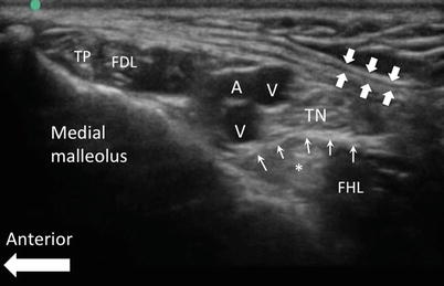
Fig. 66.5
Sonography of the tibial nerve at the level of medial malleolus. The flexor retinaculum is indicated by the block arrows. From anterior to posterior, the structures are tibialis posterior tendon (TP), flexor digitorum longus tendon and muscle (FDL), posterior tibial artery (A) and veins (V), tibial nerve (TN), and flexor hallucis longus (FHL). Please note the tibial nerve is usually seen resting on the fascia overlying FHL (line arrows). The tendon of the FHL (*) is deep to the fascia and should not be mistaken as the tibial nerve (Reproduced with permission from Philip Peng Educational Series)
The patient is in the supine position with hip in external rotation, so that the medial side of the ankle is facing upward. The ultrasound probe is placed in a transverse orientation just proximal to the prominence of the medial malleolus. The main landmark is the posterior tibial artery and is usually accompanied by two venae comitantes. The tibial nerve is a round hyperechoic structure usually posterior to the artery and rest on the fascia of flexor hallucis longus. By extending and flexing the big toe, the tibial nerve can be seen moving up and down, a maneuver to help identifying the tibial nerve.
Related posts:


Stay updated, free articles. Join our Telegram channel

Full access? Get Clinical Tree


