Fig. 71.1
Anatomy of the brachial plexus. A anterior divisions, P posterior divisions
In children, upper limb blocks are performed under general anesthesia or heavy sedation, although in cooperative and older children, it may be possible to perform these blocks awake or under light sedation. Since monitoring paresthesia is precluded by general anesthesia/heavy sedation or the child is too young to verbalize paresthesia, the use of nerve stimulation is essential for localization of nerve and avoiding intraneural injection. In addition, ultrasound allows direct visualization of the needle and neural structures and has further improved the success rate and safety of regional blocks in children.
Several approaches for brachial plexus block have been described, namely, interscalene, supraclavicular, infraclavicular, and axillary. Blockade of a single peripheral nerve is possible distal to the axilla. Different approaches should be used depending on the site of the surgery. For example, interscalene block provides the best coverage for shoulder surgery, while more distal techniques are indicated for surgery of the forearm and hand. General complications of brachial plexus block include infection at the needle insertion site, bleeding/hematoma formation, nerve injury, and local anesthetic toxicity. For more proximal blocks (e.g., interscalene), specific complications, such as phrenic nerve palsy, Horner’s syndrome, recurrent laryngeal nerve palsy, epidural/intrathecal injection, and vertebral artery injection can occur. Pneumothorax is also a potential complication with supraclavicular and infraclavicular blocks; however, the increasing use of ultrasound has lowered the risk of these complications significantly.
Interscalene Block
Introduction, Indications, and Complications
The interscalene block targets the roots and proximal trunks of the brachial plexus between the anterior and middle scalene muscles at the level of the cricoid cartilage (C6) (Fig. 71.2). It is indicated for surgery of the shoulder and upper arm since it reliably anesthetizes the axillary and musculocutaneous nerves, although the hand and fingers are often spared. Phrenic nerve block is a major complication in infants and young children due to greater dependence on diaphragmatic function. Other risks include vertebral artery puncture and injection, epidural or intrathecal injection, Horner’s syndrome, recurrent laryngeal nerve blockade, and hematoma formation in the neck due to arterial or venous puncture. A combined nerve stimulation and ultrasound-guided approach should be used to minimize complications.
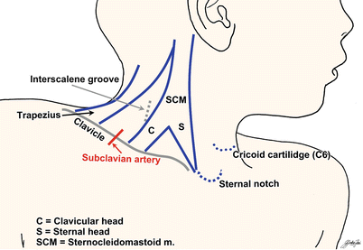
Fig. 71.2
Patient positioning and surface landmarks for interscalene brachial plexus block
Patient Positioning, Preparation, Equipment, and Dosage
The patient is placed with the head turned approximately 45° away from the side to be blocked. For parascalene block, a rolled towel is placed underneath the shoulders. The skin is cleaned with antiseptic solution. If ultrasound guidance is used, prepare the probe surface by applying a sterile adhesive dressing.
A 50-mm, 22-G insulated short-beveled needle is used. Recommended local anesthetics are 0.125–0.25 % bupivacaine or 0.1–0.2 % ropivacaine. Using 0.25 % bupivacaine at 0.2–0.3 mL/kg is a reasonable option for this block. The duration of sensory block averages 15 ± 4.5 h, independent of the type and concentration of the local anesthetic used. Epinephrine 1:200,000 may be added to detect intravascular injection.
Nerve Stimulation Technique
Different approaches have been described for this block. For interscalene approach, the point of needle insertion is at the level of cricoid cartilage (C6) within the interscalene groove (Fig. 71.2). The interscalene groove is located between the anterior and middle scalene muscles posterior to the lateral border of the sternocleidomastoid muscle (SCM), slightly above the point where the sternal and clavicular heads of the SCM separate. The line between the point of needle insertion and the cricoid cartilage should cross the Chassaignac’s tubercle (anterior tubercle of the transverse process of the C6 vertebra), which can be felt easily. The needle is inserted at 60° to skin and directed medially, posteriorly, and caudally to prevent inadvertent puncture of the vertebral artery or epidural/intrathecal space. The initial current is set at 0.8–1.0 mA (2 Hz, 100–300 μs) and then gradually reduced to a threshold current of 0.2–0.4 mA (0.1–0.2 ms) after eliciting appropriate motor responses. Response at a current >0.4 mA indicates that the needle is too far away from the plexus; a current ≤0.2 mA signifies intraneural placement.
Another approach to this block is the parascalene approach [1]. A line is drawn between the midpoint of clavicle and the Chassaignac’s tubercle. The needle is inserted two thirds of the way down this line and advanced posteriorly until twitches are seen. The risk of vascular puncture, Horner’s syndrome, and phrenic nerve block is lower with this technique, but the external jugular vein may be penetrated.
Ultrasound-Guided Technique
A high-frequency (13-6 MHz) hockey stick or linear transducer probe is suitable for this block. The probe is placed on the neck in an axial oblique view at the level of the cricoid cartilage (C6) (Fig. 71.3). The anechoic great vessels (common carotid artery and internal jugular vein) and the overlying triangular-shaped sternocleidomastoid muscle are identified first. If necessary, color Doppler can be used to locate the vessels. Move the probe proximally and distally to identify the roots/trunks of the brachial plexus (which commonly seen as three round or oval-shaped hypoechoic structures) in the interscalene groove between the anterior and middle scalene muscles. Occasionally, the vertebral artery can be seen deep to the plexus and anterior to the C6 transverse process. Extra caution should be exercised not to confuse the artery with a nerve and inject into it. Visualization of the neural structures can be difficult in small children, so a “traceback” approach is recommended. In the traceback approach, the probe is placed in a coronal oblique plane at the upper border of the clavicle. The brachial plexus at this point appears as “a bunch of grapes,” superolateral to the subclavian artery. The plexus is then traced back to the interscalene region by scanning in a cephalad direction (Fig. 71.4).
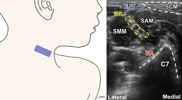
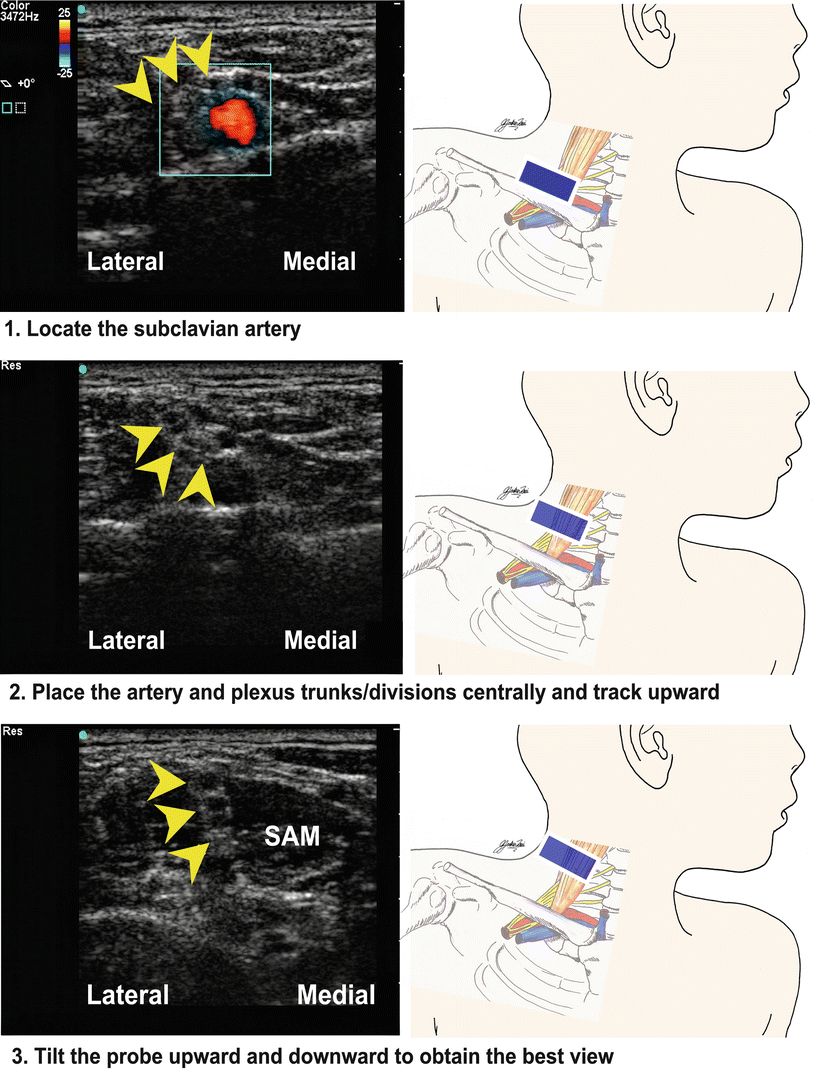
Fig. 71.3
Ultrasound image of the brachial plexus at the interscalene groove. EJV external jugular vein, SCM sternocleidomastoid muscle SAM scalenus anterior muscle SMM scalenus medius muscle, VA vertebral artery, BP brachial plexus, C7 C7 vertebra
Fig. 71.4
Ultrasound traceback approach for interscalene block. SAM scalenus anterior muscle; arrowheads show location of brachial plexus
The needle is inserted either in-plane or out-of-plane, although the in-plane approach is preferred to ensure visualization of the needle, thereby minimizing the risk of complications. For the in-plane approach, the needle is inserted in a lateral-to-medial fashion into the interscalene groove. When using the out-of-plane approach, the plexus is centered in the middle of the screen, and the needle is inserted cranial to the probe at the midline. Direct the needle tip, which appears as bright dot on the screen, in a “walk-down” manner (see Ref. [1]) in proximity to the nerves. Nerve structures can be confirmed by nerve stimulation. A test dose with D5W is useful to visualize the spread and confirm nerve localization. After negative aspiration for blood or CSF, local anesthetic is deposited to achieve good spread surrounding the nerves within the interscalene groove. The depth should be less than 1–2 cm, even in teenage adolescents.
Supraclavicular Block
Introduction, Indications, and Complications
This block targets the trunks and/or divisions of the brachial plexus where they are located cephaloposterior to the subclavian artery above the first rib (Fig. 71.5). The rapid onset of this block offers the most reliable blockade of the brachial plexus for anesthesia and analgesia of the entire upper extremity, especially the elbow, forearm, and hand. Because of the high risk of pneumothorax due to the proximity of the apex of the lung to the brachial plexus, an ultrasound-guided approach is strongly recommended.
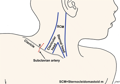
Fig. 71.5
Patient positioning and surface landmarks for supraclavicular brachial plexus block. X indicates the point of needle entry
Patient Positioning, Preparation, Equipment, and Dosage
The head of the patient is turned to the contralateral side. The arm is placed on the side, and the shoulder is pushed backward on to the mattress and down toward the feet (Fig. 71.5). The skin is cleaned with antiseptic solution. If the ultrasound approach is used, prepare the probe surface by applying a sterile adhesive dressing.
A 50-mm, 22-G insulated needle is used. Depth of insertion is related to the age and weight of the patient in a nonlinear manner. For a 10-kg child, the depth of insertion is about 10 mm. For every 10 kg increase in weight, the depth of insertion increases 3 mm until the child reaches 50 kg. After that, advance 1 mm for every 10 kg increase in weight. The maximum depth should not exceed 35 mm. The required depth of penetration is usually less than 1 cm for children and 1–2 cm for teenagers.
Recommended local anesthetics are 0.25–0.5 % bupivacaine, 0.2 % ropivacaine, and 2 % lidocaine. Blockade at this level can be achieved with volumes as low as 0.15–0.2 mL/ kg.
Nerve Stimulation Technique
The needle insertion point is located 1 cm above the midpoint of the clavicle posterolateral to the subclavian artery. The subclavian artery pulsation serves as the landmark for localization of the plexus. The current is initially set at 0.8 mA (2 Hz, 100–300 μs) and then gradually reduced to a threshold current of 0.2–0.4 mA (0.1–0.2 ms) after obtaining appropriate response. Motor response at a current ≤0.2 mA indicates intraneural placement, and the needle should be withdrawn. The spread of local anesthetic solution in children may be greater than for adults since, in children, the fascia adheres less to the nerve trunks. This increases the likelihood of a successful block with any motor response.
Ultrasound-Guided Technique
A high-frequency (13-6 MHz) hockey stick probe is ideal for small children. For older and/or obese children, a small footprint curved array probe (8-5 MHz) is a better option. The probe is first placed in a coronal oblique plane at the lateral end of the upper border of the clavicle. It is then moved medially until the subclavian artery is seen. The subclavian artery is anechoic, hypodense, pulsatile, and round; its identity can be confirmed by color Doppler. The plexus is located superior and lateral to the artery above the first rib and appears as a “bunch of grapes” outlined by a hyperechoic fascia sheath (Fig. 71.6). Below the artery, the first rib appears as a hyperechoic structure with a hypoechoic acoustic shadow, while the lung pleura is accompanied by a hyperechoic shadow due to air artifacts.
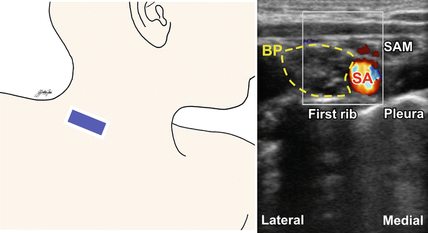
Fig. 71.6
Ultrasound image of the brachial plexus at the supraclavicular level. BP brachial plexus, SAM scalenus anterior muscle, SA subclavian artery
The needle is inserted immediately above the clavicle in a lateral-to-medial direction at a shallow angle. An in-plane approach is strongly recommended to ensure visualization of the needle tip at all times so as to minimize the risks of pneumothorax and vascular puncture. A test dose of D5W is used to visualize the spread and confirm nerve localization. Local anesthetic is first deposited into the “corner pocket” (the corner between the subclavian artery and the first rib). This way, the plexus is often lifted up and away from the pleura so as to reduce the chance of pleural puncture upon subsequent injection. The needle may then be repositioned to achieve good local anesthetic spread around the nerves within the fascia.
Related posts:


Stay updated, free articles. Join our Telegram channel

Full access? Get Clinical Tree


