Fig. 13.1
Acute abdominal pain differential diagnosis for female individual in the emergency setting
Diagnosis of an OB/GYN emergency can take place in a hospital or other urgent care facility. The physician needs to take the patient’s medical history and perform a general and pelvic physical examination. It is crucial that on any suspicious case of either an obstetric or gynecologic emergency a specialist must be consulted. Aside from that, any patient that presents herself with signs of hemorrhagic shock must be treated accordingly with life support measurements before searching for any diagnosis.
On the following sections, authors will focus at the main important OB/GYN chief complaints in the emergency room (ER) which basically belongs to two big groups: (1) vaginal bleeding and (2) lower abdominal or pelvic pain.
13.2 Vaginal Bleeding
Gynecologic bleeding usually presents into two main cohorts: those who have abnormal uterine hemorrhage (AUB) and those who have a cause based on ovulation, which is not necessarily characterized as an emergency situation. Obstetric causes of vaginal bleeding can include trauma during pregnancy, vulvar/vaginal trauma, ectopic and molar pregnancy, abruptio placentae, placenta previa, uterine rupture, and abortion.
13.2.1 Abnormal Uterine Bleeding (AUB)
In general AUB is not a life-threatening cause of bleeding, except for ruptured ectopic pregnancy. It may occur as menorrhagia – bleeding greater than 7 days presenting at regular intervals – or metrorrhagia – uterine bleeding presenting at irregular intervals. As AUB is treated based on the root cause, the emergency physician and acute care surgeon must have in mind an organized mechanism to quickly perform the diagnosis. Figure 13.2 exhibits a flowchart to assist nonspecialist physician in breaking down the diagnosis into its compartment causes. As each cause can be different from the others, a diagnostic direction is proposed.
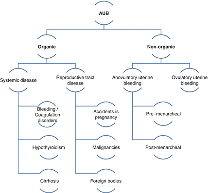
Fig. 13.2
Abnormal uterine bleeding causes
Many patients suffering of AUB end up developing symptomatic anemia due to blood loss. It is relevant to emphasize that normal menstrual cycle has an interval of 28 days (±7 days). When menstrual cycle occurs either before 21 days or after 35 days it is consider abnormal. Nonetheless, the average length of menses is 4 days; thus, menses lasting than 7 days are also considered abnormally long [1–4].
Once finding the cause of patient’s bleeding, laboratory evaluation should proceed including complete blood count, coagulation panel, liver function tests, thyroid-stimulating hormone, and pregnancy test. OB/GYN specialist must be consulted for specific examination of the vagina; if a foreign body is present, such as an intrauterine device, it should be removed and sent for culture. In regard to AUB treatment, acute care surgeon or emergency physician should follow the steps proposed on Fig. 13.2. Adolescent patients who present with AUB at the emergency room are at high risk for having a coagulation defect. Labs must be obtained including von Willebrand factor and evaluation for prothrombin deficiency. Beyond less common causes seen at the emergency room, pregnancy is a common cause of AUB. It can be presenting as a threatened, incomplete, or missed abortion or either on those who have an ectopic pregnancy. Many of these patients can be followed by the specialist with serial beta-hCG levels and transvaginal ultrasound. Nevertheless, the acute care surgeon may face a case of a young lady in hemorrhagic shock with positive history for pregnancy. In such cases, ACS has to be prepared for a definitive treatment through exploratory laparotomy. If there is concern that the patient may also have a gestational trophoblastic neoplastic process, counseling on the risk for hysterectomy is advised [2–5].
Over again, patients presenting with hemodynamic instability, medical supportive care must be taken in place including ventilatory support and blood product transfusion – sometimes requiring massive transfusion protocol. Emergency room health care providers may be aware that in severe AUB cases high-dose estrogen for stabilization of the endometrial lining may be needed as a life-saving measure although patient may only respond to the hormonal therapy within 6–12 h. It is worth to remember that this specific treatment may increase risk for deep vein thrombotic events which could be a great issue in severe cases of AUB coursing with hemorrhagic shock as the doctor may not be able to employ anticoagulant therapy. In this example, compressive leg stockings and intermittent self-compression devices may play a good prophylactic step [3, 4, 6].
When a pregnant patient presents to the ER with vaginal bleeding, the risk of abortion may be real. In Fig. 13.3, we propose a flowchart to facilitate decision making in emergency room.
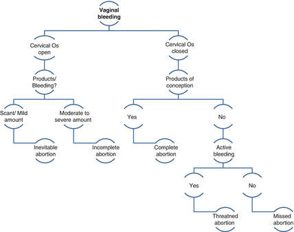
Fig. 13.3
Vaginal bleeding and pregnancy at the ER
In regard to ectopic pregnancy (EP), specifically, it worth to remember that young women with past history of salpingitis or any other inflammatory gynecologic or pelvic disease are on risk for EP. When fallopian tube presents with acute or chronic inflammation, its luminal diameter decreases and the oocyte may have difficulty navigating the tubal length into the intrauterine environ favoring fallopian tube embryo implantation rather than in the uterus. Patients who have ectopic pregnancy are found with abdominal pain, amenorrhea, and/or irregular vaginal bleeding most prevalently as well as ipsilateral adnexal tenderness and eventually adnexal mass. Not all patients present with rebound tenderness or peritoneal signs. Diagnosis of an ectopic pregnancy must be made based upon past/actual history, physical examination, lab exams – including CBC, quantitative beta-human chorionic gonadotropin (β-hCG) on admission and 48 h later (if the diagnosis remain unclear), blood type – and transvaginal ultrasound (TVUS). Association of TVUS and β-hCG has proven to allow earlier diagnosis of ectopic pregnancy and abnormal gestations. After initial assessment, an OB/GYN must be consulted for further investigation and direct treatment, unless ruptured ectopic pregnancy is diagnosed and surgical treatment is immediately required. In this last scenario, laparoscopic approach is possible on a hemodynamically stable patient and has shown to have fewer postoperative pelvic adhesions, less estimated blood loss, decreased hospital length of stay, and improved recovery time. Salpingectomy or salpingostomy may be performed as indicated [7].
13.2.2 Vulvar and Vaginal Trauma
Vulva and vaginal bleeding are usually secondary to local trauma such as fall on a bicycle, sports-related injuries, and automobile crashes [8]. Injuries not involved in blunt trauma can also be due to rupture of a varicosity in the late antepartum period, during labor or postpartum period when venous pelvic congestion is installed. Sex abuse should be strongly considered when pelvic examination includes hymeneal abnormal findings or labial tearing in a pediatric or adolescent patient [9].
If patient is admitted with involvement in recent trauma mechanism, pelvic fractures should be ruled out initially. In the setting of blunt or penetrating injury, especially with associated pelvic fractures, trauma to the bladder, bowel, and peritoneal cavity must also be considered. If gross or microscopic hematuria is found, a CT scan with a voiding cystourethrogram should be obtained before placement of a Foley catheter, as already defined by ATLS®. Figure 13.4 illustrates a case admitted on our institution with severe open book pelvic trauma associated with vulvar trauma. In this specific case, a young lady involved on a motor vehicle crash sustaining a pelvic fracture (Tile B), left femur and ankle fractures presented in hemorrhagic shock and was treated according to the ATLS® principles at the emergency room [10–12].
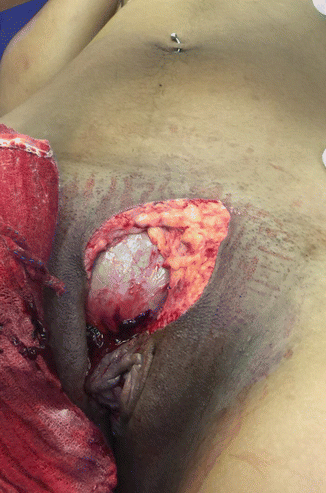
Fig. 13.4
Young lady admitted on our institution after blunt trauma (MVC), sustaining a Tile B pelvic fracture, vulvar trauma, bladder exposition, femur and ankle fractures
Before examination, intravenous, topical, or regional anesthesia may be required. Careful notation of the hematoma size and location should be made. Rectovaginal examination is important to assess extension into the retroperitoneal spaces. In the pediatric patient, the parent’s assistance is recommended to help position the child for the examination. Visualization is best obtained with the child either in a frog-leg position or lying down in a knee-to-chest position. The vaginal vault should be well irrigated with warm saline and any foreign bodies removed.
A CBC may be drawn in the setting of hemodynamic instability or rapidly expanding hematoma. For the vast majority of patients, little benefit is found in obtaining laboratory or radiographic data unless the injury is traumatic and there is concern for ureteral or sigmoid injury.
Management of vulvar and vaginal hematomas is usually conservative unless the hematoma exceeds 10 cm in diameter or if it is rapidly expanding. Most hematomas are venous in origin presenting as consistent dark bleeding; however, a rapidly expanding hematoma could indicate an arterial injury and attempts should be made to identify this vessel for control and ligation. Venous hematomas are typically self-limiting in nature and more difficult to control surgically than arterial hematomas [3, 8].
Of note, vaginal hematomas have the ability to expand into the retroperitoneal pelvic spaces allowing for significant blood loss. Most patients respond to ice packs and compression. In cases wherein the bleeding site cannot be identified, interventional radiology is useful and treatment can be performed through angioembolization. If necessary, a cystoscopy can be performed to evaluate function of the ureters. A Foley catheter should be placed because urinary retention can occur with significant hematoma expansion. Before placing a Foley’s catheter, careful examination is recommended, and in any doubtful case a retrograde cystourethrogram should be obtained prior to inserting the catheter. In the setting of a chronic, expanding hematoma, debridement and placement of a drain is recommended. The patient should be aware that complete resolution may take several weeks. Once healed, there is usually minimal scarring or sequelae.
13.2.3 Trauma in Pregnancy
Trauma is highly prevalent and taken as the third cause of death worldwide. It complicates 6–7 % of all pregnancies and is the leading nonobstetric cause of maternal morbidity and mortality. Trauma also is the leading cause of non-pregnancy-related death in women under 40 years of age in the United States, accounting for 46 % of maternal deaths. Blunt abdominal trauma specifically is associated with 3–38 % of fetal mortality and may be the result of motor vehicle crash (MVC) – leading cause of maternal trauma cases, falls, pedestrian hit by car (PHBC), direct abdominal trauma and assault [13–15]. All these reported trauma mechanisms are related to maternal and fetal death when pregnant women present to the ER on hypovolemic shock due to traumatic bleeding. When high-energy mechanism is involved and blunt trauma and hemorrhagic shock is not the cause of mother and/or fetal death, abruption is the next leading cause of fetal mortality followed by uterine disruption. In regard to penetrating trauma, maternal mortality occurs in less than 5 % of the cases. Visceral trauma is also lower in pregnant woman when compared to nonpregnant population. Gunshot wounds (GSW) to the pregnant woman abdomen result in up to 70 % of fetal injury or death (which includes direct fetal trauma or preterm delivery).
Treatment guidelines for blunt or penetrating injuries are similar to those for nonpregnant patients [5, 14, 16, 17]. Management options include immediate surgical exploration, FAST (Focused Assessment Sonography for Trauma), laparoscopy, CT imaging, local wound exploration, and/or observation. Radiographic studies are helpful in localizing a missile or shrapnel that has not exited. Management often is individualized and should involve a multidisciplinary team, including ERP, trauma surgeons, and obstetricians.
The incidence of burns in pregnancy is relatively low and is difficult to determine. Maternal and fetal mortality are directly related to the type, location, and severity of the burn sustained, in addition to the presence of complications. Fetal outcome also is related to gestational age. The fetal loss rate is approximately 56 % when patients sustain burns over 15–25 % of the total body surface area (TBSA). Fetal mortality is as high as 63 % when 25–50 % of the TBSA is burned and approaches 100 % when burns exceed 50 % of TBSA. Burns are managed aggressively using treatment protocols for nonpregnant victims [15, 17–21].
In regard to falls, it accounts for 3–31 % of injuries. The morbidity associated with falls is modest and typically is associated with a less than 10 % incidence of maternal or fetal complications. The gravid female has an increase in spinal lordosis that allows the shifting of her center of gravity over her legs. This change in the center of gravity contributes to more falls as pregnancy progresses. As such, overly aggressive, high-impact activity should be avoided as pregnancy advances. The degree of injury is related to the distance and high of the fall and the specific body part involved. When patients fall, they fall primarily on their buttocks, side, or onto their abdomen. The nature of injuries includes bruises, cuts, ankle sprains, strains, and fractures. Associated complications include preterm labor, abruption, uterine rupture, low birth weight neonates, and stillbirths [16, 17, 22–25].
The prevalence of assault and domestic violence ranges from 10 to 30 % and is associated with a 5 % risk of fetal death. Abused pregnant women have a threefold higher risk of being victims of attempted and completed homicide than do nonabused controls. Most often the abuser is the patient’s boyfriend or partner. Pregnancy and the postpartum period may escalate the incidence and severity of the abuse, and the uterus and fetus may sustain the brunt of the force. Common sites of abuse include the face, head, breasts, and abdomen. Patient under suspicious involvement on an assault or domestic violence event must be clinically evaluated on privacy and interrogated for any kind of abuse. Police intervention is necessary in positive cases [26–30].
13.2.4 Anatomical and Physiological Considerations of Pregnant Women in the Trauma Scenario
When managing the pregnant victim of trauma, appreciation of the anatomic and physiologic changes associated with pregnancy is critical. Before 13 weeks’ gestation the uterus has not yet become an abdominal organ and is protected by the bony pelvis. Fetal loss in the first trimester is less likely the result of direct trauma (occurring less than 1 % of the time) but instead is likely to be caused by uterine hypoperfusion resulting from maternal hypotension or death. As the uterus enlarges, it displaces the bowel to cephalad position, thereby protecting these structures, but rendering the fetus more vulnerable to injury. Thinning of the uterine wall with fetal growth and the relative decrease in amniotic fluid volume also contribute to fetal vulnerability. In regard to amniotic fluid, specifically, attention to amniotic fluid embolism (AFE) is advised in trauma case scenarios. AFE is a rare but potentially fatal complication of pregnancy [14, 17].
Back to anatomic considerations, the bladder is displaced cephalad by the enlarging uterus, making it susceptible to injury; therefore, hematuria cases after traumatic injuries must be evaluated aggressively. Splenic injuries occur most commonly in the third trimester and may occur after even apparently mild trauma. Engorgement of the spleen renders it vulnerable to injury and excessive blood loss. Injuries to the liver or spleen may result in abdominal pain, shoulder pain, and elevated transaminases [13, 14]. It is important to highlight that nowadays most solid organs trauma, are eligible for non-operative management (NOM) [31–33] as well some inflammatory urgencies including in pregnant population with safety [34].
Pelvic fractures commonly are associated with blunt trauma and are the most common trauma resulting in direct fetal injury manifested by skull fractures and brain injury, particularly when the head is engaged into the bony pelvis. Fetal mortality can approach 25 % in these cases [17, 35]. Attention is advised to pelvic trauma-associated injuries to the bladder, urethra, and rectosigmoid colon [36, 37]. Pelvic radiographs required as part of the ATLS® protocol for assessing trauma in the ER must be interpreted with caution, because there is a normal widening of the sacroiliac joints and symphysis pubis with pregnancy.
Physiologic changes begin as early as the first trimester and can alter or mimic maternal response to trauma, thereby confounding ERP or trauma surgeon evaluation. The pregnant woman is physiologically prepared for the blood loss associated with delivery. Blood volume increases by approximately 50 %, and there is a 30 % increase in erythrocyte volume. With that said, a pregnant trauma patient can bleed up to 2000 mL of blood (or 30–40 % of her blood volume) before manifesting shifts in heart rate or blood pressure.
Cardiac output increases up to 50 % beginning in the first trimester and peaks somewhere between 20 and 30 weeks’ gestation. Cardiac output remains at third trimester values for the first 48 h postpartum and then decreases gradually to nonpregnant values over the following 2 weeks. Uterine blood flow comprises about 20 % of cardiac output, increases up to 600 mL/min during pregnancy, and can serve as a significant source of hemorrhage in the face of trauma. Uterine blood flow has no autoregulation and, therefore, is dependent on maternal mean arterial blood pressure. As such, changes in blood pressure can impact uterine blood flow negatively, thereby compromising fetal perfusion and oxygenation. By 20 weeks’ gestation the enlarging uterus is capable of aortocaval compression, which can compromise venous return to the heart and reduce cardiac output. When the pregnant woman is supine, aortocaval compression reduces cardiac output by approximately 30 % and can result in maternal pallor, sweating, nausea, vomiting, hypotension, tachycardia, and mental status changes. Blood pressure is lower in pregnancy secondary to decreased systemic vascular resistance. Lower vascular resistance is the result primarily of the vasodilatory effects of progesterone and the low resistance of the placental bed. Also, the increase of plasma volume over red blood cell volume results in hemodilution and, therefore, a physiologic anemia. Systolic and diastolic blood pressures decrease by 5–15 mmHg, reaching a nadir at 28 weeks’ gestation and gradually returning to normal at term. Central venous pressure drops slowly from 9 mmHg to about 4 mmHg by term. Estrogen-mediated increases in myocardial alpha receptors result in heart rate increases of approximately 15 beats per minute [14]. ECG changes during pregnancy are common. Normal findings include sinus tachycardia, ectopic beats, left axis deviation, inverted or flattened T waves, and a Q wave in lead III. The increased metabolic demands of pregnancy increase oxygen consumption by 15–20 %. Minute ventilation increases by 50 %, largely as a result of increased tidal volume, because respiratory rate changes minimally. The diaphragm moves approximately 4 cm upward in pregnancy, and the thoracic anteroposterior diameter increases. Intra-abdominal pressure as well intra-thoracic pressure increases. These anatomic alterations, in addition to the enlarging uterus, cause decreased expiratory reserve and residual lung volumes, thereby decreasing functional residual capacity. The 20 % decrease in functional residual capacity coupled with increased oxygen consumption of pregnancy results in rapid maternal desaturation in the face of depressed respiration or apnea. The regular pregnant mother compensates in a state of mild respiratory alkalosis. Elevated progesterone levels act on the medullary respiratory center stimulating ventilatory drive causing a decrease in PaCO2 values to levels approaching 25–30 mmHg.
There is a compensatory renal excretion of bicarbonate resulting in serum levels of 17–22 mEq/L, thereby maintaining an arterial pH of 7.40–7.45. The increased minute ventilation results in PaO2 levels that is higher than nonpregnant values, ranging between 104 mmHg and 108 mmHg. Maternal oxygen saturation should be maintained at 95 % to maintain a PaO2 greater than 70 mmHg, optimizing oxygen diffusion across the placenta. Fetal oxygenation is maintained when maternal PaO2 remains above 60–70 mmHg, and it is compromised immediately at lower levels. The smooth muscle relaxation effects of progesterone contribute to decreased gastric tone and motility as well as reduced lower esophageal sphincter tone. These changes, in addition to the cephalad displacement of the stomach, result in an increased risk of aspiration when the trauma victim is unable to protect her airway. Normal chest radiographic findings in a pregnant patient include mild cardiomegaly, a widened mediastinum, an increased anteroposterior diameter, and prominence of the pulmonary vasculature [14, 16].
Related posts:


Stay updated, free articles. Join our Telegram channel

Full access? Get Clinical Tree


