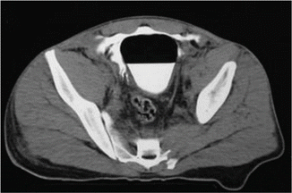
Fig. 14.1
Intraperitoneal bladder injury: (a) tomography and (b) VCUG

Fig. 14.2
Extraperitoneal bladder rupture
Cystoscopy is a minimally invasive procedure that can help identify the location and size of the bladder rupture as well as provide additional information regarding the lesion and the position of the ureters and bladder neck. Also, cystoscopy can be used to diagnose foreign bodies inside the bladder [11].
14.1.3 Management
14.1.3.1 Nonoperative
The nonoperative management of extraperitoneal bladder rupture is the standard treatment for patients that present with uncomplicated extraperitoneal bladder injuries [3, 7, 8]. Almost all blunt extraperitoneal bladder ruptures can be treated nonoperatively. Nonoperative management consists of continuous bladder drainage with a catheter, antibiotic prophylaxis, and clinical observation [3].
Orthopedic surgeons often choose to stabilize pelvic fractures with open reduction and internal fixation. Extraperitoneal bladder injuries should be repaired while pelvic fractures are surgically treated to reduce the risk of infection [3, 4]. If laparotomy is performed to treat another abdominal injury in patients with extraperitoneal bladder rupture, latter complications can be avoided if the bladder injury is sutured during the same operation [7].
14.1.3.2 Operative Treatment of Bladder Ruptures
Bladder injuries are operatively managed in patients with intraperitoneal bladder ruptures, penetrating injuries, or for patients with extraperitoneal bladder ruptures with concomitant bladder neck involvement, bones fragments inside the bladder wall, concomitant rectal injury, or entrapment of the bladder wall [3, 9].
Bladder penetrating injuries should be treated with exploration, debridement of devitalized tissue, and primary wall suture [12]. Penetrating injuries caused by gunshot wounds require special attention. If a gunshot wound to the bladder is identified, the surgeon must assess the presence of enter and exit wounds. A midline cystotomy should be performed to inspect the bladder, treat the lesions, and evaluate the ureters bilaterally. A careful inspection for foreign bodies, fecal material, and bone fragments needs to be done before closing the bladder [9, 12].
Overall, approximately 40 % of penetrating bladder injuries is associated with rectal injuries. The diagnosis of concomitant rectal injury is challenging and only 25 % of rectal injuries can be identified during ordinary physical examination. If a rectal injury is suspected, proctoscopy or even proctosigmoidoscopy should be performed [13]. When the colon or the rectum adjacent to the bladder lesion is damaged, a fecal diversion may be necessary in up to 67 % of patients [13].
Laparoscopic management of blunt intraperitoneal ruptures can be safely performed in well-selected patients with no other intra-abdominal injuries or intracranial pressure issues. A single layer of suture can be performed, offering the patients the benefits of minimally invasive procedures such as faster recovery and better cosmetic results [14].
Urinary catheter insertion is necessary to prevent elevated intravesical pressure by reducing the leakages and allowing bladder healing [15]. Bladder injuries treated conservatively should be followed-up with a cystography, usually within 7–14 days. If no contrast extravasation is identified, the catheter can be safely removed [15]. If a leakage is found during the control cystography, radiological reevaluation can be performed 7 days later or, depending on the extent of the extravasation, size of the laceration, and presence of complications, surgeons can perform a surgical repair.
Simple injuries that required surgical repair do not need to undergo cystography before catheter removal. The catheter can be safely removed after 7–10 days [7, 15]. On the other hand, cystography is highly recommended before removing the catheter in cases of complex bladder injuries, lesions at the bladder neck, concomitant ureteral reimplantation, or high risk factors for wound healing (steroids, previous radiation, malnutrition, infection) [15].
14.2 Difficult Urinary Catheterization
Indwelling urethral catheters are often used for bladder drainage in the hospital setting. The indications for urethral catheter placement are broad and include patients with acute urinary retention, unstable patients in the emergency department or intensive care unit, patients undergoing major surgeries, and patients who present with hematuria. Placement of urethral catheters is usually performed by health care partners but adequate training is required [16].
Some patients may present with difficult urinary catheterization (DUC). In men, the most frequent causes of DUC are urethral strictures, benign prostatic hyperplasia, phimosis, meatal stenosis, penile edema, and bladder neck sclerosis following urologic procedures. Women can present with difficult urinary catheterization too, mostly caused by morbid obesity, atrophic vaginitis, intravaginal retraction of the urethral meatus, and previous pelvic procedures [17].
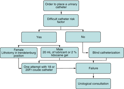
Several potential complications of difficult urinary catheterization (DUC) can cause acute and chronic complications including rectal perforation, meatal and urethral erosions, infection, false passages, and chronic urethral strictures. In order to avoid complications, the absolute need for placement of an indwelling urethral catheter must be assessed because unnecessary catheterization rates can be as high as 50 % [16]. Proper sterile technique with lubrication of the urethra using lidocaine gel is a key step to achieve successful catheterization and decrease the chances of urinary tract infection. Contrary to common perception, larger diameter catheters (18–22 French) are preferable to smaller catheters due to the firmness and better success rates. Finally, when facing a difficult catheterization, application of excessive pressure or force is not recommended as it may cause or increase the magnitude of iatrogenic injury to the urethra. It is crucial that urethral trauma be considered a contraindication for urethral catheterization by the novice. Urology consultation is mandatory in such cases [18] (Fig. 14.3).


Fig. 14.3
Urology consultation flow chart
14.2.1 Difficult Female Catheterization
Morbidly obese females may require lithotomy with slight Trendelenburg position for better exposure. If the urethra is not yet visible, a vaginal speculum can be used to press the posterior vaginal wall downwards while an assistant retracts the labia majora to expose the meatus. Similarly, in older postmenopausal women, the meatus may be retracted posteriorly toward the vagina. In these cases, insertion of the first and second fingers into the vaginal introitus may guide the insertion of the Foley catheter by locating the urethral meatus above the introitus and about 2–3 cm below the clitoris. Female meatus stricture can be secondary to previous instrumentation, injury, and female circumcision. In these complex cases, urethral dilation with endoscopic approach is often necessary and is usually performed by the urology team [18].
14.2.2 Difficult Male Catheterization
Male patients admitted for heart failure, sepsis, hepatic failure, and trauma can present foreskin edema, making it difficult to visualize the urethral meatus. In this case, hand compression of the distal half of the penis with gauze or application of loose fitting elastic dressings for 10–20 min can reduce the swelling and a Foley catheter can be placed using standard sterile technique. Phimosis can also make it difficult or impossible to visualize the meatus. Pulling the foreskin outward can open a passage that allows visualization. In severe cases of phimosis, blind or endocopic approach may be necessary to introduce the catheter by aiming toward the 6 o’clock. Rarely, a dorsal slit is indicated [18].
To avoid complications caused by inflating the balloon inside the urethra or the prostate, the Foley retention balloon should only be inflated after the Foley catheter is introduced into the bladder up to the Y-hub bifurcation of the catheter and drainage of urine is visualized. Bedside bladder ultrasound is recommended when in doubt of catheter’s placement [18].
14.2.3 Catheter Selection
Multiple urethral catheters are commercially available. The most used in common practice are latex or silicone catheters, three-way Foley for continuous irrigation of the bladder, and Coudé (curved tip) catheter. Catheter size is measured using the French scale, where 1 French equals 1/3 mm in diameter [17].
If no complications are known or anticipated, 16 or 18 French catheters are generally used for catheterization. Coudé catheters are used when patients are known to have a large prostate or history of difficult catheterization with clearance at 12 o’clock (high bladder neck). The tip should be directed anteriorly and upward in order to facilitate bypassing obstruction or false passages. Council tip catheters are designed to be placed over a stiff guidewire, preplaced under cystoscopic guidance by the urologist [18, 19].
14.2.4 Flexible Cystoscopy and Dilatation
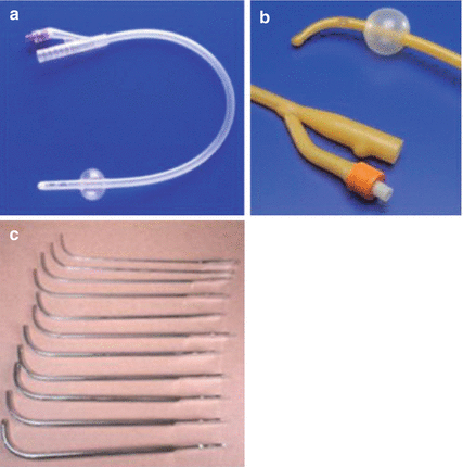
Flexible cystoscopy under local anesthesia can assist the placement of Council tip catheters or suprapubic catheters under direct visualization. During the cystoscopy, a stiff guidewire is placed through the stenosis or false passage. The scope is then removed and a Council tip catheter is placed using the wire as a guide. If needed, “S” curved urethral dilators can be used to dilate the urethral stricture before urethral catheter placement [17] (Fig. 14.4).


Fig. 14.4
(a) Silicon catheter, (b) Coude catheter, (c) S-curved dilators
Urethral dilation has traditionally been done blind with metal Van Buren sounds. The use of such sounds is encouraged only for very distal strictures or meatal stenosis. Similarly, filiforms and followers can also be used but false passages may occur. Currently, the use of sequential urethral dilators over the guidewire and under fluoroscopic guidance is often practiced [17].
14.2.5 Suprapubic Catheter
A suprapubic catheter (SP) can be inserted percutaneously in cases of catheterization failure or when total urethral disruption is likely. SP tubes are placed using local anesthesia with or without the use of ultrasound placed 2 inches above the pubic bone in the midline [20].
Contraindications for percutaneous suprapubic tube placement include bladder cancer, unavoidable bowel injury due to previous surgery, uncorrected coagulopathies, and active anticoagulation, and abdominal wall abscess or cellulitis [18].
Stay updated, free articles. Join our Telegram channel

Full access? Get Clinical Tree


