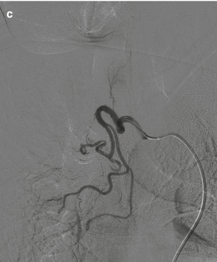
Fig. 20.1
Massive hemoptysis (a) CT Scan shows a bronchial artery of 2.3 mm in diameter (arrow). (b) Angiogram of the pathologic bronchial arteries in a chronic inflammatory disease. (c) Angiogram after embolization with particles
Before of BAE, the bronchial artery number and origin must be carefully evaluated.
Always, other vessels that can be involved in hemoptysis must be investigated: intercostals, subclavian, phrenic, and mammary arteries.
The decision of which arteries need embolization is carried out on the base of CT, bronchoscopic and angiographic findings.
The most used embolic material in BAE is polyvinyl alcohol particles, usually 350–500 μm in diameter, as they are nonreadsorbable (Fig. 20.1c). Smaller particles can lead to pulmonary infarction as bronchopulmonary anastomosis of 325 μm are demonstrated in human lung [7].
Gelatin sponge may lead to recanalization and therefore is less used than particles.
Coils are indicated in mammary artery and in bronchial artery aneurysm.
Isobutyl-2 cyanoacrylate could be used in neovascularization and AV malformation.
BAE is very effective in acute massive hemoptysis. Long-term recurrence rate, however, has been reported to be from 10 to 52 % [8]. Recurrence of bleeding is more frequent in tuberculosis, aspergilloma, and neoplasms.
Complications of BAE are usually transient: more frequent complications are chest pain and dysphagia. The prevalence of spinal cord ischemia is reported to be 1.4–6.5 % and embolization, when Adamkiewicz is visualized at angiogram of target vessel, must be avoided.
20.3 Anticoagulation-Related Soft Tissue Spontaneous Hemorrhages
In the last decade, the widespread use of anticoagulation therapy in elder patients has led to an increase in soft tissue spontaneous hemorrhage [9, 10].
While conservative management (anticoagulation withdrawal or reversal, volume expansion, transfusion of fresh frozen plasma and packed RBCs, and vasoactive drugs) is recommended in hemodynamically stable patients [11], the treatment of life-threatening hemorrhage is endovascular embolization [12].
Embolization preserves the tamponade effects, if in a muscular compartment, of the already developed hematoma, is repeatable and allows selective closure of target vessel.
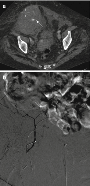
A CT is normally performed before embolization: usually, if there is an active bleeding, the vessel where hemorrhage originates can be identified (Fig. 20.2a).


Fig. 20.2
Spontaneous rectus sheet bleeding: (a) CT scan shows large hematoma and contrast media blush (arrow); (b) selective angiogram of the inferior epigastric artery in the same patient
The embolization materials in these cases are mainly gelatin sponge, as adsorbable material allows the recovery of vascularization after the coagulopathy is solved.
Precautionary embolization with readsorbable materials of arteries feeding tha anatomical region of the hematoma should be performed also if absence of active bleeding at angiography: bleeding is often intermittent and to restrict embolization to angiographic evident blush leads to more frequent reintervention needs.
20.4 Aorta
Acute rupture of the thoracic and abdominal aorta (AAR) is a life-threatening condition that had an extremely high mortality rate before the introduction of thoracic and abdominal endovascular aortic repair (TEVAR/EVAR). Emergency surgical repair of the aorta consisted of thoracotomy or laparotomy, replacement of the diseased aorta, and interposition of a prosthetic graft.
Stay updated, free articles. Join our Telegram channel

Full access? Get Clinical Tree


