Neuraxial Anesthesia


Neuraxial Anesthesia


Sorin J. Brull

Roy A. Greengrass


How Do Complications of a High Spinal Manifest Clinically?


▪ OVERVIEW

With very few exceptions, the effects of spinal anesthesia on the cardiovascular system are almost entirely because of block of the preganglionic sympathetic fibers (white rami communicans or type B fibers) by the local anesthetic injected in the subarachnoid space. The local anesthetic used for spinal anesthesia does not have any systemic effects after absorption by the vascular system.1 Although absorption of local anesthetics from the cerebrospinal fluid (CSF) into the circulation does occur, the plasma concentration is too low to produce significant hemodynamic effects in most patients.2

Physiologic trespass is directly related to the intrathecal level of sympathetic denervation. The degree to which the spinal anesthetic alters the normal hemodynamic status, however, varies considerably. Differences may be due to many factors, including the general state of health, age, intravascular fluid status, and concurrent medications. In general, more extensive sympathetic block produces more profound hemodynamic changes. The effects of sympathetic denervation are extensive, both on the arterial (afterload) and venous (preload, capacitance) vessels.


Arterial Circulation (Afterload)

Afterload is the measure of resistance against which the left ventricle must eject blood. It may be measured as the stress (or tension) that is developed in the ventricular wall during systole. Neuraxial anesthesia decreases afterload by producing arterial vasodilation.3 This vasodilatation, however, is not equivalent in all vascular beds. For instance, muscle and skin blood flow may be decreased by sympathectomy, whereas the total blood flow to the same extremity may be more than quadrupled.4 Additionally, the extent to which afterload is decreased by sympathetic denervation varies considerably from one patient to another. Therefore, patients with equivalent sympathetic denervation do not necessarily demonstrate equal changes in afterload.5 Young and robust patients are able to maintain peripheral resistance better than elderly or cachectic patients. The extent to which vascular tone is maintained in various organs is also variable. It is retained most effectively in renal and splanchnic vasculature, less so in skeletal muscle, and least effectively in skin vessels.

When arterial vasodilation results from sympathetic denervation, a simultaneous, compensatory reflex
vasoconstriction occurs in areas of the body in which the sympathetic nervous system is intact, usually in areas cephalad to the site of spinal or epidural anesthetic block. The effectiveness of this reflex vasoconstriction in maintaining normotension is a function of the extent of the sympathetic block. If, for instance, sympathetic denervation reaches the fourth thoracic dermatome (T4) or higher, the intact upper limb vasculature may contribute only 5% of the total cardiac output (CO). Even maximal vasoconstriction will be insufficient to compensate for the profound arterial vasodilation in the rest of the body. Reflex cerebral vasoconstriction does not occur, because of the intrinsic cerebral autoregulatory mechanisms.


Venous Circulation (Preload)

Preload is a measure of the volume of venous blood returned to the right ventricle from the periphery. Sympathetic denervation associated with central neuraxial block dilates not only the arterial and postarteriolar circulation (afterload), but also the venous circulation. Neural innervation of the venous circulation is similar to that of the arterial circulation, and areas of vasodilation will be equivalent. However, the degree of vasodilation in the arterial and venous sides of the circulation is markedly different. Because the arterial walls contain more smooth muscle fibers and supporting structures (media) than their venous counterparts, sympathetic denervation results in less vasodilation in afterload vessels, because they maintain their own intrinsic vascular tone. In contrast, the preload (venous) vasculature undergoes maximal dilatation, and venous capacitance increases maximally. This change results in the rapid pooling of an abnormally large volume of blood in the periphery and a marked decrease in the blood volume returning to the right ventricle, resulting in a significant decrease in CO and a decrease in BP.


Cardiac Function


Heart Rate

Slowing of the HR is characteristically associated with neuraxial anesthesia, and the extent of bradycardia correlates well with the extent of sympathetic denervation. However, the relation between denervation and the degree of bradycardia may be modified by a number of other factors including age, coadministration of intravenous drugs, and the position of the patient on the operating room table. Importantly, bradycardia during high (thoracic) levels of spinal or epidural anesthesia is due to two main factors: (i) denervation of preganglionic cardiac accelerator fibers (T1-4) and (ii) diminished venous return to the right ventricle because of decreases in preload. In extreme cases, the rapid decrease in venous return to the right ventricle (due to massive hemorrhage or assumption of head-up positioning during epidural or spinal anesthetic-induced near-total sympathectomy) may result in activation of mechanoreceptors and chemoreceptors in the ventricle. This activation results in severe bradycardia or asystole due to further increases in parasympathetic activity and inhibition of sympathetic activity (Bezold-Jarisch reflexinduced asystole).


Cardiac Output

Reduced CO following spinal and epidural anesthesia is one of the most consistent findings reported in the literature and is the sine qua non of neuraxial anesthesia. The extent of CO decrease also is a function of the degree of sympathetic denervation. Because one of the many determinants of CO is the amount of blood in the ventricle (preload), and preload is exquisitely sensitive to the effects of gravity, marked changes in CO may be induced by patient positioning. Placing patients undergoing neuraxial anesthesia in the horizontal position while elevating the legs will facilitate venous return to the heart and tends to maintain CO and BP. Conversely, assumption of an even slight head-up position during neuraxial anesthesia with high levels of sympathetic denervation (in the misguided attempt to prevent further extension of the spinal or epidural block) may have catastrophic consequences such as profound bradycardia, cerebral hypoperfusion, and cardiac arrest.6,7 Reports of severe complications related to improper positioning of patients (i.e., head-up) during high levels of spinal or epidural anesthesia have spanned the last six decades.8,9


Myocardial Work

Because high levels of sympathetic denervation are associated with decreased afterload (and preload), the amount of work performed by the heart per unit time is decreased. Over half a century ago, Eckenhoff demonstrated, in dogs with a spinal-induced total sympathectomy, a 66% decrease in left ventricular work.10 The significant decrease in myocardial work is due primarily to three factors: (i) Decrease in HR, (ii) decrease in arterial/total peripheral resistance (afterload), and (iii) decrease in stroke volume of the left ventricle secondary to the decreased preload.


Myocardial Irritability

It would seem unlikely that myocardial irritability should be increased during high levels of sympathetic denervation produced by neuraxial anesthesia. Indeed, the development of tachyarrhythmia has not been reported. There are, however, reports of sinus bradycardia and even asystole in patients with sick sinus syndrome undergoing spinal anesthesia.11


Coronary Perfusion

Perfusion is determined by the difference between the driving force (mean aortic pressure) and the coronary vascular resistance. The sympathectomy-induced decrease in mean aortic pressure does not have a deleterious effect on coronary perfusion because of coronary circulation autoregulation. The decrease in coronary perfusion pressure is compensated by several factors: (i) decreased ventricular intramural pressure during diastole, when coronary perfusion takes place; (ii) decreased rate of contraction of the ventricle, which decreases myocardial oxygen demand; and (iii) coronary autoregulation. Half a century ago, Hackel reported that in patients with high spinal anesthetic-sympathetic denervation, the mean arterial
pressure decrease (48%) was compensated by a relatively larger decrease in myocardial work and oxygen utilization (53%).12

Neuraxial anesthesia also induces favorable changes in the distribution of coronary blood flow. Following myocardial infarction, sympathetic blockade associated with high thoracic epidural anesthesia increases subendocardial perfusion more than epicardial flow by decreasing left ventricular end-diastolic pressure and left ventricular wall tension.13 Similar beneficial effects of thoracic epidural anesthesia were reported in patients with unstable angina pectoris.14


▪ DIAGNOSIS

Neuraxial anesthesia-induced hypotension usually develops in the first 15 to 20 minutes. Many factors influence the time course of hypotension, including:


  • Speed of injection


  • Total dose and/or volume and concentration of local anesthetic


  • Intravascular volume status


  • Patient positioning and


  • Patient comorbidities (hypertension, diabetes and autonomic dysfunction, chronic diuretic or β-blockade therapy, etc.)

Severe hypotension from spinal anesthesia may be predicted reliably by analyzing HR variability, which is an indirect measure of autonomic control.15


▪ TREATMENT

If the patient’s baseline systolic BP decreases by more than 25% following injection, treatment should include:


  • Increasing total circulating blood volume (by the administration of intravenous fluids)


  • Facilitating venous return to the heart (by placing the operative patient in the horizontal position while elevating the legs)


  • Augmenting preload (by administering vasoactive agents), and increasing HR (by administering vagolytic agents)


Vasopressors

Understanding the pathophysiology of sympathectomy-induced hypotension is extremely important in the selection of vasopressors for treatment. CO may be increased by increasing HR, increasing stroke volume, or both. Atropine may increase CO through its chronotropic effects on HR. However, it is rarely effective by itself during sympathectomy-induced bradycardia and hypotension because of its lack of vasoconstrictive properties. In such instances, drugs that provide both chronotropic and venoconstrictive effects are preferred.

Because severe hypotension and associated bradycardia in high sympathetic denervation result from the marked increase in venous capacitance, vasoactive substances should increase preload preferentially. A vasoconstrictor that predominantly increases afterload (on a background of low preload) may increase peripheral BP toward normal, but will further decrease perfusion pressure to the core organs because of arterial vasoconstriction. Mixed adrenergic agonists (such as ephedrine) correct hypotension more effectively than either pure α-adrenergic (phenylephrine) or β-adrenergic (isoproterenol) agonists.16 Ephedrine provides both venoconstriction and chronotropy, thereby reversing the denervation of the T1-4 cardioaccelerator fibers. On the other hand, an agent such as phenylephrine may increase afterload by increasing arterial vasoconstriction and further induce reflex bradycardia. Of the currently available sympathomimetic amines, norephinephrine has the most venoconstrictive properties, followed by metaraminol, ephedrine, mephentermine, and phenylephrine.


Intravascular Fluids

Sympathectomy-induced hypotension may be treated with intravenous infusion of crystalloids (usually 1.0 to 1.5 L per 70 kg) administered rapidly (10 minutes or less).17 In most cases, balanced electrolyte solutions are preferred over noncrystalloid solutions (such as dextrose in water) or hypertonic solutions (as they produce osmotic diuresis). Whenever large volumes of crystalloid solutions are used, either prophylactically or as treatment of hypotension, other factors should be considered:


  • Large volumes of crystalloids produce hemodilution and a decrease in blood viscosity, resulting in increased perfusion and flow in previously shunted areas, and leading to slightly decreased central venous pressure and increased CO. At the same time, hemodilution decreases the blood oxygen-carrying capacity. If hemodilution is excessive, decreased oxygen content may offset the improved blood flow.


  • The rapid administration of large volumes of crystalloid may be tolerated poorly by patients with decreased myocardial function or with valvular heart disease.


  • Large volumes of crystalloids will increase the possible complications associated with postoperative urinary retention, because the parasympathetic block of the urinary bladder far outlasts the sympathetic block.


  • Large volumes of crystalloid may increase the coagulability of blood, leading to an increased incidence of deep venous thrombosis.18


Supplemental Oxygen

Supplemental oxygen should be administered during neuraxial anesthesia, especially if the intended surgical procedure requires thoracic levels of sensory denervation or if intrathecal narcotics are used. The purpose of supplemental oxygen is to assure that tissue oxygenation is maintained, despite decreases in CO and peripheral blood flow.


Other Treatment Methods


Cerebrospinal Lavage

In addition to the supportive measures designed to minimize the hemodynamic changes induced by inadvertently high sympathetic denervation, cerebrospinal lavage was reported to be effective. The authors describe the successful treatment of a total spinal anesthetic from an inadvertent, intrathecally placed epidural catheter. Following the intrathecal injection of up to 200 mg lidocaine and 61 mg bupivacaine, replacement of 20 mL of CSF with 20 mL of crystalloid solution through the catheter resulted in return to spontaneous respirations 5 minutes after CSF exchange, and tracheal extubation 30 minutes later.19


Unilateral Spinal Anesthesia

In an effort to minimize the hemodynamic responses to total or near-total sympathectomy that may follow high spinal anesthesia, researchers investigated the feasibility of inducing unilateral sympathetic (and therefore sensory) block. Placement of patients in the lateral decubitus position during injection, and maintenance of this position for a minimum of 20 to 30 minutes before assuming the supine position, resulted in better maintenance of hemodynamics and faster recovery compared to patients with equivalent bilateral sympathetic denervation.20


▪ PREVENTION

The literature is mixed with regard to the effectiveness of fluid preloading to avoid sympathectomy-induced hypotension. Some investigators have found the incidence of hypotension not to be affected by crystalloid preloading,21 whereas others have reported preloading to be of value, especially when the level of sympathectomy extends above T6.17 Data in obstetric patients are more consistent in documenting the effectiveness of fluid preloading. However, only colloids have been shown to be consistently effective.22


What Are the Mechanisms for Sudden Intraoperative Asystole?


▪ OVERVIEW

Sudden severe bradycardia or asystole during central neuraxial anesthesia has been an enigma for a number of years. Cardiac arrest is more common during spinal (0.07%) than epidural (0.01%) anesthesia,23 whereas cardiac arrest from any cause during noncardiac surgery is 0.03%.24 Increased awareness of this problem followed a closed claims report of 14 otherwise normal patients undergoing minimal risk procedures who suffered sudden intraoperative cardiac arrest with significant morbidity and mortality.25 Additional case series were contributed to the literature.26,27 In all cases, the patients were hemodynamically stable before the event, as were their blocks. Onset of severe bradycardia or asystole occurred 5 minutes to 3 hours after initiation of the block. Resuscitation drugs included atropine, ephedrine, and epinephrine. Cardiopulmonary resuscitation was initiated during asystole. In the initial reported series,25 all 14 patients had difficult resuscitation and either died or survived with significant neurologic damage.

Respiratory insufficiency associated with sedation may have been etiologic in the cardiac arrest, but a subsequent reported series documented cardiac arrest in spite of adequate oxygenation as determined by pulse oximetry.23,27,28,29 Reflex cardiac causes were postulated in these arrests. Supportive evidence came from physiologic experiments in nonsedated volunteers who experienced bradycardia and cardiac arrest in settings mimicking spinal anesthesia.30,31 Decreased venous return enhances cardiac vagal activity. Significant decreases in right atrial pressure and central venous pressure associated with spinal anesthesia or experimental phlebotomy can result in enhanced vagal tone and asystole.28

Epidemiologic studies have shown that a baseline HR <60 bpm is associated with a fivefold increase in the incidence of severe bradycardia during spinal anesthesia, whereas β-blockade is associated with a threefold increased risk.29 Young, active, ASA I (American Society of Anesthesiologists physical status classification I) patients also have a threefold increased incidence of developing significant bradycardia during spinal anesthesia. Preexisting cardiac conduction abnormalities such as sick sinus syndrome have been associated with asystole after spinal anesthesia,32 and the extent of the sympathetic denervation was not directly related to with the occurrence of severe bradycardia.29

Depressor reflexes associated with low filling pressures were initially reported by von Bezold33 and were later confirmed by Jarisch.34 The afferent limb of the Bezold-Jarisch reflex consists of nonmyelinated cardiac C fibers ascending through the vagus nerve. Experimental studies of rapid hemorrhage in animals demonstrated increased cardiac receptor activity, leading to enhanced vagal activity.35 Decreased venous return is common to all physiologic mechanisms resulting in bradycardia and asystole.


▪ TREATMENT

Asystolic cardiac arrest mandates immediate implementation of advanced cardiac life support protocols, including cardiopulmonary resuscitation and administration of epinephrine and adjuvant drugs. Although investigators in the first series of severe bradycardia/asystole events recommended the early administration of epinephrine for bradycardia, subsequent series reported successful outcomes with the administration of atropine or ephedrine. For bradycardia associated with spinal anesthesia, atropine is the antimuscarinic drug
of choice, because glycopyrrolate has been shown to be ineffective in this setting.28 Treatment consists of pharmacologic treatment of bradycardia, rapid administration of fluids, and physical maneuvers to enhance venous return such as head-down positioning.36


▪ PREVENTION

Almost all cases of sudden intraoperative asystole occurred in the absence of significant surgical stimulation or exaggerated blood loss. Experimental creation of decreased preload by head-up positioning, phlebotomy, or application of negative pressure to the lower extremities has resulted in bradycardia and hypotension.37 Maneuvers to maintain and enhance venous return should be utilized whenever possible. Fluid deficits should be corrected, and standard guidelines for colloid and blood administration should be followed.

During performance of a central neuraxial block, it may be prudent to prophylactically treat bradycardia (HR <50), particularly in patients with a strong resting vagal tone in whom enhanced vagal activity can lead to cardiac arrest.27


What Are the Origins of Epidural Hematomas?


▪ OVERVIEW

Before introduction of the newer, low molecular weight anticoagulants, epidural hematoma in patients receiving central neuraxial block was very rare. Between 1906 and 1994, 61 cases of epidural hematoma were reported. In 53 of these cases, either difficult needle placement or a clotting abnormality was present. Fifteen patients received spinal anesthesia, and 46 received epidural anesthesia (of whom 32 had an indwelling catheter). In 15 of the 32 patients, epidural hematoma occurred after the catheter was removed. Nine catheters were removed during therapeutic levels of heparinization.38


Clinical Presentation

Epidural hematoma presents as progressive motor/sensory block with bladder and bowel dysfunction. Severe back pain often is not the heralding symptom.


Low Molecular Weight Heparin

In 1993, the introduction of low molecular weight heparin (LMWH) was associated with at least 60 more reported cases of epidural hematoma in the ensuing 13 years. The median time interval between initiation of LMWH therapy and neurologic dysfunction was 3 days, whereas median time from onset of symptoms to laminectomy was more than 24 hours. Less than one third of patients had fair or good neurologic recovery.39

Of note, significantly more epidural hematomas have been reported in American versus European literature, which may reflect the different dosing regimens used. In the United States, enoxaparin 30 mg twice daily is the usual therapy, whereas in Europe, enoxaparin is administered as a single daily dose of 20 to 40 mg. Dalteparin recently has been released, with recommended doses of 2,500 units administered 8 hours postoperatively and 5,000 units 24 hours later. The European dosing of LMWH has been suggested by some to allow the safe use of indwelling epidural catheters.40 The American Society of Regional Anesthesia convened two consensus conferences on neuraxial anesthesia and anticoagulants and provided recommendations for patients receiving LMWH, oral anticoagulants, and antiplatelet agents41 (see Tables 61.1 to 61.4).


Oral Anticoagulants

Oral anticoagulants have been associated with epidural hematomas. Epidural catheters may theoretically be safer in patients initiating oral anticoagulant treatment than in patients who have discontinued oral treatment. During initiation of treatment, factor VII activity is reduced, whereas other clotting factors (II, IX, X) remain active. After discontinuing treatment, the opposite occurs.42 Nonsteroidal anti-inflammatory drug use in the absence of other clotting disorders is generally safe.43 The newer thienopyridine antiplatelet agents (clopidogrel and ticlopidine) have a profound effect on platelet function, and central neuraxial block is not recommended while these agents are at therapeutic levels.44

Epidural hematomas in pregnant patients are extremely rare. A retrospective study of 505,000 patients receiving epidurals for labor and delivery reported only one such case.45 Other applications of epidural injection have been associated with epidural hematoma, including epidural steroids used for chronic pain therapy.46


TABLE 61.1 Risk Factors Associated with Spinal Hematoma during Low Molecular Weight Heparin Thromboprophylaxis


Patient Factors


Female gender


Increased age


Anesthetic Factors


Traumatic needle/catheter placement


Epidural (compared with spinal) technique


Indwelling epidural catheter during LMWH administration


Low Molecular Weight Heparins Dosing Factors


Immediate preoperative (or intraoperative) LMWH administration


Early postoperative LMWH administration


Concomitant antiplatelet or anticoagulant medications


Twice-daily LMWH administration


LMWH, low molecular weight heparin.


Adapted from: Horlocker TT, Wedel DJ, Benzon H, et al. Regional anesthesia in the anticoagulated patient: Defining the risks (The Second ASRA Consensus Conference on Neuraxial Anesthesia and Anticoagulation). Reg Anesth Pain Med. 2003;28:172.


TABLE 61.2 Management of Patients Receiving Low Molecular Weight Heparin


Monitoring of the anti-Xa level is not recommended.


Avoid concomitant antiplatelet or oral anticoagulant medications during LMWH thromboprophylaxis.


The presence of blood during needle and catheter placement does not necessitate the postponement of surgery.


PREOPERATIVE LMWH: Needle placement should occur at least 10 to 12 hours after LMWH doses associated with thromboprophylaxis (enoxaparin 40 mg or dalteparin 5000 U every 24 hours). Higher doses of LMWH (enoxaparin 1 mg/kg every 12 hours, enoxaparin 1.5 mg/kg every 24 hours, dalteparin 120 U/kg every 12 hours, dalteparin 200 U/kg every 24 hours, or tinzaparin 175 U/kg every 24 hours) require delays of at least 24 hours


POSTOPERATIVE LMWH (TWICE DAILY DOSING): The first does of LMWH should be administered no earlier than 24 hours postoperatively, regardless of anesthetic technique and only in the presence of adequate hemostasis. Indwelling catheters should be removed before initiation of LMWH thromboprophylaxis. If a continuous technique is selected, the epidural catheter may be left indwelling overnight and removed the next day, with the first dose of LMWH administered 2 hours after catheter removal.


POSTOPERATIVE LMWH (SINGLE DAILY DOSING): This dosing regimen approximates the European application. The first postoperative LMWH dose should be administered 6-8 hours postoperatively. The second postoperative dose should occur no sooner than 24 hours after the first dose. Indwelling neuraxial catheters may be safely maintained. However, the catheter should be removed a minimum of 10-12 hours after the last dose of LMWH. Subsequent LMWH dosing should occur a minimum of 2 hours after catheter removal.


LMWH, low molecular weight heparin.


Adapted from: Horlocker TT, Wedel DJ, Benzon H, et al. Regional anesthesia in the anticoagulated patient: Defining the risks (The Second ASRA Consensus Conference on Neuraxial Anesthesia and Anticoagulation). Reg Anesth Pain Med. 2003;28:172.


▪ DIAGNOSIS

When clinical symptoms of progressive motor/sensory block in a patient who has received a central neuraxial block are unexplained, urgent magnetic resonance imaging (MRI) is the diagnostic procedure of choice. MRI findings are specific (see Fig. 61.1A and 61.1B). On sagittal sections, the hematoma appears as a biconvex mass, dorsal to the thecal sac, clearly outlined with tapering superior and inferior margins.47 It usually extends over 2 to 4 vertebrae (T2-T4), but may extend up to 11 segments. Differential diagnosis includes subdural hematoma, epidural neoplasm, and epidural abscess.


TABLE 61.3 Management of Patients Receiving Oral Anticoagulants


For patients on chronic oral anticoagulation, the anticoagulant therapy must be stopped (ideally 4-5 days before the planned procedure), and the PT and INR measured before initiation of neuraxial block. Early normalized ratio reflects predominantly factor VII levels, and despite acceptable factor VII levels, factors II and X levels may not be adequate for normal hemostasis.


The concurrent use of medications that affect other components of the clotting mechanisms may increase the risk of bleeding complications for patients receiving oral anticoagulants, and do so without influencing the PT/INR ratio. These medications include aspirin and other NSAIDs, ticlopidine and clopidogrel, unfractionated heparin, and LMWH.


For patients receiving an initial dose of warfarin before surgery, the PT/INR ratio should be checked before neuraxial block if the first dose was given >24 hours earlier or if a second dose of oral anticoagulant has been administered.


Patients receiving low-dose warfarin therapy during epidural analgesia should have their PT/INR ratio monitored on a daily basis and checked before catheter removal, if initial doses of warfarin are administered >36 hours preoperatively.


Neuraxial catheters should be removed when the INR is <1.5. This value was derived from studies showing that excellent hemostasis was obtained during surgery when the PT/INR ratio values are within 20% of the normal range.


Neurologic testing of sensory and motor function should be performed routinely during epidural analgesia for patients on warfarin therapy. The type of analgesic solution should be tailored to minimize the degree of sensory and motor blockade.


An INR >3 should prompt the physician to withhold or reduce the warfarin dose in patients with indwelling neuraxial catheters. Clinical judgment must be exercised in making decisions about removing or maintaining these catheters.


PT, prothrombin time; INR, international normalized ratio; NSAID, nonsteroidal anti-inflammatory drug; LMWH, low molecular weight heparin.


Adapted from: Horlocker TT, Wedel DJ, Benzon H, et al. Regional anesthesia in the anticoagulated patient: Defining the risks (The Second ASRA Consensus Conference on Neuraxial Anesthesia and Anticoagulation). Reg Anesth Pain Med. 2003;28:172.


▪ TREATMENT

Once MRI has confirmed the presence of an epidural hematoma, emergent laminectomy and decompression are indicated. Prognosis worsens as the time increases from onset of symptoms to definitive surgical decompression.


TABLE 61.4 Management of Patients Receiving Antiplatelet Agents


There is no universally accepted test, including the bleeding time, which will guide antiplatelet therapy. Careful preoperative assessment of the patient to identify alterations of health that may contribute to bleeding is crucial.


The use of NSAIDs alone does not create a level of risk that will interfere with the performance of neuraxial blocks.


At this time, there does not seem to be specific concerns as to the timing of single dose or catheter techniques in relation to the dosing of NSAIDs, postoperative monitoring, or the timing of neuraxial catheter removal.


The increase in perioperative bleeding in patients undergoing cardiac and vascular surgery after receiving ticlopidine or clopidogrel warrants concern regarding the risk of spinal hematoma. The recommended time interval between discontinuation of thienopyridine therapy and neuraxial blockade is 14 d for ticlopidine and 7 d for clopidogrel.


Data on the combination of antiplatelet agents with other forms of anticoagulation are lacking. However, the concurrent use of other medications affecting clotting mechanisms, such as oral anticoagulants, unfractionated heparin, and LMWH, may increase the risk of bleeding complications in these patients.


Cyclooxygenase-2 inhibitors have minimal effect on platelet function and should be considered in patients who require anti-inflammatory therapy in antithrombotic therapy.


NSAID, nonsteroidal anti-inflammatory drug; LMWH, low molecular weight heparin.


Adapted from: Horlocker TT, Wedel DJ, Benzon H, et al. Regional anesthesia in the anticoagulated patient: Defining the risks (The Second ASRA Consensus Conference on Neuraxial Anesthesia and Anticoagulation). Reg Anesth Pain Med. 2003;28:172.


▪ PREVENTION

A high level of vigilance for possible complications should be maintained in patients receiving continuous central neuraxial anesthesia. The current practice of continuous epidural analgesia utilizes multimodal analgesia with small doses of local anesthetic and opioids, allowing a motor/sensory differential block. Any increase in the density or extent of the block should raise suspicion of either subdural or intrathecal migration of an epidural catheter, or formation of a space-occupying lesion such as epidural hematoma or abscess. In a patient at low risk for epidural hematoma, the infusion may be discontinued, and the patient reassessed hourly for block regression. In anticoagulated patients, more urgent consideration of immediate diagnostic investigation should be entertained.


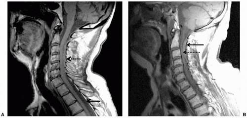
FIGURE 61.1 A: Epidural hematoma. Note the loss of normal cerebrospinal fluid-spinal cord interface (solid arrow) below the level of the hematoma. In the cervical region, blood has replaced the cerebrospinal fluid around the spinal cord (dotted arrow). B: Note the presence of blood in the epidural space (solid arrow) and compression of the spinal cord by the hematoma (dotted arrow).

Central neuraxial anesthesia generally should be avoided in patients with inherited bleeding disorders due to coagulation factor abnormalities, platelet dysfunction, or thrombocytopenia. Acquired bleeding disorders due to
liver failure, leukemia, and other entities also preclude the use of central neuraxial block. Cutaneous angiomas may be associated with spinal arterial venous abnormalities.48 Hydraulic pressure changes induced by epidural anesthesia have been associated with rupture of spinal angiomas.49 Therefore, performance of central neuraxial block in these patients should be approached with caution.

Only gold members can continue reading. Log In or Register to continue

Stay updated, free articles. Join our Telegram channel

Jul 15, 2016 | Posted by in ANESTHESIA | Comments Off on Neuraxial Anesthesia

Full access? Get Clinical Tree

Get Clinical Tree app for offline access