Invasive Monitoring Complications


Invasive Monitoring Complications


Gregory M. Janelle

Nikolaus Gravenstein


▪ INTRODUCTION

All invasive monitors have associated complications that fall into three main categories: (a) placement, (b) interpretation, and (c) maintenance complications, and may be further subdivided into those which occur early versus late. This chapter examines arterial, central venous, and pulmonary arterial catheterization and their respective complications, as well as complications of transesophageal echocardiography (TEE).


PART I INTRA-ARTERIAL CATHETERS

Arterial catheters are the most commonly placed invasive monitors in the operating room. Major complications related to arterial cannulation occur in a relatively low percentage of patients.1 The prevalence of arterial catheterization, however, makes many such complications frequent occurrences. Complications range from minor to life-threatening, and are based in part on the anatomic insertion point. Complications may be related to placement, interpretation, or maintenance as described in Table 52.1.


TABLE 52.1 Arterial Catheter Complications


Placement


Interpretation


Maintenance


Pain


Calibration error


Thrombosis


Hematoma


Zeroing error


Infection


Hemorrhage


Underdamped


Embolization


Embolization


Overdamped


Skin necrosis


Nerve injury


Hemorrhage


Ischemia


What Are the Most Significant Complications Seen with Arterial Catheters?


▪ PAIN

Pain associated with arterial cannulation is decreased by local infiltration of plain 1% lidocaine. The infiltration of anesthetic beneath the dermis causes less discomfort than an intradermal injection. Epinephrine as an additive is specifically avoided.


▪ HEMATOMA

Hematoma formation is a complication of placement, attempted placement, and catheter removal. It is more frequently associated with multiple attempts, as well as transfixation of the artery (puncturing both the anterior and posterior vessel walls) compared to direct threading of the catheter through an anterior puncture. The application of direct pressure for 10 minutes to the puncture site or a temporary noncircumferential pressure dressing may limit hematoma formation.2 A hematoma may not be problematic by itself, but appears to predispose the underlying vessel to thrombosis. The combination of poor aseptic technique with hematoma predisposes the patient to infection, because static blood is a culture medium for bacteria.


▪ HEMORRHAGE

Hemorrhage may occur from bleeding around an indwelling catheter, during placement, or after decannulation. Blood loss in small infants may represent a significant amount of the child’s blood volume. Hemorrhage is typically controlled with the application of direct pressure. Bleeding may also occur from the inadvertent disconnection of the connecting tubing from the patient or malposition of a stopcock. Luer-locking connections are preferable to friction-fit ones, and occlusive caps should be used on stopcocks. Bleeding is particularly troublesome when it is not readily evident, as may occur from a femoral arterial access site which, if punctured proximal to the inguinal ligament, may bleed into the retroperitoneal space.3


▪ EMBOLIZATION

Embolization during arterial pressure monitoring may be anterograde or retrograde and may include particulate matter (such as thrombus or sheared catheter tip) or air. A continuous flush solution is used to maintain catheter patency and to prevent thrombosis of the radial artery while the catheter is in place. Embolization of thrombotic material to the hand4 or to the central circulation5 has been reported with intermittent flushing of radial artery cannulae. Rapid manual flushing of radial arterial catheters at rates faster than 1 mL per second produces retrograde flow (i.e., toward the brain and heart) in the proximal axillary artery.6 Because of the risk of retrograde embolization to the cerebral circulation with as little as a 3 mL hand injection of flush solution, radial artery cannulae should be flushed slowly and with small volumes (1 to 3 mL) of solution.5 In a primate model, it has been shown that, in the sitting position, when using as little as 2 to 2.5 mL of air given at rates as slowly as 0.6 mL per second, air travels retrograde from the radial artery to the brain.7 The demonstrated propensity for embolization into the central circulation needs to be considered in the context of the frequency, volume, and velocity of irrigation of arterial catheters. The risk of retrograde embolization to the central nervous system (CNS) is greatest for arterial catheters placed in the right upper body (especially brachial, axillary, or temporal). Right-sided catheters pose a higher risk because an air embolus or thromboembolus traverses the origin of the carotid and vertebral arteries.8 Temporal artery and right axillary artery catheters should be avoided, if possible.9

Emboli that travel antegrade may cause distal ischemia in the affected extremity (e.g., fingertips). Preventive measures for distal embolization include aspirating the catheter before irrigating it, and also aspiration during catheter removal to remove as much of the thrombus surrounding the catheter as possible.


▪ NERVE INJURY

Nerve injury during arterial cannulation is unlikely if the radial artery is used. Ulnar, brachial, axillary, and femoral arteries are anatomically close to a nerve(s) that might be injured by the needle during placement or compressed by a hematoma. If a paresthesia is encountered, the catheter and needle should be redirected or an alternate site chosen. Leaving the cannulated wrist in a hyperextended position predisposes to a median nerve injury, particularly if a hematoma is present.


What Delayed Complications Occur from Arterial Catheters?

All of the acute complications related to placement may still occur later. Additional delayed complications are catheter-related thrombosis and infection.


▪ THROMBOSIS

Thrombosis, or reduced flow accompanying or following radial artery cannulation, is common and occurs in >20% of patients.1,10 The likelihood of thrombosis increases with the duration of cannulation and with the percentage of the vessel lumen occupied by the catheter.11


Diagnosis

Polyethylene catheters have been shown to be associated with more frequent thrombus formation than Teflon catheters of the same size.12

The incidence of thrombus formation is less frequent when using a continuous irrigation (2 to 3 mL per hour) flush apparatus than if the catheter is kept clear by intermittent irrigation. Alternative anticoagulants, such as bivalirudin and argatroban, have been added to the flush solution in low concentrations to prevent thrombus formation, particularly in patients with heparin-induced thrombocytopenia.

The incidence of arterial occlusion increases linearly as the ratio of cannula diameter-to-vessel diameter increases.11 These data validate the use of smaller catheters, that is, 20 gauge. By extension, it is reasonable to use smaller catheters in small adults and children. Further support for this inference comes from repeated observations that radial artery thrombosis is much more common in women (presumably smaller arteries) than men.13 It has been postulated that if the relation between wrist circumference and radial artery size is linear, the critical wrist circumference for a 20 g catheter is 15 cm.14

The duration of radial artery occlusion, that is, time to recanalization, is longest for the smallest vessels. In those sporadic cases where permanent ischemic damage was related to arterial catheterization, it has almost always occurred in a setting of significant coexisting disease such as hyperlipoproteinemia, prolonged shock, use of vasopressors, inadvertent drug administration through the catheter, or emboli of separate origin. The overall incidence of distal digital ischemia is approximately 0.01% or less.


Treatment

Radial artery thrombosis is usually asymptomatic, resolves spontaneously over days or weeks, and requires no special care. If signs of distal ischemia occur, aggressive therapy is warranted. If ischemia does not resolve following removal of the arterial catheter, a vascular or hand surgeon should be immediately consulted. Workup and therapy may include angiography, embolectomy, regional sympathetic blockade, and even surgical exploration.


Prevention

In spite of the extremely small risk of permanent ischemic damage associated with arterial cannulation, it remains the practice of some to assess the adequacy of the collateral circulation when arterial catheters are placed in the wrist. This assessment can be made by Allen’s test or a modification of it. In the cooperative patient, the examiner occludes ulnar and radial arteries simultaneously, while the patient exsanguinates the hand by making a clenched fist. After the hand is opened, the ulnar artery is released while the radial is left occluded. If a blush returns to the palm, especially the thenar eminence within 7 seconds, then ulnar collateral circulation is considered adequate. If it takes between 7 and 15 seconds, it is abnormal. If it requires longer than 15 seconds, it is considered absent.15 The procedure is repeated releasing the opposite artery. By comparing relative times to thenar blush for each artery, the examiner can establish which vessel is dominant (shortest time to blush). With an uncooperative patient, the hand can be exsanguinated passively and the test completed as mentioned in the preceding text, or a plethysmograph or pulse oximeter may be placed on the thumb to objectively demonstrate the qualitative presence of pulsatile flow, that is, perfusion, while each artery is alternately occluded.16 Another method is that of occluding the radial artery at the intended cannulation site and palpating for a pulse distal to the point of occlusion. If present, it suggests retrograde perfusion from the ulnar artery.

Although none of these methods are well correlated with ischemic outcomes, they do serve to document collateral circulation. Despite the recommendation of the Allen’s test by some clinicians, or a modification thereof, as a standard of care, it is well documented that an abnormal test does not reliably predict ischemic complications, nor does a normal one preclude them.1,17

Factors to consider in decreasing the incidence of arterial catheter-related thrombosis include cannulation of short duration with a small, nontapered Teflon catheter with continuous irrigation after verifying collateral perfusion.


▪ INFECTION

Arterial catheter-related infection is uncommon if catheters are left in place <96 hours.18 Most infections appear to be caused by invasion of the skin flora into the intracutaneous catheter tract. Hematogenous seeding of organisms from other sites and contaminated flush solutions are other mechanisms of infection. In view of the prevalence of thrombosis, it is easy to imagine a thrombus serving as a nidus for colonization and then infection.18


Diagnosis/Treatment

Culturing the catheter is the definitive way to diagnose a catheter-related infection. However, the wait time for culture results does not provide helpful information needed in the intraoperative period. A causal relation has been shown to exist between clinical signs of local inflammation of catheter wounds and infection.18 If inflammation or purulence is noted at the cannulation site, the catheter should be removed.


Prevention

Changing flush solution and tubing at least every 96 hours virtually eliminates the catheter system as a source of infection. Aseptic site preparation (preferably a chlorhexidine-containing solution) and catheter
placement (wearing sterile gloves) prevents the contamination of the access site at the outset. In a random sampling, arterial catheter system stopcocks had a contamination rate as high as 38%.19 Occlusive caps or syringes kept on all stopcock ports, as well as irrigating them to clear blood (bacterial culture medium) after any sampling, are appropriate preventive measures.

Although it is unusual for patients to manifest arterial catheter-related sepsis during anesthesia, there can be no doubt that some contamination and infections occur perioperatively during placement or use of an intra-arterial catheter. A number of factors have been identified which are thought to predispose to catheterrelated infections. These include the following:


  • Catheter placement by cutdown rather than percutaneously


  • Catheters left in place for longer than 4 days


  • Catheters left in patients who have experienced sepsis


  • Glucose-containing flush solution

Avoidance of these factors when possible and adherence to the guidelines for care of arterial catheters should make arterial catheter-related infections even less common20 (see Table 52.2).


TABLE 52.2 Guidelines for Care of Arterial Catheters


1.


Observe proper hand hygiene procedures either by washing hands with conventional antiseptic-containing soap and water or with waterless alcohol-based gels or foams. Observe hand hygiene before and after palpating catheter insertion sites, as well as before and after inserting, replacing, accessing, repairing, or dressing an intravascular catheter. Palpation of the insertion site should not be done after the application of antiseptic, unless aseptic technique is maintained.


2.


Wear sterile gloves when inserting arterial catheters.


3.


Disinfect the site with an appropriate antiseptic (2% chlorhexidine-containing solution is preferred to iodine, an iodophor, or 70% alcohol).


4.


Insert the catheter by percutaneous puncture rather than by surgical cutdown whenever possible.


5.


Use sterile gauze or sterile, transparent, semipermeable dressing to cover the catheter site.


6.


Do not use topical ointment on the insertion site.


7.


Record the time and date of insertion of the catheter on the dressing and in the patient’s narrative record. Inspect the insertion site every 24 h. Change the dressing at least weekly or as visual inspection dictates.


8.


Promptly remove all catheters that are deemed nonessential. Replace catheters in patients with severe bacteremia.


9.


Keep all components of the pressure monitoring system (including calibration devices and flush solution) sterile. Do not administer dextrose-containing solutions or parenteral nutrition fluids through the pressure monitoring circuit. The pressure monitoring system and the irrigation solution should be changed every 96 h.


10.


Minimize the number of manipulations of and entries into the pressure monitoring system. Use a closed flush system (i.e., continuous flush), rather than an open system (i.e., one that requires a syringe and stopcock), to maintain the patency of the pressure monitoring catheters.


11.


When the pressure monitoring system is accessed through a diaphragm rather than a stopcock, wipe the diaphragm with an appropriate antiseptic before accessing the system.


12.


Evaluate each patient daily with regard to catheter-related infection. Local pain or inflammation, embolic lesions distal to the catheter, unexplained fever or especially bacteremia without an obvious source should prompt removal of the entire infusion apparatus as well as the catheter; both the catheter and the sample of remaining infusate should be cultured. Any purulent material that can be expressed from the catheter wound should be gram-stained and cultured. At least three blood cultures should be obtained by separate venipuncture.


13.


Remove the catheter if the patient is septic. It is the single, most important therapeutic maneuver in the management of catheter-related septicemia. Patients who are clinically septic, with purulent material that can be expressed from the wound, and, certainly, those who have positive blood cultures, should also receive systemic antimicrobial therapy.


Adapted from: O’Grady NP, Alexander M, Dellinger EP, et al. Centers for Disease Control and Prevention. Guidelines for the prevention of intravascular catheter-related infections. Prevention. MMWR Recomm Rep. 2002;51 (RR-10):1-29.


What Are the Common Errors Made in Data Acquisition from Arterial Catheters?

Proper attention to the zero reference, the zero and gain adjustments, and the use of a system free of ringing and damping are necessary to obtain an accurate reflection of intra-arterial pressure. Because both underestimation and overestimation of arterial pressure are possible, clinical management may be biased in either direction.


▪ SELECTION OF ZERO REFERENCE

The zero reference describes the vertical location relative to the patient at which the clinician measures pressure. In the supine patient, the organs of greatest interest (brain, heart, kidneys) are all at the same level. In this case, the proper zero reference is the midaxillary line in the fourth intercostal space. Erroneous data are recorded when the vertical distance between the transducer and the patient
is not kept constant. If, for example, the patient’s bed is raised 30 cm while the transducer is kept mounted on an intravenous (IV) pole, then the height difference between the zero reference (midaxillary line) and the transducer adds the weight of the fluid column in the pressure monitoring tubing (30 cm H2O) to the measured pressure. This raises the apparent pressure by the change in height between transducer and zero reference, that is, 30 cm H2O × 0.7 mm Hg per cm H2O = 21 mm Hg overestimation. The same effect is noted when the transducer is mounted on the head of the bed and the bed is moved into a Trendelenburg position, or when the transducer is patient-mounted to the wrist and the bed is tilted to the side of the transducer. The opposite effect occurs if the patient’s zero reference position is lowered in relation to the transducer.

A relevant example of patient positioning is the case of the patient who is operated on in a sitting position. There is a large difference (e.g., 30 cm) in height between the level of the heart and the brain. For patients in a headelevated position, the appropriate zero reference for blood pressure (BP) is the brain (external auditory meatus) and not the heart. Failure to zero reference to the brain after sitting a patient up would, in this example, lead to a 21 mm Hg overestimation of the true cerebral perfusion pressure (see Fig. 52.1).


Diagnosis

Monitoring of a proper zero or zero reference is made by opening a stopcock to air at the desired zero reference level, and verifying that it reads zero at that location at the time monitoring is begun and each time the position of the patient or transducer changes.


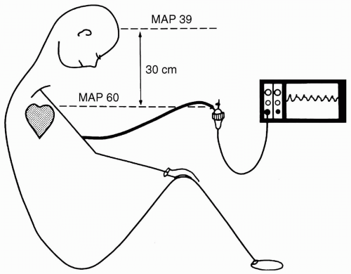
FIGURE 52.1 The relation of the transducer to the pressure that is to be measured. Placing the transducer or opening the fluid path to air at the level of the heart and not the head in this patient would cause the MAP in the brain to be overestimated by the height difference converted to mm Hg. It makes little difference which artery is cannulated for arterial pressure monitoring. MAP, mean arterial pressure. (Modified from: Gravenstein JS, Paulus DA. Arterial pressure. In: Clinical monitoring practice, 2nd ed. New York: JB Lippincott Co; 1987:54.)


▪ ZERO SETTING

Zeroing the pressure transducer has been described as the single, most important step in setting up a pressure monitoring system.21 Errors in the zero adjustment result in a fixed offset (high or low) from the true value, just as with an improper zero reference. Changes in BP will still be apparent, but the measured values will differ from the actual pressures by the offset. Proper zeroing requires opening the fluid path of the arterial catheter system to air at the level of the heart (midaxillary line) in a supine patient, or at the level of the brain in a patient who is in a head-elevated position. The zero value may drift over time, especially if there is any moisture in the electric connections, and therefore, periodically rechecking the zero is advisable.


▪ SETTING GAIN

Once zeroed, the next source of error is improper calibration or gain of the transducer and monitor. Although this is typically taken for granted, the gain adjustment on the monitor may be off, in which case recalibration is easily performed. Ideally, calibration is:


  • A three-point calibration to verify that the system is linear


  • Done in the range of pressures expected to be monitored, for example, arterial pressure 200, 100, and 50 mm Hg


  • Done using a mercury manometer or equivalent as reference

One method is to connect a piece of fluid-filled extension tubing of known length to the transducer, filling it with solution and elevating it. If the gain is correct, the monitor should display a value that is the height (length) of the tubing × 0.7 mm Hg per cm.


▪ RINGING AND DAMPING

Ringing and damping are two other common sources of “bad” data and are associated with problems of dynamic accuracy. They reflect the capability of the system to “respond accurately to rapidly changing pressure waveforms.”22 An analysis of the physical properties of catheter systems reveals that, to accurately measure a pressure, the system must be able to respond to frequencies at least 10 times the heart rate. Therefore, accurate monitoring is more difficult in infants than in β-blocked adults. Because a pressure monitoring system has resistance, inertia, and compliance, it can oscillate or ring, that is, exaggerate the systolic and underestimate the diastolic pressures. The frequency at which this is most evident is called the natural frequency. The counterpart of the natural frequency is the damping coefficient which reflects how quickly the system comes to rest after a change occurs.23 In contrast
to a system which is ringing (underdamped) and results in higher systolic and lower systolic pressures, a damped system gives lower systolic and higher diastolic pressures. Typically, systolic pressure is affected more than diastolic pressure by both ringing and damping. Mean pressure is the best-preserved and most accurate value in the face of either ringing or damping.


Diagnosis

If the dynamic response of a system is questionable, it can be clinically tested by determining the natural frequency and damping coefficient using a fast flush from a pressurized infusion (see Fig. 52.2).


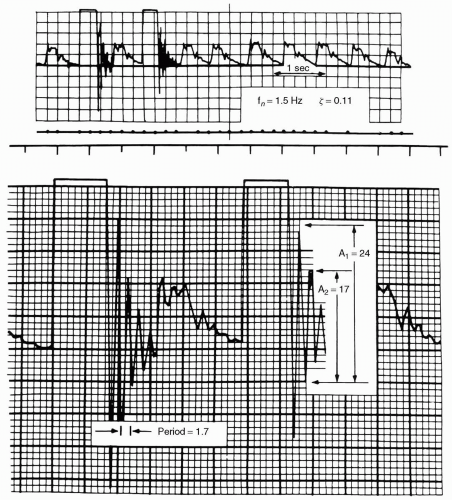
FIGURE 52.2 The upper panel shows an arterial pulse waveform with two flushes. The natural frequency and damping coefficient can be determined from either flush. The lower panel shows the flush segment enlarged to illustrate the method. The natural frequency (fn) of the system is estimated by taking the period of one cycle (period), in this case 1.7 mm, and dividing this into the paper speed, 25 mm/s fn = 25/1.7 = 15 Hz. The damping coefficient is determined by taking the amplitude ratio of successive peaks of the oscillations, in this case A2/A1, = 17/24 = 0.71. (From: Gardner RM. Direct blood pressure measurements—dynamic response requirements. Anesthesiology. 1981;54:233.)


Prevention/Treatment

In general, the accurate dynamic response of physiologic pressure monitoring systems is best achieved by using a technique that strictly eliminates all bubbles. Bubbles will seem to appear de novo as gas comes out of solution from increases in room temperature, from a fluid pressure decrease (as occurs between the pressure bag and the catheter), or following vigorous sampling of flush solution where air can be brought out of solution or entrained from a loose connection. Using short (<3 ft.), stiff, large bore connecting tubing with as few stopcocks as possible is important. The use of the fewest possible stopcocks limits them as air bubble reservoirs and as constrictors in the
fluid path. Utilizing T-connectors is discouraged because they are compliant and serve as air bubble reservoirs or point of air entrainment during blood sampling if they have a needleless access. It is impossible to predict the relative contributions of each of these factors; therefore, if there is a clinically significant discrepancy between invasive and noninvasive pressures, the flush test provides a simple way to assess the system’s dynamic response characteristics. This, coupled with zero reference, and zero and calibration verification assures the accuracy of the system in use. If the test demonstrates inadequate dynamic response, each of the potential causes should be ruled out. If dynamic response, zero, and gain are correct, consider other causes discussed in the subsequent text.


▪ AORTA-RADIAL PRESSURE GRADIENTS

Several reports describe significant disparities between aortic, femoral, and radial artery pressures.24,25 Although these reports are specific for patients post cardiopulmonary bypass, the analogous circumstances also exist in other clinical settings. In both studies, the radial artery pressure underestimated the aortic or femoral pressures by at least 10 mm Hg in most patients. The proposed mechanisms for this phenomenon include vasodilatory steal or, conversely, peripheral vasoconstriction. The appreciation of this phenomenon after cardiopulmonary bypass, or in the setting of hypovolemia (vasoconstriction), sepsis (vasodilation), or vasodilator therapy requires the suspicion that there is a difference. Diagnosis is made either by obtaining a brachial cuff pressure (generally much less vulnerable to changes in peripheral resistance) or placing a femoral or axillary artery catheter. No preventive measures are known for this phenomenon.


▪ CLAIMS AND LITIGATION

The American Society of Anesthesiologist (ASA) Closed Claims Project database uses a standardized summary of data collected from a group of professional liability insurance carriers.26 In the period of time from 1970 through 2001, only 2.1% of the 6,894 claims were related to peripheral catheters, with only 9% of these 140 claims related to arterial catheters. Of these 13 claims, thrombosis and iliac artery puncture represented the vast majority (31% each).

Of the seven cases involving radial artery catheterization, two involved problems with retained catheters or wires, two involved radial nerve damage (associated with multiple punctures), two were associated with arterial occlusion with resulting hand ischemia, and one resulted in carpal tunnel syndrome from a hematoma. Each of the two involving arterial insufficiency occurred in patients with significant risk factors for malperfusion (Raynaud’s disease and severe peripheral vascular disease). Within the remainder of patients, there were two pediatric claims related to femoral arterial catheterization, with one involving leg ischemia necessitating amputation and another that resulted in massive hemorrhage due to iliac artery laceration, causing hypotension, cardiac arrest, and subsequent profound neurologic deficits.26


PART II CENTRAL VENOUS CATHETERS

Injuries related to central venous catheters (CVCs) are associated with a higher severity of injury than other anesthesia-related complications.27 Overall, it is estimated that 15% of patients in whom CVCs are used will suffer some form of mechanical, infectious, or thrombotic complication.28,29


What Types of Complications Can Be Expected from Central Venous Catheters?


▪ ACUTE COMPLICATIONS

Complications related to CVC placement are listed in Table 52.3. The complication rates of the two most commonly used sites, internal jugular veins (IJVs) and the subclavian artery, are significant. The findings of a large prospective study by Sznajder highlight the importance of operator experience.30 In this study of 714 placement attempts, the experienced physicians (50 or more previous catheterizations) were much less likely to fail in their catheterization attempt or to have a complication. Although complications and failures were only about half as common with experienced physicians, they still occurred with an approximately 6% frequency with placement at both sites.


Arterial Puncture

Arterial puncture is associated with approximately 3% of subclavian and 4% to 10% of IJV placements, depending on practitioner experience.30,31,32 Carotid artery puncture is, by far, the most common complication associated with IJV cannulation. It is usually without sequelae, but may result in a hematoma sufficient to effect tracheal compression or a significant extrapleural or mediastinal hematoma. Stroke has also been reported from punctures of the carotid artery as well as from its
cannulation, occasionally resulting in death.33 Vertebral artery puncture during CVC placement has similarly been associated with fatal stroke.34 Hemothorax is reported following subclavian artery puncture. If CVC placement is required in a patient with a coagulopathy, the subclavian approach is to be avoided. Unlike with carotid artery puncture, it is difficult to apply direct pressure to the subclavian artery or to observe it for hematoma formation. A series of 1,000 IJV placements in patients with coagulopathies resulted in no complications referable to a 7% incidence of carotid artery puncture.32 One patient with a goiter required surgical decompression of a venous bleeding site.32 These authors suggest that IJV cannulation does not result in severe complications in patients with a coagulopathy. Despite these findings, the external jugular route may be preferable in patients with coagulopathy because it avoids the danger of arterial puncture.


TABLE 52.3 Complications due to Central Venous Catheter Placement


Arterial puncture


Pneumothorax


Hemothorax


Arrhythmias


Malposition


Air embolism


Nerve injury


Thoracic duct injury


Perforation


-Hydro- or hemothorax


-Hydro- or hemomediastinum


-Hydro- or hemopericardium


Infection


From: Gravenstein N, ed. Manual of complications during anesthesia. Philadelphia: JB Lippincott Co; 1991:271, (Table 7-4).


Diagnosis of arterial puncture is made by the appearance of pulsatile blood, bright red blood, direct measurement of intraluminal pressure, or a pulsating needle. The absence of one of these signs does not reliably exclude an arterial puncture, as highlighted by a very bothersome observation where, in a series of 1,021 attempted IJV cannulations, 5/43 arterial punctures went unrecognized, and an 8F catheter sheath was placed into the carotid artery and one patient died.35


Prevention

Traditionally, it is taught that the neck should be palpated to identify the location of the carotid pulse before IJV puncture. A newer approach is to use an ultrasound device to identify the precise location of the target vein. Ultrasonographic guidance has been shown to significantly reduce complications associated with IJV cannulation. In a prospective, randomized intensive care unit (ICU) study of 900 IJV central venous catheterizations, the traditional use of anatomic landmarks resulted in puncture of the carotid artery in 10.6% of patients, hematoma in 8.4%, hemothorax in 1.7%, pneumothorax in 2.4%, and CVC-associated blood stream infection in 16%, which were all significantly higher than in the group in which ultrasound guidance was used (p < 0.001).36 Average access time (skin to vein) and number of attempts were also reduced in the ultrasound group compared with the landmark group (p < 0.001). Other studies have found similar results, although a learning curve has been noted with the use of ultrasound to facilitate access.36,37,38,39,40 However, Augustides et al. published rates of carotid puncture of 4.2% with or without ultrasound-assisted needle guidance across differences in level of training,40 with carotid puncture rates of 0% in the hands of experienced attendings. Complication rates are known to increase with repeat punctures, with complications as high as 54% when more than two punctures are necessary.41 With a trend toward more frequent use of laryngeal mask airways for general anesthesia, it should be noted that the laryngeal mask airway has been shown to alter the normal anatomic location of the IJV with respect to the carotid artery. It has been found that at the middle and more cephalad approach points to the IJV, the overlap of the IJV over the common carotid artery rendered a statistically significant increase of the overlap index (percentage of carotid overlapped by the IJV), whereas the index at low access points was unchanged.42 Rotating a patient’s head <40 degrees also decreases the amount of internal jugular carotid overlap during IJV CVC placement.43

Subclavian artery punctures secondary to jugular venous cannulation, although less common than carotid punctures, have also been reported.44,45,46 It has been hypothesized that due to anatomic variations between the right-sided and left-sided arterial structure (the right subclavian artery branches from the brachiocephalic trunk medial to the IJV), this is possibly a right-sided phenomenon and may be a consequence of either direct needle puncture or inadvertent advancement of the dilator into the subclavian artery.47 Verterbral artery puncture,48 dissection, and creation of iatrogenic arteriovenous (AV) fistulae have also been reported with IJV approaches.49

With respect to subclavian artery puncture during a subclavian vein access attempt, one should avoid placing a subclavian catheter lateral to the juncture of the middle and distal thirds of the clavicle due to the anatomic location of the subclavian artery behind the vein at this level. If there is any question about which vessel was entered with any central venous catheterization access site, even using ultrasonographic guidance, the intraluminal pressure should be transduced to identify an arterial puncture (not itself a significant complication; treated by direct, but not blood flow-obstructing, pressure for 5 to 10 minutes) before placing a larger catheter or sheath (a significant complication if placed intraarterially). This can be easily accomplished with a disposable length of sterile tubing attached to the access needle. The tubing is first held below the level of the access point to fill it with blood. Then the tubing is lifted above the level of the vessel to verify that the blood column descends (venous). This method of “air transduction” should not be considered in spontaneously breathing patients or patients in whom the Trendelenburg
position is avoided (risk of air embolism), nor is it reliable when CVCs are placed in extremely hypotensive patients, because the central venous pressure (CVP) and arterial pressure may be comparable.


Pneumothorax

Pneumothorax may occur with as many as 6% of subclavian vein CVC placements. The incidence following jugular CVC placement is <0.5% for IJV and 0% for external jugular vein placements.


Diagnosis

Pneumothorax is confirmed by chest radiograph. Ideally, the film is taken during end expiration and in an upright view. Clinical signs of pneumothorax include tachypnea, hyperresonance to percussion, decreased breath sounds, and, if under tension, contralateral tracheal deviation. A supine chest radiograph may fail to reveal a small pneumothorax because the air is anterior with lung behind it. When reviewing a supine film for pneumothorax, the medial costophrenic sulcus should be carefully inspected, because air will tend to collect in that area. If the diagnosis remains unclear, a lateral decubitus film with the affected side up is helpful. Pneumothorax should be suspected if air is aspirated during needle localization of the vein. Absence of air aspiration does not, however, preclude the appearance of a pneumothorax. The index of suspicion regarding the possibility of pneumothorax should be increased if the clinician is inexperienced (i.e., <50 previous CVC placements by that route), and if placement or attempted placement requires multiple needle passes.31


Treatment

Treatment of pneumothorax is by tube thoracostomy. Awake, spontaneously breathing patients may be treated conservatively, with repeated observation if the pneumothorax is minimal (<20%) and asymptomatic. Any patient being mechanically ventilated or anticipated to require positive pressure ventilation should be treated by chest tube drainage. If a chest tube is not immediately available, and pneumothorax is symptomatic, a large bore IV catheter can be placed in the midclavicular line, typically in the second or third intercostal space. If severe hemodynamic or respiratory compromise is related to CVC placement, the immediate decompression of a suspected pneumothorax takes precedence over radiographic confirmation.


Prevention

Prevention of CVC placement-related pneumothorax is by choosing the IJV over the subclavian approach. A chest radiograph following any subclavian catheter placement is a priority; this allows early assessment for pneumothorax as well as verification of catheter tip position. The extremely low incidence of pneumothorax following IJV catheter placement makes a chest radiograph a much lower priority, typically taken after leaving the operating room, because the radiograph is used primarily for assessing catheter tip position. If air is aspirated during IJV placement, an immediate chest radiograph is indicated. The use of nitrous oxide should be limited if possible after any intraoperative subclavian or difficult jugular catheter placement because:


  • Nitrous oxide diffuses into a pneumothorax much more rapidly than air diffuses out, and therefore the size of the pneumothorax may double in <10 minutes in the presence of 50% N2O.50


  • Not all pneumothoraces are evident immediately on postplacement chest radiograph.


  • The use of intraoperative positive pressure ventilation can increase the size of a pneumothorax.

In patients with severe chronic obstructive pulmonary disease (COPD) or ventilated with high airway pressures, the external jugular route is the least likely to result in a pneumothorax.


Hemothorax

Hemothorax is a complication of subclavian artery puncture or laceration. It may also be associated with perforation of an intrathoracic vein or the vena cava by a needle, guidewire, dilator, or catheter.


Diagnosis

Hemothorax is diagnosed by chest radiograph, which should always follow subclavian catheter placement.


Treatment

Treatment is by chest tube drainage. If bleeding persists, surgical repair of the laceration is indicated. In the case of a catheter-induced venous perforation, the catheter should be removed and the effusion drained if it is symptomatic.


Prevention

The preferential use of the jugular approach avoids the subclavian artery and lung. Selecting the right side for all CVC placements, regardless of route, makes venous perforations less common because the guidewire, dilator, and catheter path is more direct, and less likely to result in impinging at an acute angle on the innominate vein or superior vena cava (SVC) wall (see Fig. 52.3). Guidewires with flexible tips are less prone to perforation. It should be noted that many flexible J-tipped guidewires have an inflexible straight tip at the other end. When rigid dilators are placed, they should only be inserted deep enough to dilate the skin and subcutaneous tissue. There is no additional benefit, and there is increased risk of perforation when dilators are inserted further because they are more likely to impinge on vessel walls (Fig. 52.3).


Arrhythmias

Arrhythmias are commonly noted during CVC placement.51 They are the result of mechanical irritation of the atrium or ventricle and can be caused by either guidewire
or catheter. Treatment consists of withdrawing the wire or catheter. Mechanically induced arrhythmias resolve upon removal of the stimulus. The lengthy guidewire is most commonly the culprit. A prospective study demonstrated that rigorous control of the depth of guidewire insertion markedly reduced the incidence of arrhythmias during pulmonary artery catheter (PAC) introducer insertion from 58% to 15%.51 An additional benefit was that limiting guidewire insertion depth to <22 cm from the right IJV approach virtually abolished the more hemodynamically severe, ventricular arrhythmias.51


FIGURE 52.3 Central venous access sites. (From Cook. Triple lumen central venous catheter package insert. Cook, Critical Care; 1986.)

Jul 15, 2016 | Posted by in ANESTHESIA | Comments Off on Invasive Monitoring Complications

Full access? Get Clinical Tree

Get Clinical Tree app for offline access