Fig. 39.1
Anatomy of the thoracic spinal nerves. (1) Spinal ganglion, (2) spinal nerve, (3) ganglion of the sympathetic trunk, (4) dorsal and ventral branch, (5) dorsal and ventral root, (6) white and gray communicating branches (With permission from Danilo Jankovic)
The dorsal rami of the thoracic nerves pass between the two transverse processes to their area of distribution and divide into the two typical branches, the medial and lateral branches; they give off muscular branches (back muscles) and cutaneous branches (spinous processes, posterior wall of the thorax, and lumbar region). The ventral rami of the thoracic nerves are also termed intercostal nerves, and they are distributed segmentally (Fig. 39.2).
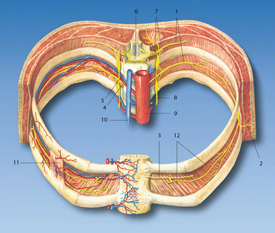
Fig. 39.2
Intercostal nerves. (1) Ventral branches (intercostal nerves), (2) lateral cutaneous branch, (3) anterior cutaneous branch, (4) posterior intercostal artery, (5) posterior intercostal vein, (6) spinal cord, (7) spinal nerve, (8) sympathetic trunk, (9) thoracic aorta, (10) azygos vein, (11) external intercostal muscles, (12) internal intercostal muscles (With permission from Danilo Jankovic)
The 11 upper nerves are (relative to the thoracic ribs) genuinely intercostal because the nerves at least partially run in the intercostal space. The twelfth, however, lies caudal to the twelfth rib and is known as the subcostal nerve. The six upper intercostal nerves run entirely in the intercostal spaces, as far as the edge of the sternum; the six lower ones reach the area of the linea alba. All of the intercostal nerves, with the exception of the twelfth, run in the relevant intercostal space in front of the superior costotransverse ligament and on the inner surface of the external intercostal muscles.
The internal intercostal muscles are absent from the spine far as the costal angle, replaced by internal intercostal membrane (Fig. 39.3). Over this area, the intercostal nerves lie over the endothoracic fascia and costal or parietal pleura. Approaching the angle of rib, the nerves lie between the internal intercostal muscles and the innermost intercostal muscles, and they are accompanied by the intercostal vessels (the intercostal artery and vein). They lie caudal to the vessels. Special care needs to be taken during procedures, as due to the proximity of blood vessels to the nerves, toxic concentrations of local anesthetic can easily be reached.
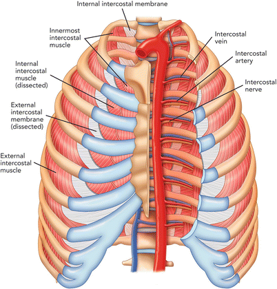
Fig. 39.3
Intercostal muscles in the chest wall (Reproduced with permission from Philip Peng Educational Series)
The intercostal nerves give out various branches: muscular branches for various chest wall muscles such as serratus posterior and rectus abdominis muscles; lateral cutaneous branches supplying the skin of lateral sides of the thorax and abdomen, as well as the skin of the axilla (first intercostal nerve); anterior cutaneous branches supplying the anterior side of the thorax; and pleural and peritoneal branches supplying the pleura and thoracic wall and the peritoneum of the lateral and anterior abdominal wall, as well as the pleural and peritoneal covering at the origin of the diaphragm.
Sonoanatomy
The ideal location for ultrasound-guided injection is at the level of angle of rib because the internal intercostal muscle is formed from this point onward and the lateral cutaneous branch is still incorporated in the intercostal nerve.
The patient lies on the contralateral side or in prone position with arm abducted to bring the scapula away from midline (Fig. 39.4). A high-frequency ultrasound probe is used. The key landmarks are the upper and lower ribs of each intercostal space, the intercostal muscles, and the pleura. The probe is placed 90° to the course of the ribs so that upper and lower ribs, intercostal muscles, and pleura can be visualized. The target is the intercostal nerve deep to the internal intercostal muscle just caudal to the cranial rib and is usually less than 5 mm from the pleura (Fig. 39.5). To enhance the visualization of the internal intercostal muscle, the cranial end of the ultrasound probe is tilted laterally to align with the muscle (Fig. 39.6). If an in-plane approach is used, the transducer is positioned on the intercostal space, parallel to the upper and lower ribs. It is advisable to measure the distance between the skin and the pleura, and the needle should never be introduced more that this distance.
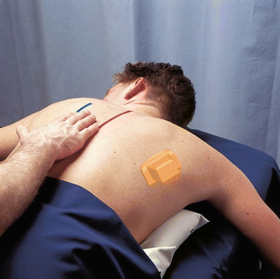
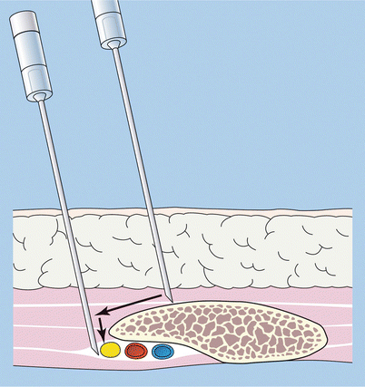
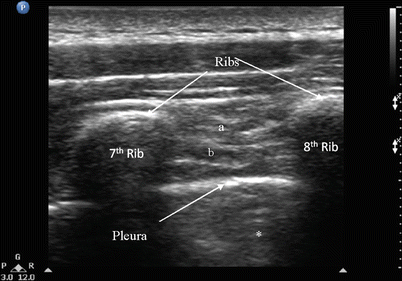
Fig. 39.4
Prone position. The red line indicates the midline and blue indicates line joining the angle of ribs. The ultrasound probe position is indicated on the right side
Fig. 39.5
The needle position with landmark technique (With permission from Danilo Jankovic)
Fig. 39.6
Ultrasonographic image showing the intercostal muscles and pleura at the angle of rib. a external intercostal muscle, b internal intercostal muscle, * reverberation artifact. The pleura appears as a hyperechoic line (Reproduced with permission from Philip Peng Educational Series)
Indications
Surgical
1.
Superficial surgery in the innervation area
2.
Insertion of a chest drain
Diagnostic
1.
Differential diagnosis of somatic and autonomic pain conditions
Therapeutic
1.
Pain in the intercostal area (neuralgia and causalgia)
2.


Pain after rib fractures or contusions of the thoracic wall and pleuritic pain
Related posts:


Stay updated, free articles. Join our Telegram channel
Full access? Get Clinical Tree
