Fig. 67.1
This figure shows the hip joint and the labrum (left) and the ligaments in the anterior hip joint (Reproduced with permission from Philip Peng Educational Series)
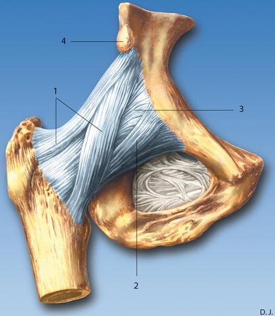
Fig. 67.2
Ligaments of the hip joint (ventral view). (1) Iliofemoral ligament, (2) pubofemoral ligament, (3) articular capsule, (4) anterior inferior iliac spine (Reproduced with permission from Danilo Jankovic)
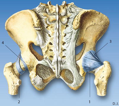
Fig. 67.3
Ligaments of the hip joint (dorsal view). (1) Ischiofemoral ligament, (2) orbicular zone, (3) iliofemoral ligament, (4) acetabular labrum (Reproduced with permission from Danilo Jankovic)
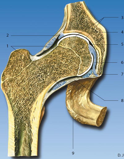
Fig. 67.4
Dissection through the hip joint. (1) Orbicular zone, (2) acetabular labrum, (3) articular cartilage, (4) os coxae, (5) cavum articulare, (6) ligament of head of femur, (7) transverse acetabular ligament, (8) joint capsule, (9) tuber ischiadicum (Reproduced with permission from Danilo Jankovic)
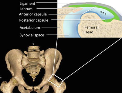
Fig. 67.5
Figure shows the anterior synovial recess (***). Under normal circumstances, the amount of synovial fluid in the recess is kept at a minimum. This figure shows a hip with effusion for demonstration (Reproduced with permission from Philip Peng Educational Series)
The capsule is reinforced by the extracapsular hip ligaments: iliofemoral, ischiofemoral, and pubofemoral ligaments (Figs. 67.2 and 67.3). In the anterior hip regions, the structures from medial to lateral are pectineus muscle, femoral neurovascular bundle, iliopsoas muscle and tendon, rectus femoris, and sartorius muscle (Figs. 67.6, 67.7, 67.8, and 67.9).
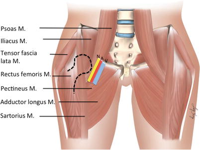
Fig. 67.6
Muscles (M) around hip joint. The femoral head and neck (in dotted line) and the schematic of femoral neurovascular bundle are shown here for reference. V femoral vein, A femoral artery, N femoral nerve (Reproduced with permission from Philip Peng Educational Series)
Fig. 67.7
Frontal dissection through the pelvis. (1) Obturator internus muscle, (2) iliacus muscle, (3) gluteus minimus muscle, (4) gluteus medius muscle, (5) gluteus maximus muscle, (6) greater trochanter, (7) obturator externus muscle, (8) iliopsoas muscle, (9) pectineus muscle, (10) deep femoral artery, (11) vastus lateralis muscle, (12) adductor brevis muscle, (13) pubic bone (inferior ramus), (14) medial femoral circumflex artery, (15) iliofemoral ligament, (16) acetabular labrum, (17) ligament of head of femur (Reproduced with permission from Danilo Jankovic)
Fig. 67.8
Transversal dissection through the hip joint. (1) Femoral vein, (2) femoral artery, (3) femoral nerve, (4) sartorius muscle, (5) rectus femoris muscle, (6) tensor fasciae latae muscle, (7) iliopsoas muscle, (8) obturator nerves with vasa obturatoria, (9) greater trochanter, (10) ischial spine, (11) sciatic nerve, (12) rectum, (13) bladder, (14) neck of the femur, (15) orbicular zone, (16) fovea capitis, (17) ligamentum of head of femur, (18) acetabular labrum, (19) pubic bone, (20) pectineus muscle (Reproduced with permission from Danilo Jankovic)
Fig. 67.9
Transversal dissection. (1) Sartorius muscle, (2) rectus femoris muscle, (3) tensor fasciae latae muscle, (4) iliofemoral ligament, (5) vastus lateralis muscle, (6) iliotibial tractus, (7) gluteus maximus muscle, (8) iliopsoas muscle, (9) sciatic nerve (Reproduced with permission from Danilo Jankovic)
The greater trochanter (GT) comprises of four facets: anterior, lateral, superoposterior, and posterior (Fig. 67.10). The anterior and posterior tendons of the gluteus medius insert into the lateral and superoposterior facets respectively. The tendon of the gluteus minimus inserts into the anterior facet. The muscles in the lateral region are divided into two layers. The superficial layer comprises the tensor fascia lata, and gluteus maximus muscle with the triangular interval between these two muscles is filled with fascia lata (Fig. 67.11). The iliotibial (IT) tract is a thickening of the fascia lata commencing at the level of greater trochanter, where three quarter of gluteus maximus muscle and tensor fascia lata are inserted into it. The deep layer comprises of gluteus medius and minimus muscle. Three bursae are described consistently in this region (Fig. 67.12), namely, the subgluteus maximus bursa (SMaB), the submedius bursa (SMeB), and the subminimus bursa (SMiB). The function of the bursae is to serve as a cushion against friction between tendons and fascia lata. The tendons of gluteus minimus and medius can be considered as the rotator cuffs of the hip joint with the IT band and fascia lata as the coracoacromial arch. In both situations, the tendons are covered with bursa against friction.


