, Corinna Eleni Psomadakis2 and Bobby Buka3
(1)
Department of Family Medicine, Mount Sinai School of Medicine Attending Mount Sinai Doctors/Beth Israel Medical Group-Williamsburg, Brooklyn, NY, USA
(2)
School of Medicine Imperial College London, London, UK
(3)
Department of Dermatology, Mount Sinai School of Medicine, New York, NY, USA
Keywords
Molluscum contagiosumVirusPoxvirusContactSkin-to-skinChildhoodPediatricPapuleGenitalSexual transmissionSexually transmittedInfectionSTDSTICantharidinImiquimodCryotherapyCryosurgeryLiquid nitrogenCurettageGenital wartsCondylomata acuminataAnogenital wartsHPVHuman papillomavirusCancerPodophyllotoxin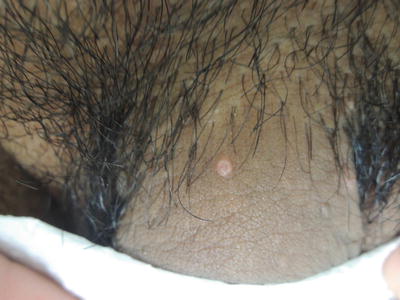
Fig. 32.1
Variably umbilicated pink shiny 3–4 mm papules on penile shaft

Fig. 32.2
Flesh-toned flat topped verrucous papules on penile shaft
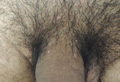
Fig. 32.3
Note here the difference between molluscum and warts found here concurrently on penile shaft
Primary Care Visit Report
A 27-year-old male with past medical history of genital warts presented with new lesions in his genital area. The wart-like lesions had been present for about 1 year. The smaller pink lesions had been present for 2–3 weeks. All were non-itchy. The patient had been to a urologist 3 weeks prior, who advised the lesions were both molluscum contagiosum and genital warts. The urologist gave the patient a prescription for cryo sticks to use at home, but he had not been able to find these in any pharmacy.
Vitals were normal. On examination, there were approximately 20 lesions scattered on his pubic mound, shaft of penis and medial thighs. The smallest lesion was 1–2 mm, the largest was about 7–8 mm. Some lesions were irregularly textured flesh-colored papules, and other lesions were smooth, erythematous papules with central umbilication.
About 20 lesions were treated with cryotherapy , treating with two sets of 30 s of iceball formation per lesion (with complete thawing in between each set). The patient was also given Aldara (imiquimod ) 5 % cream to use three times per week after the lesions had healed from cryotherapy.
The patient returned 2 weeks later and said all lesions that were frozen with cryotherapy had fallen off but he also noticed that some new lesions had formed. Cryotherapy was again performed on all remaining lesions. The patient never used the Aldara cream.
Discussion from Dermatology Clinic
Dermatology was in agreement with the urologist that the penis featured both molluscum contagiosum and genital warts . Both conditions are discussed separately below.
Molluscum Contagiosum
Differential Dx
Molluscum contagiosum
Verruca
Favored Dx
The lesions at the base and proximal shaft of the penis have a smooth, pearly appearance consistent with molluscum contagiosum.
Overview
Molluscum contagiosum (MC) is a skin infection caused by the Molluscum Contagiosum virus (MCV), a type of poxvirus . It is spread by person-to-person contact. MC is a common infection in children, with incidence rates estimated between 5.6 and 7.4 %, and much higher during outbreaks in closed communities [1]. Children with atopic dermatitis are more susceptible to MC, and have a higher likelihood of developing multiple lesions and a prolonged course of illness [1–3]. Adulthood MC primarily affects the genital region and is considered a sexually transmitted disease [4].
MC is usually benign and self-limiting in healthy individuals; however, it may take 6–12 months to resolve [1]. Immunocompromised persons, including HIV-infected individuals, can develop very large lesions (>15 mm), overlaying bacterial superinfections, and a prolonged and more severe course of disease [2]. The prevalence in HIV-infected people may be as high as 18 % [1]. Patients with HIV/AIDS are more likely to develop lesions on extragenital sites.
Presentation
MC lesions typically present as multiple, skin-colored or pearly-white dome-shaped papules that are 2–5 mm in size. Lesions often feature a central depression, referred to in the literature as an umbilication. There may be surrounding erythema and eczematous patches, sometimes called molluscum dermatitis [3]. Typically 1–20 lesions present on a healthy patient; however, hundreds can appear in some cases. Children can develop lesions anywhere, while adults typically present with lesions on the genitals, perineum, inner thighs, mons pubis, and lower abdomen. MC lesions may appear in a linear arrangement if patients have scratched and spread them. Scratching can also cause vulnerability to secondary infections, which may be noted.
Workup
MC is typically a clinical diagnosis. A biopsy is indicated if a deep fungal infection such as histoplasmosis is on the list of differential diagnoses for any severely immunocompromised people.
Treatment
MC is typically a self-limiting infection; however, aesthetic and contagion concerns, or prolonged presence of lesions may prompt individuals to seek treatment. Immunocompromised patients with MC should be referred to dermatology or an infectious disease specialist.
MC lesions are treated by physical destruction. This may be achieved by applying cantharidin , salicylic acid, or retinoids. Physical destruction may also be achieved by cryosurgery , or curettage ; however, there are considerations for associated pain and a slight risk of scarring with those procedures [2]. A recent Cochrane Database review did not find enough evidence to support any single therapy against molluscum, and treatment is not necessary due to the benign, self-limiting nature in most cases [1]. Treatment options should be thoroughly discussed with the patient.
For pediatric patients, cantharidin 0.7 % may be used to treat individual lesions. This is often applied using the wood end of a cotton-tipped applicator. It must be carefully applied only to lesions and not to healthy skin, and rinsed off after 4 h. Cantharidin causes blistering within 1–2 days, and the lesion typically resolves within 1 week [5]. Clearance has been reported in up to 90 % of patients [6]. Rarely it may cause severe blistering that results in scarring [3]. It is not recommended for use on the face or genital area [4]. Imiquimod 5 % cream is an immune response modifier that may be used to treat MC in adults; however, it is not approved as a treatment for MC in children. Clearance in 50 % of adult genital MC lesions has been noted [7]. It may cause erythema and pruritus in some patients.
The preferred treatment for adult genital MC is curettage or cryotherapy [2]. Topical anesthetic should be used prior to curettage as it can be painful. One study found that use of imiquimod combined with destruction led to lower recurrence rates [7]. Cryosurgery is a reliable mode of lesion destruction; however, it may be uncomfortable for patients with many lesions, and it may involve multiple treatments due to the appearance of new lesions. Both curettage and cryotherapy may cause scarring.
Follow-Up
If patients choose to treat MC, they may require multiple visits. Lesions should be reevaluated after 1–2 weeks to monitor healing, and patients should be examined for new lesions. Scratching the lesions should be avoided, as it makes the skin more susceptible to secondary bacterial infection. MC can spread by autoinoculation so patients should avoid touching lesions. There is no need to keep children away from school if they have MC. If there is concern of spreading the virus, the lesions may be covered with bandages. This also aids in avoiding touching the lesions. Adults should avoid intimate contact until the genital lesions have resolved.
Genital Warts
Differential Dx
Genital warts (AKA condylomata acuminata )
Molluscum contagiosum
Pearly penile papules
Condylomata lata of secondary syphilis
Premalignant or malignant tumor (e.g., squamous cell carcinoma, penile carcinoma in situ, Bowen disease of the penis)Related posts:

Stay updated, free articles. Join our Telegram channel

Full access? Get Clinical Tree


