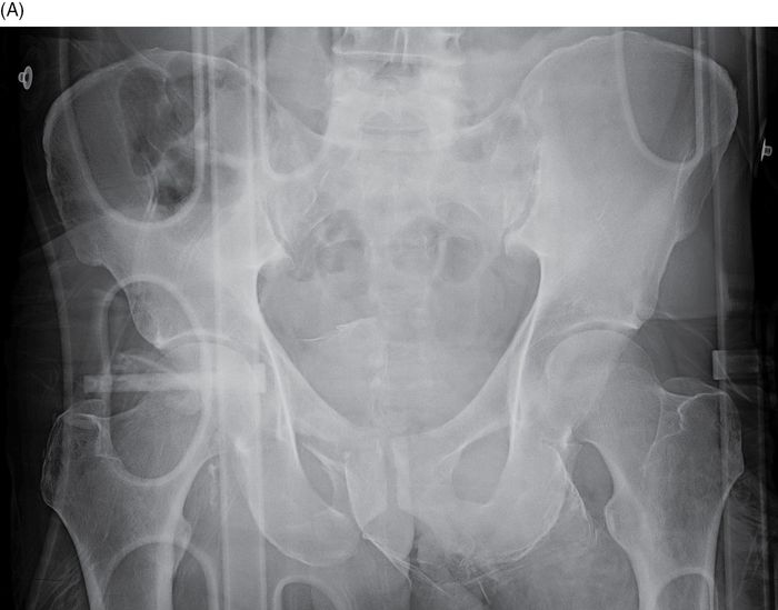
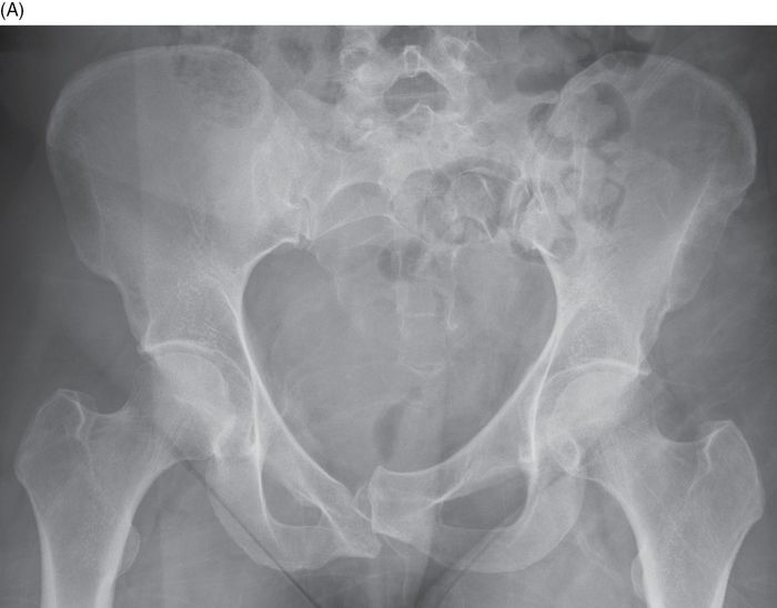
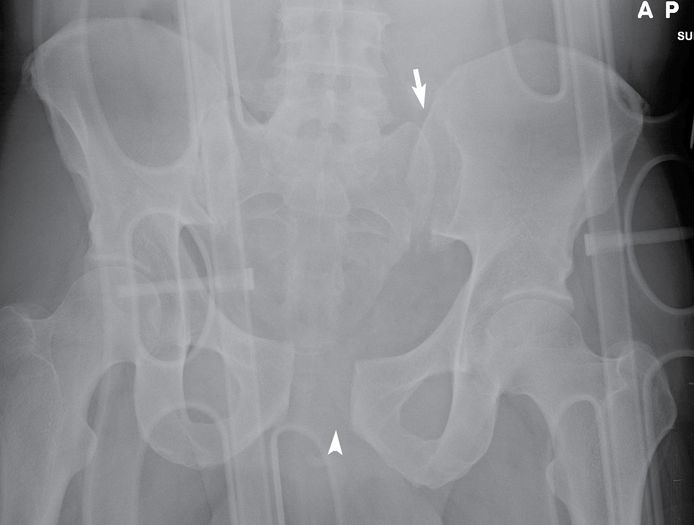
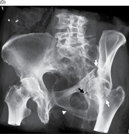
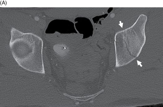
45-year-old man who was the passenger on a motorcycle that crashed into the back of a parked truck. AP radiograph of the pelvis. There is mild widening of the right sacroiliac joint and gross diastasis of the symphysis pubis.
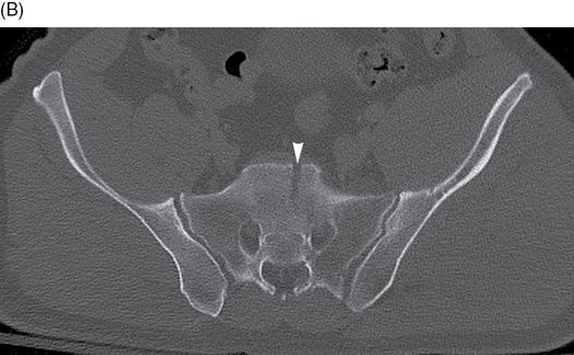
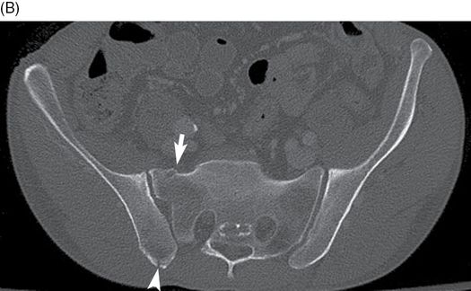
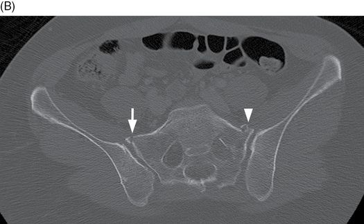
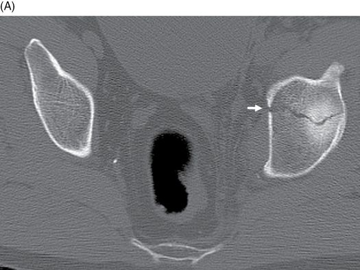
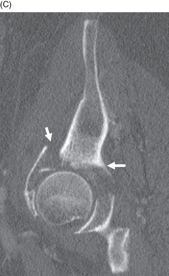
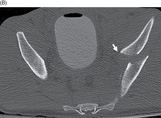
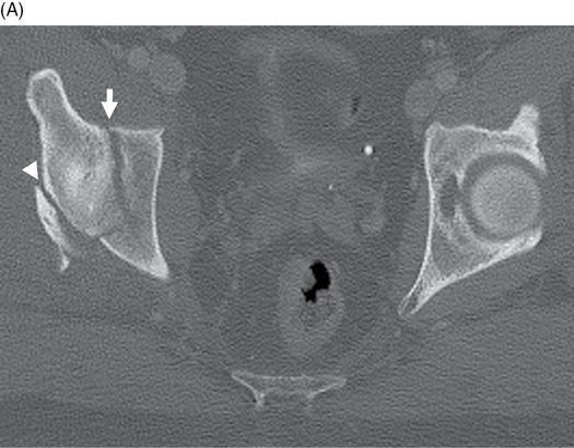
Axial CT of the pelvis, superior (B) to inferior (C). There is mild widening of the anterior aspect of the right SI joint (arrow), and a fracture of the sacrum that begins near the midline and extends through the left neural foramina (arrowheads). With severe anterior compression, the pelvis is flattened. The iliac wings rotate externally, resulting in disruption of the anterior and posterior arches of the pelvis under tension [1–2]. The anterior arch injury is typically diastasis of the symphysis pubis, vertical fractures of one or both obturator rings, or some combination; the posterior arch injury may be diastasis of one or both SI joints, fractures of the sacral wing, or some combination. Anterior compression injury is commonly sustained in automobile vehicle crashes in which the victim’s car hits head-on.
Case 9–2
Pelvis anterior compression injury
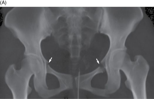
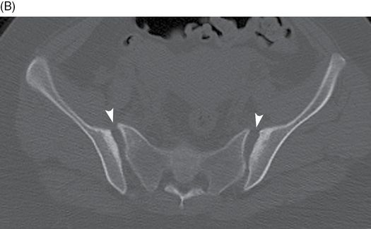
59-year-old man who was injured in a horseback riding accident. 3D volume-rendered CT of the pelvis. There is subtle bilateral sacroiliac joint diastasis and marked symphysis pubis diastasis. There are no fractures.
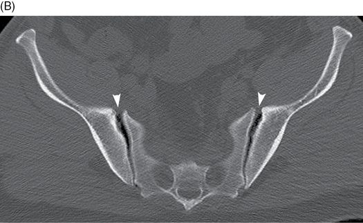
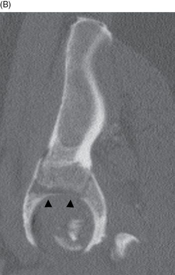
Axial CT of the pelvis. There is mild, symmetric widening of the anterior sacroiliac joints bilaterally (arrowheads). This pattern of injury is that of AP compression, sometimes called open-book injury.
Case 9–3
Pelvis straddle fractures
25-year-old woman in high-speed motor vehicle crash. 3D volume-rendered CT of the pelvis. There are fractures of the right and left superior (arrows) and right and left inferior pubic rami. This combination of fractures is descriptive of the appearance, but not of the mechanism. The straddle fracture is generally caused by tensile loading of the anterior pelvic arch, as occurs in anterior compression of the pelvis.
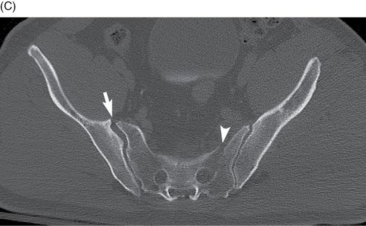
Axial CT of the pelvis. There is posterior dislocation of both SI joints. The bilateral SI joints are widened (arrowheads) and the iliac wings are posteriorly translated relative to the sacrum.
Case 9–4
Pelvis lateral compression injury
60-year-old man who fell 20 feet off a ladder, landing on his right side. AP radiograph of the pelvis. There are fractures of the right sacral wing and right obturator ring.

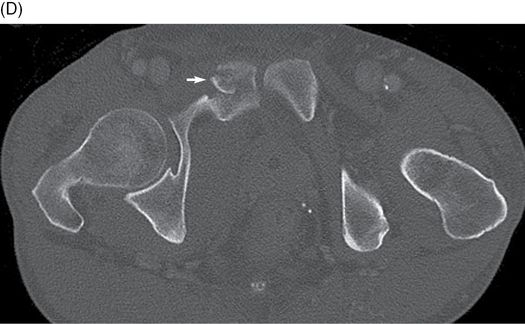
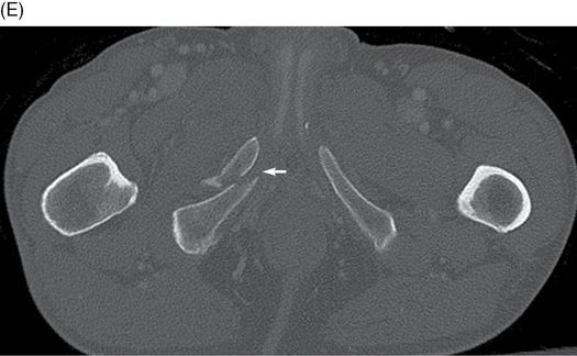
Axial CT of the pelvis, superior (B) to inferior (E). (B) There is a compression fracture of the right sacral wing (arrow), and a posterior distraction fracture of the right ilium (arrowhead). (C) At the level of the hip joints, the right acetabulum is intact but there is slight internal rotation of the right hemi-pelvis (long arrow shows direction of rotation). (D-E) There are fractures of the right pubis and right interior pubic ramus (small arrows). With lateral compression of the pelvis, the outboard iliac wing is rotated inward, typically resulting in some combination of an ipsilateral iliac wing fracture, impacted ipsilateral sacral wing fracture, or ipsilateral posterior sacroiliac joint distraction. The posterior sacroiliac may have sprains or avulsion fractures that are caused by the iliac wing rotating on the fulcrum of the sacrum. Fractures from a shearing mechanism will typically involve the anterior pelvic arch as it is compressed together [1–2].
Case 9–5
Windswept pelvis from lateral compression injury
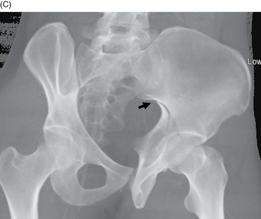
Woman injured in a motor vehicle crash. AP radiograph of the pelvis. The symphysis pubis has been disrupted, with overlap of the right and left pubic bones in the axial plane. There are fractures of the left sacral wing and subtle diastasis of the right sacroiliac joint. There is inward rotation of the left iliac wing and outward rotation of the right iliac wing.
Axial CT of the pelvis. At the upper sacrum (B), there is an impaction fracture (arrowhead) of the left sacral wing that involves the neural foramen. There is an avulsion fracture of the right sacral wing (short arrow).
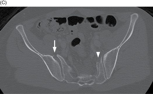
At the lower sacrum (C), the right sacroiliac joint is widened anteriorly (long arrow), but not posteriorly. The impaction fracture of the left sacrum (arrowhead) is seen. There is inward rotation of the left iliac wing, and outward rotation of the right iliac wing.
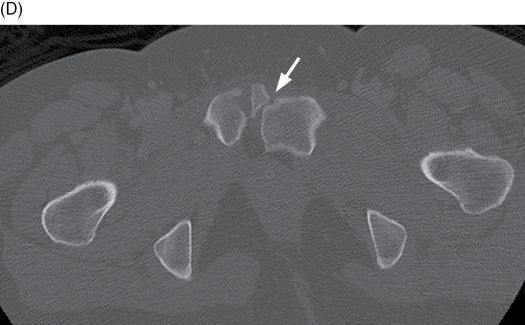
At the symphysis pubis (D), there is diastasis with the left pubis posterior to the right pubis, and a fragment sheared off the right pubis anteriorly (angled arrow).
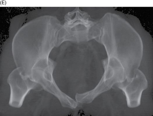
3D volume-rendered CT of the pelvis. 3D volume-rendered CT inlet view shows inward rotation of the left hemi-pelvis and outward rotation of the right hemi-pelvis. The windswept pelvis is the result of severe lateral compression [1–2].
Case 9–6
Pelvis vertical shear injury
36-year-old man who fell 40 feet and landed on his buttocks. AP radiograph of the pelvis. There are fractures through the left superior and inferior pubic rami (arrowhead), and superior dislocation of the left hemi-pelvis through the left sacroiliac joint (arrow). In vertical shear injuries, one hemi-pelvis is typically displaced superiorly relative to the other, with disruptions involving both the anterior and posterior arches. The anterior disruption may be through the symphysis pubis or either obturator ring, and the posterior disruption may be through either sacroiliac joint, either sacral wing, either iliac wing, or some combination [1–2].
Case 9–7
Left hemi-pelvis dislocation
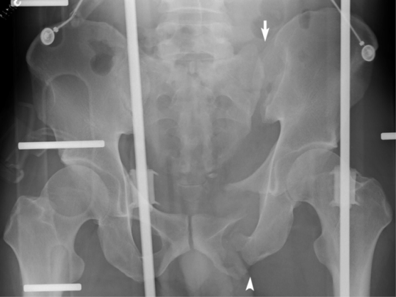
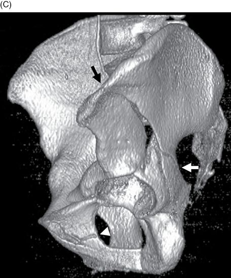
38-year-old man bucked from a horse. AP radiograph of the pelvis. The left sacroiliac joint (arrow) and symphysis pubis (arrowhead) are widely diastatic with lateral displacement of the otherwise intact left hemi-pelvis. There is a vertical fracture through the left L5 transverse process. In general, the outcome of pelvic ring injuries is less dependent on the morphology of the injury and more dependent on the associated visceral injuries that the patient has sustained [3].
Case 9–8
Anterior column acetabular fracture
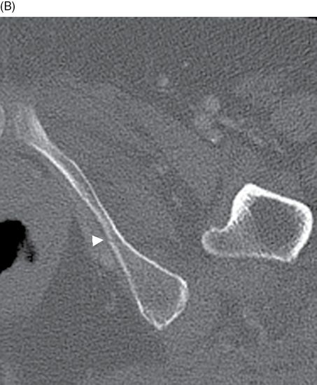
54-year-old man who fell from a ladder. Axial (A), sagittal (B), and 3D surface-rendered (C) CT of the left hip. Axial CT image shows a transversely oriented fracture line involving the anterior column (white arrow). Sagittal CT image shows a vertically oriented fracture line involving the anterior column (black arrows). 3D surface-rendered CT shows anterior column fracture extending to the left ilium (black arrowhead). Anatomically, the acetabulum consists of anterior and posterior columns, configured like an inverted Y. The anterior column extends from the upper sacrum to the anterior pubic ramus, forming two limbs of the Y, and the posterior column extends from the posterosuperior iliac spine to the ischial tuberosity, forming the third limb of the Y. Each column has an articular surface or wall. Acetabular fractures may be described according to involvement of columns and walls and direction of fracture [4–5].
Case 9–9
Anterior column acetabular fracture
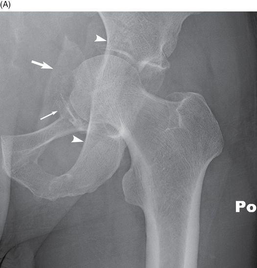
54-year-old man in motor vehicle crash. AP radiograph of the left hip. The femoral head appears as if it had been punched through the acetabulum into the pelvic cavity. The iliopubic line is interrupted where there are displaced fractures of the anterior column (long arrow) and anteromedial displacement of the femoral head. A large fragment of the medial wall (arrow) has been displaced medially with the femoral head. The ilioischial line (arrowheads) has not been interrupted indicating that the posterior column is intact. In all likelihood, this patient was the driver of a car that was struck by another vehicle from the left side.


Axial CT of the pelvis, superior (B) to inferior (D). The medial wall of the acetabulum is fractured and displaced medially (arrowhead). The femoral head is dislocated medially from the posterior acetabulum (long arrow), which is still attached to the pelvis, but the articular surfaces of the displaced medial and anterior acetabular fragments remain in contact with the femoral head (short arrow).
Case 9–10
T-type acetabular fracture
76-year-old woman who fell on her left hip while golfing. Multiplanar (A, B, C) and 3D volume-rendered (D) CT of the left hip. (A) Axial CT shows a sagittally oriented fracture line at the level of acetabular roof (white arrows). There is a coronally oriented second fracture line (dotted white arrow). (B) Axial CT at the level of ischium shows a non-displaced fracture at left ischium (white arrowhead). (C) Sagittal CT at the columnar junction shows that the fracture involves anterior and posterior columns as well as the quadrilateral plate (white arrows). (D) 3D volume-rendered CT shows that the fracture has a transverse fracture (white arrows) and a second fracture (black arrow) with a component extending into the obturator ring (white arrowhead).
Case 9–11
Both column acetabular fracture
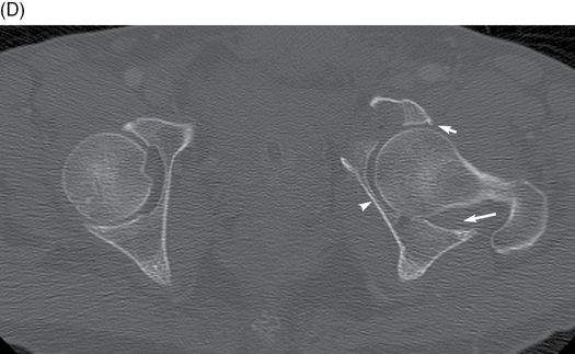
20-year-old man with motor vehicle crash injuries. Axial (A, B) and 3D surface-rendered (C) CT of the left hip. (A) Axial CT at the level of the quadrilateral plate shows comminuted fracture involving anterior and posterior columns of left acetabulum. (B) Axial CT at the level of the ilium shows a spur sign. A spur sign is only seen in both column fractures, and it is a differential point from T-type fracture (white arrow). The spur sign is shown only on the ipsilateral obturator oblique view and is created by medial translation of the distal fragment. The visualized spur fragment is simply the inferior end of the weight-bearing strut of bone in communication with the axial skeleton. On CT, this fragment is easier to see because one can trace down the weight-bearing strut of bone in continuity with the axial skeleton from the sacroiliac joint across the sciatic buttress. The fragment of bone never descends down to the acetabular surface in the setting of a both-column fracture, thus creating the “CT spur” sign. (C) 3D surface-rendered CT shows fracture involving anterior column (white arrow), posterior column (black arrow), and the obturator ring (white arrowhead).
Case 9–12
Posterior column acetabular fracture
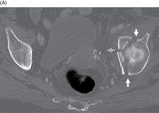
21-year-old man with injuries from a high-speed motor vehicle crash. Multiplanar (A, B) and 3D volume-rendered (C) CT of the pelvis and left hip. (A) Axial CT at the level of the sciatic notch shows sagittally oriented fracture line involving the posterior column (white arrows). (B) Sagittal CT at the level of columnar junction shows the fracture extending to the sciatic notch (black arrow). (C) 3D volume-rendered CT shows fracture extending to the sciatic notch (black arrow). The fracture extends inferiorly to involve the obturator ring (not shown).
Case 9–13
Transverse and posterior wall acetabular fracture
Related posts:


Stay updated, free articles. Join our Telegram channel

Full access? Get Clinical Tree


