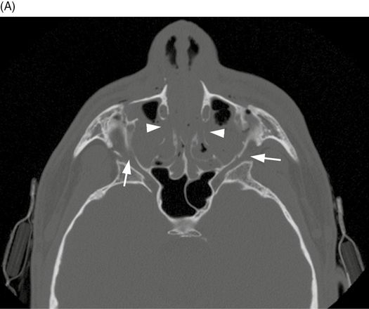
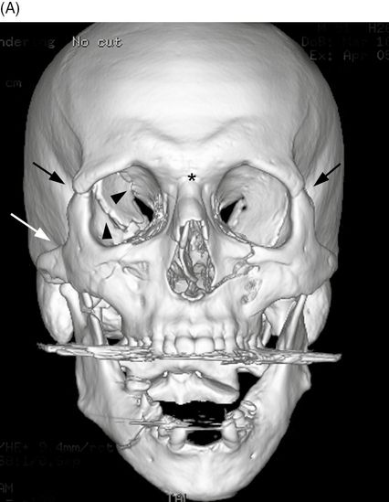
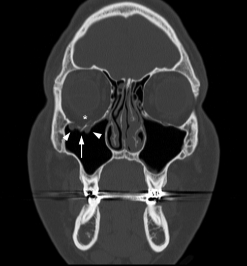
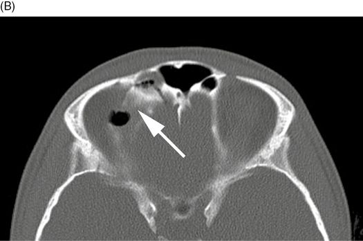
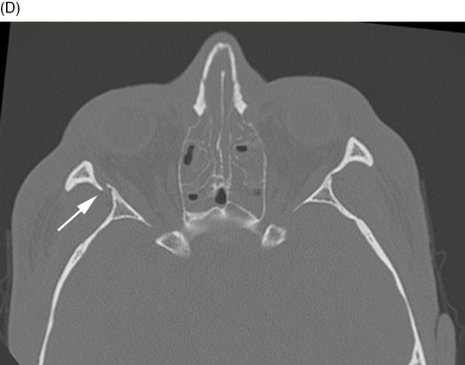
44-year-old man who fell. Axial CT of the face. The tripod fracture is the most common zygomatico-orbital fracture. The most common cause is being struck in the face during an altercation. As might be expected, mandibular fractures often occur in association with tripod fractures [1]. Three fractures comprise the tripod fracture pattern as shown. Fractures of the left zygomatic arch (long white arrow), anterior (*), and lateral walls (short white arrow) of the left maxillary sinus are seen in the axial CT image through the level of the maxillary sinuses.
Coronal CT of the face. Coronal CT image through the orbits shows a fracture of the lateral wall of the left orbit (arrow).
Case 15–2
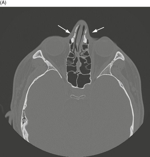
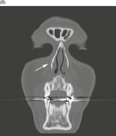
Le Fort fracture, Type I
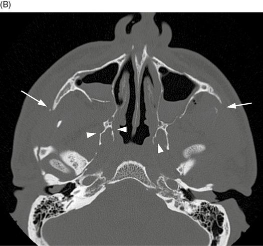
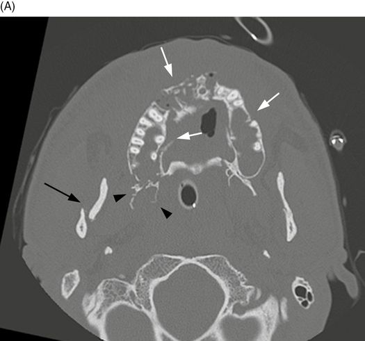
Adolescent male who fell off his bicycle. Axial CT of the face. Le Fort I fractures separate the palate from the maxilla [2–3]. In the axial CT, the medial walls (arrowheads) and lateral walls (short white arrows) of both maxillary sinuses are fractured.

Coronal CT of the face. Bilateral pterygoid plate fractures are shown in the coronal CT (arrows). The teeth of the maxilla would move relative to the rest of the face on physical exam.
Case 15–3
Le Fort fracture, Type II
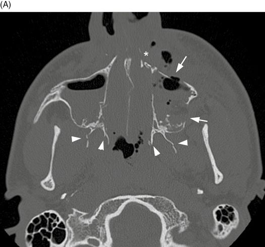
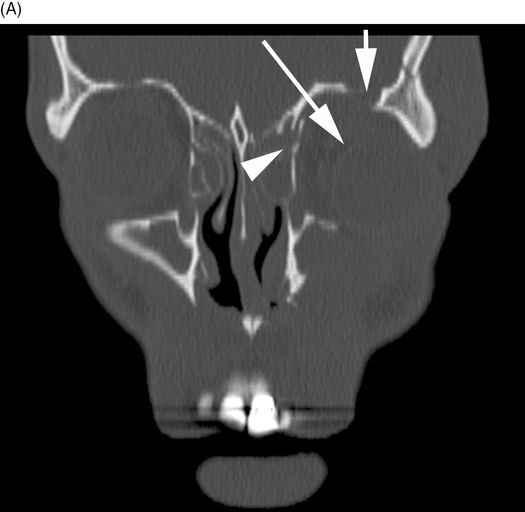
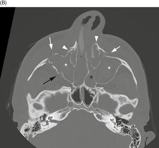
Facial injuries sustained in a motorbike crash. Axial CT of the face. In Le Fort II, or pyramidal fractures, the fractures form a pyramid around the nose. In the axial CT image through the maxillary sinuses, fracture lines pass across the left nasal bone (*), through the left anterior and posterolateral maxillary sinus walls (white arrows) and bilateral pterygoid plates (arrowheads).
3D surface-rendered CT of the face. Oblique 3D CT of the right side of the face shows the pyramidal nature of the fracture (black line) and the involvement of the medial (arrow) and inferior orbital rims (arrowheads). In these fractures, the nose and teeth would move as a unit relative to the rest of the face [2–3]. Le Fort fractures may be asymmetric, with different types of fractures on each side of the face.
Case 15–4
Le Fort fracture, Type III
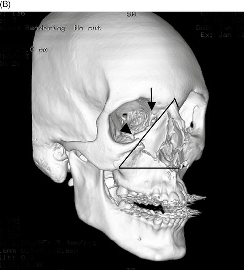
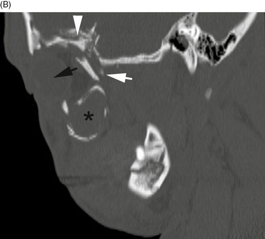
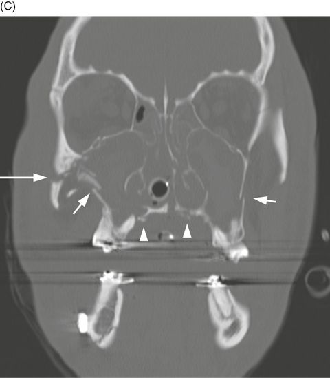
Multiple facial fractures sustained in a fall. 3D surface-rendered CT of the face. Instead of passing inferiorly through the front of the face, Le Fort III fractures continue to extend laterally through the lateral orbital wall, zygomaticofrontal suture and zygomatic arch [2–3]. This is usually a high-energy injury, and fractures will also pass through the ethmoid, vomer, and pterygoid plates. These fractures result in complete separation of the face from the rest of the skull. In this frontal 3D CT reconstruction, fractures are seen across the nasofrontal suture (*), right posterior and lateral orbit (black arrowheads), bilateral zygomaticofrontal sutures (black arrows) and right zygomatic arch (white arrow).
Axial CT of the face. Image through the maxillary sinuses shows fractures through both zygomatic arches (white arrows) and pterygoid plates (arrowheads).
Axial CT of the face. Image through the orbits shows bilateral lateral orbital wall fractures (arrows) to advantage. Comminuted nasal fractures are also seen (arrowheads).
Case 15–5
Isolated zygomatic arch fracture
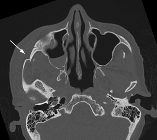
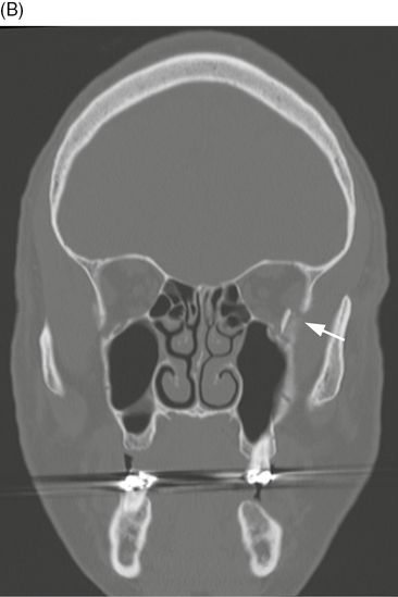
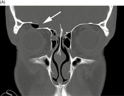
Elderly woman who fell. Axial CT of the face. Usually from a direct blow to the cheek, a zygomatic arch fracture causes flattening of the cheek contour and may affect masseter muscle activity or impinge the temporalis muscle [1]. In this axial CT through the level of the right zygomatic arch, a comminuted zygoma fracture with mild depression is demonstrated (arrow).
Case 15–6
Orbital blowout fracture
Impact injury to the right eye. Coronal CT of the face. In the orbital blowout fracture, pressure increases in the orbit and decompresses through the orbital floor. Fat often herniates into the maxillary sinus, but if the inferior rectus muscle is involved with the fracture fragments, it may become trapped and cause diplopia [4]. This patient also sustained a non-displaced right nasal bone fracture (not shown). In this coronal CT through the orbits, a comminuted fracture of the right orbital floor is shown (white arrow). Small amounts of orbital fat herniate (arrowheads) into the right maxillary sinus without additional herniation of the nearby inferior rectus muscle (*).
Case 15–7
Orbital blowin fracture
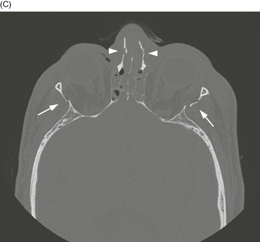
Coronal CT of the face. Orbital blowin fractures push fragments into the postseptal orbit, the opposite of the orbital blowout fracture. Characteristics include decreased orbital volume, proptosis and displacement of the globe in the orbital plane. Common complicating features of the orbital blowin fracture include restricted globe mobility (24%) and diplopia (32%). Less common complications include globe rupture, superior orbital fissure syndrome and optic nerve injury [5]. On this coronal CT image, fractures of the orbital roof (short arrow) and medial orbital wall (arrowhead) decrease the volume of the orbit, displacing the globe inferolaterally (long arrow).
Sagittal CT of the face. Sagittal CT image shows fracture fragments from the posterolateral orbit (white arrow) and orbital roof (arrowhead) causing anterior globe displacement and proptosis (black arrow). Maxillary sinus full of hemorrhage also shown (*). This case demonstrates a so-called pure fracture involving only the roof, floor, or walls. Impure fractures also involve the orbital rim.
Case 15–8
Orbital roof fracture
Blunt trauma to the face. Coronal CT of the face. Similar in mechanism to the orbital blowout fracture, orbital roof fractures occur with blunt force to the globe and orbit [6]. In this coronal CT, discontinuity of the orbital roof with a small focus of air (arrow) is identified.
Axial CT of the face. Superior ethmoid air cell origin of the air is identified on the axial CT (arrow).
Case 15–9
Facial smash fracture
High speed motor vehicle crash. Axial CT of the face. As a result of high-energy trauma such as a motor vehicle crash, comminution of most of the facial structures results. Axial CT through the maxilla. Fractures of the alveolar ridge (white arrows), right pterygoid plates (black arrowheads) and right mandible (black arrow) are present.
Axial CT of the face. Axial CT through the maxillary sinuses. The sinuses are opacified with blood (*) as a result of the fractures through the right lateral maxillary sinus wall (black arrow), bilateral anterior maxillary sinus walls (white arrows) and nasolacrimal ducts (arrowheads).
Coronal CT of the face. Coronal CT through the midface. Fractures of the lateral maxillary sinus walls (short white arrows), hard palate (arrowheads), and right zygomaticomaxillary suture (long white arrow) are present.
Axial CT of the face. Axial CT through the orbits. A right lateral orbital wall fracture is present (arrow). This patient’s injuries follow the central facial smash pattern [7–8].
Case 15–10
Nasal fracture
Young man with facial trauma. Axial CT of the face. Nasal fracture is a common result of assault or sporting injuries. For anything more than a mild, closed unilateral fracture, open reduction is frequently necessary [9]. Axial CT through the nasofrontal suture shows non-displaced right and left nasal fractures (arrows).
Related posts:


Stay updated, free articles. Join our Telegram channel

Full access? Get Clinical Tree


