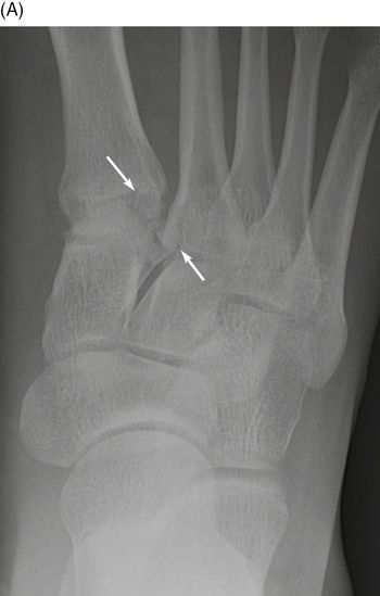
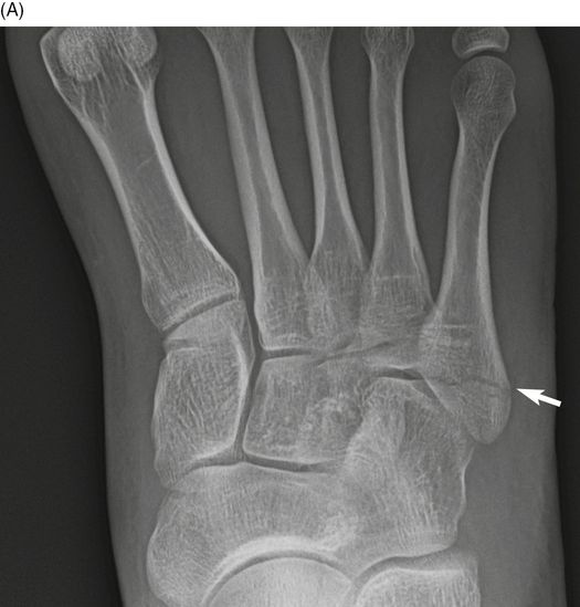
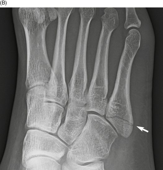
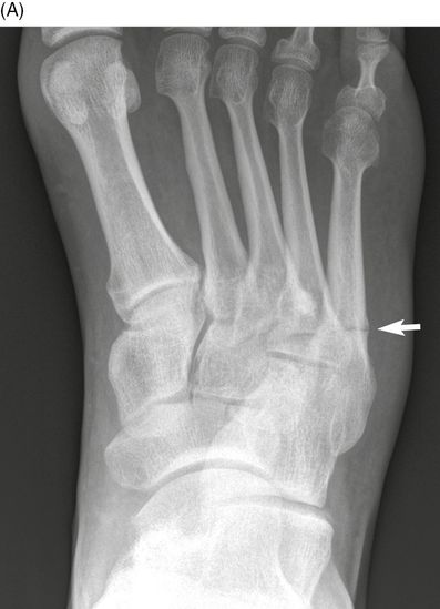
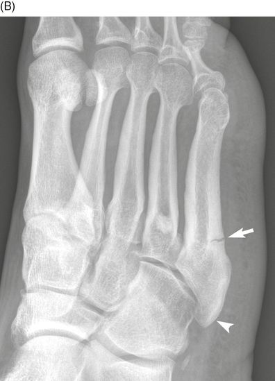
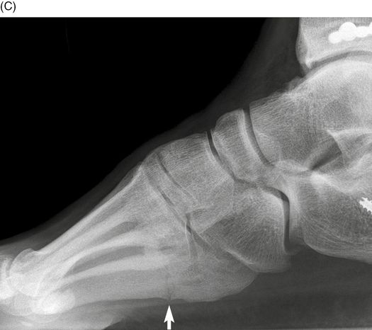
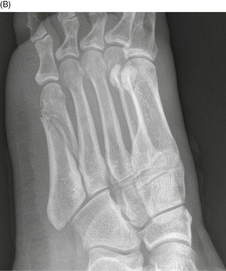
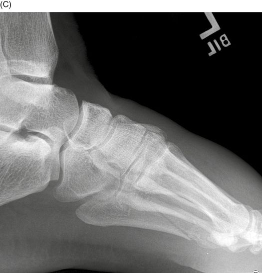
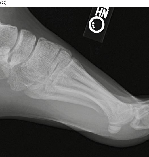
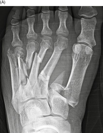
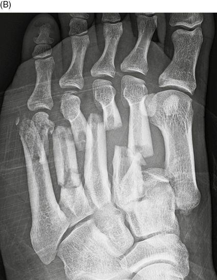
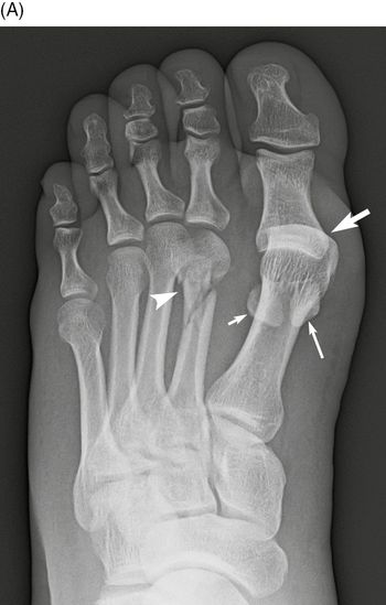
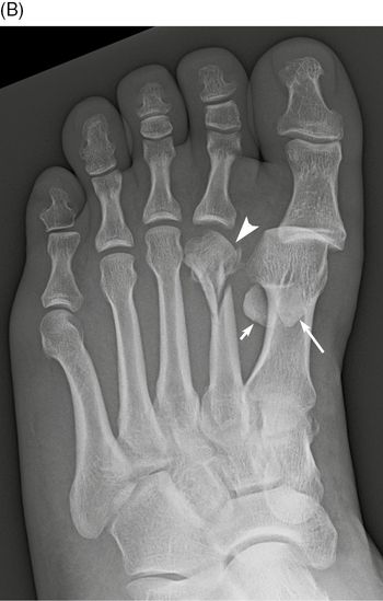
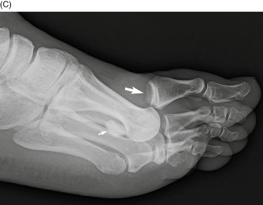
20-year-old woman who had a twisting foot injury on a trampoline. AP (A), oblique (B), and lateral (C) radiographs of the right foot. There is lateral subluxation of the first through fifth metatarsals at the tarsometatarsal (TMT) joints. The entire forefoot has been laterally displaced such that the first metatarsal is now aligned with the medial-middle cuneiform joint, and all the metatarsals are dorsally subluxated as a unit with respect to the tarsals. Small avulsed fragments are noted at the metatarsal bases on the lateral view (arrows). There is dorsal and medial soft tissue swelling around the midfoot. The Lisfranc injury is a dislocation of the TMT joints. There are three basic patterns of injury according to the Ouenu and Kuss classification [1–2]: the homolateral type is dislocation of all the metatarsals together; the divergent type is dislocation of metatarsals in separate directions; the isolated type is dislocation of some but not all the metatarsals. This case is a homolateral type, with lateral dislocation.
Case 14–2
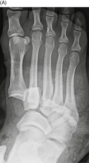
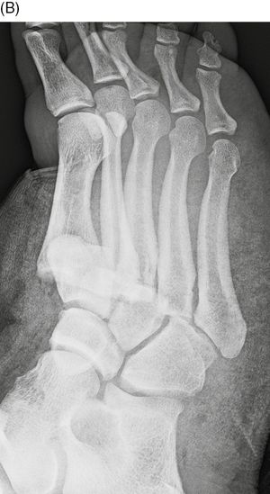
Lisfranc fracture-dislocation (divergent type)
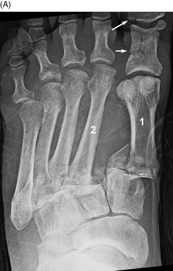
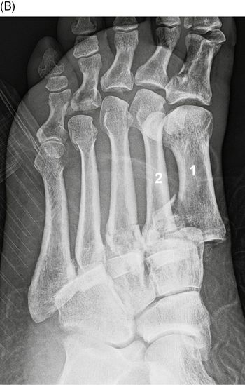
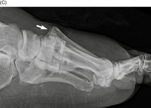
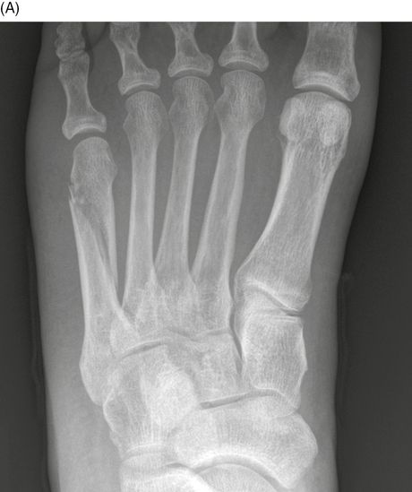
53-year-old woman injured in a motor vehicle crash. AP (A), oblique (B), and lateral (C) radiographs of the left foot. There are lateral fracture-dislocations of the second through fifth TMT joints. There is a fracture-dislocation of the first TMT joint and the metatarsal has been displaced medially and dorsally (large arrow). There are also fractures of the proximal (short arrow) and distal (long arrow) phalanges of the great toe. This case is an example of the divergent type of Lisfranc injury. The Lisfranc injury is named for a military surgeon from the Napoleonic wars who described forefoot fracture-dislocations that occurred in cavalrymen who had been unhorsed and dragged with a foot still in a stirrup. The contemporary bicyclist with feet strapped or clipped to the pedals may suffer the analogous injury.
Case 14–3
Lisfranc fracture-dislocation (homolateral type)
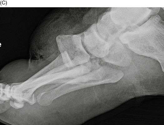
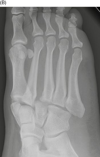
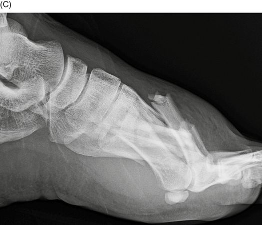
40-year-old man injured in motorcycle crash. AP (A), oblique (B), and lateral (C) radiographs of the right foot. There is plantar dislocation of all five TMT joints, with extensive degloving injury of the dorsal soft tissues. Plantar dislocation of the forefoot is unusual. This is a homolateral type of Lisfranc dislocation, but in the plantar direction rather than the lateral direction.
Case 14–4
Occult Lisfranc fracture-dislocation
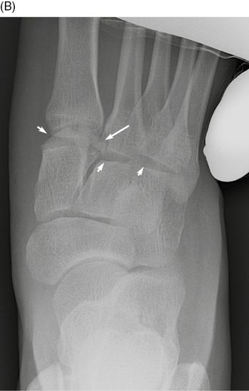
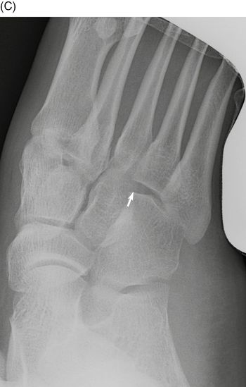
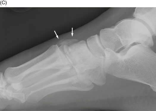
40-year-old man injured in motor vehicle crash. AP (A) and valgus stress (B-C) radiographs of the right foot. There are fracture fragments between the first and second TMT joints (long arrows) that are easy to overlook, but the joint spaces appear normal. With valgus stress, the Lisfranc joints subluxate (short arrows) and fracture fragments are more evident at the first and second TMT joints. The joint spaces should not widen under stress.
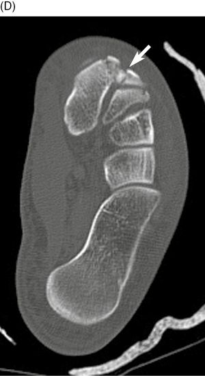
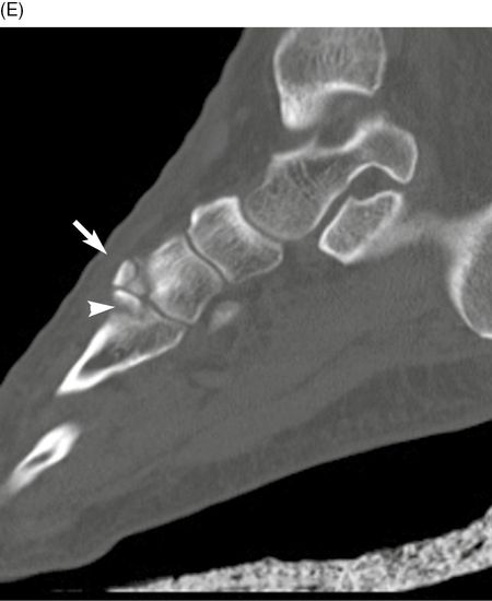
Multiplanar CT through the right midfoot, axial (D) and sagittal (E) images. There are small avulsion fractures at the TMT joints involving the medial and middle cuneiforms (arrows) and the bases of the first and second metatarsals (arrowhead).
Case 14–5
Isolated Lisfranc injury at first TMT joint
41-year-old woman who tripped on the hose at a gas station. AP radiograph of the right foot. There is widening of the space between the first and second metatarsal bases (arrow), and between the medial and middle cuneiforms (arrowhead). The first and second TMT joints appear properly aligned. The first ray was unstable on clinical exam. This case is an example of the isolated type of Lisfranc injury.
Case 14–6
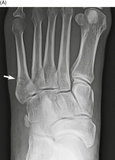
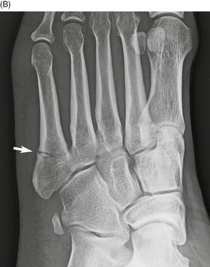
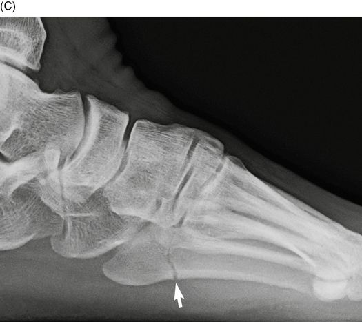
Fifth metatarsal tuberosity fracture
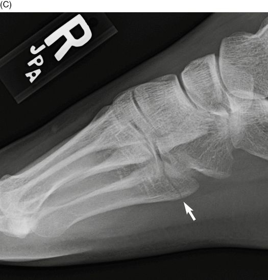
19-year-old woman who fell while walking. AP (A), oblique (B), and lateral (C) radiographs of the right foot. There is a non-displaced fracture of the base of the fifth metatarsal tuberosity (arrows). The fracture extends into the TMT joint. The fifth metatarsal is the most frequently fractured bone in the foot; the majority of fifth metatarsal fractures involve the proximal end [3–4]. The classification of proximal fifth metatarsal fractures is related to the location of the fracture relative to the fifth TMT and the fourth-fifth intermetatarsal joints [5]. The tuberosity fracture involves or is more proximal than the fifth TMT articular surface and is sometimes called a Zone 1 fracture. The size of the fracture fragment may range from one that involves the entire TMT joint to a tiny cortical avulsion fragment. These fractures generally heal well with symptomatic treatment, even when displaced [5–6]. These appear to be avulsion fractures of the lateral cord of the plantar aponeurosis [7–8].
Case 14–7
Fifth metatarsal Jones fracture
38-year-old man with a twisting injury to the left foot. AP (A), oblique (B), and lateral (C) radiographs of the left foot. There is a minimally displaced transverse fracture (arrow) of the proximal fifth metatarsal at the junction of the metaphysis and diaphysis, extending transversely into the fourth-fifth intermetatarsal joint. A fracture that involves this joint is a Jones fracture, sometimes called a Zone 2 fracture. The original description of Jones fractures designates fractures that occur 0.75 inch (2 cm) from the proximal tip of the metatarsal. There is evidence that more-proximal Jones fractures may be lumped with tuberosity fractures in terms of treatment and outcome, and perhaps be simply called metaphyseal fractures [6]. However, distal Jones fractures are at risk for complications of healing.
Case 14–8
Fifth metatarsal meta-diaphyseal fracture
34-year-old man who sustained an injury to the lateral side of his right foot playing basketball. AP (A), oblique (B), and lateral (C) radiographs of the right foot. There is an incomplete transverse fracture (arrow) at the junction of the diaphysis and metaphysis involving the inferolateral cortex of the fifth metatarsal. This fracture was treated with internal fixation by an intramedullary screw. The elongated styloid process of the base of the fifth metatarsal (arrowhead) is likely the sequel of remote trauma. Fifth diaphyseal stress fractures, sometimes called Zone 3, are located in a vascular watershed region and are at risk for complications of healing. They typically present with pain but a fracture line may not become apparent on radiographs until weeks after onset. Polzer et al. [6], who suggested combining tuberosity avulsion fractures with proximal Jones fractures, also advocate combining distal Jones fractures with diaphyseal stress fractures, calling them meta-diaphyseal fractures [6]. Unfortunately, the exact borderline does not appear to have an obvious radiographic landmark.
Case 14–9
Fifth metatarsal shaft fracture
49-year-old woman who fell while standing barefoot on furniture. AP (A), oblique (B), and lateral (C) radiographs of the left foot. There is a minimally displaced oblique fracture (arrows) at the fifth distal metatarsal shaft and neck. The morphology of the fracture suggests axial compression as the mechanism.
Case 14–10
Metatarsal shaft fractures
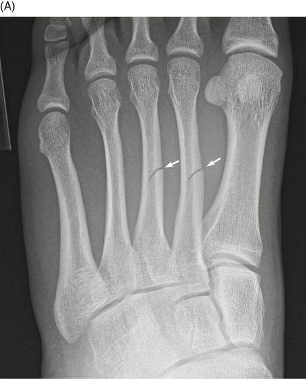
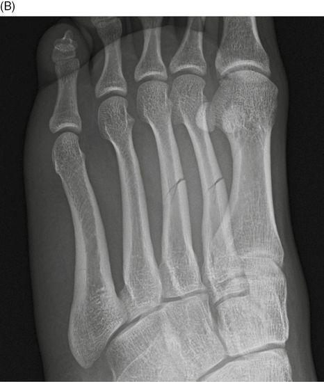
19-year-old man injured his foot playing soccer. AP (A), oblique (B), and lateral (C) radiographs of the left foot. There are transverse, incomplete fractures at the second and the third metatarsal mid-shafts (arrows). Fractures of the shaft and neck of the metatarsal bones and phalanges are often the result of heavy objects falling on the foot. Less commonly, the phalanges may be fractured as the foot strikes furniture or other objects. These fractures are transverse, oblique, or comminuted. Rarely a longitudinal linear fracture is encountered.
Case 14–11
Metatarsal shaft fractures
26-year-old man who injured his foot in a motorcycle crash. AP (A), oblique (B), and lateral (C) radiographs of the left foot. There is a fracture-dislocation of the first TMT joint with medial dislocation of the first metatarsal. There is a comminuted transverse fracture of the second metatarsal shaft. There is a transverse fracture of the third metatarsal shaft. There is a segmental fracture of the fourth metatarsal shaft and an osteochondral fracture of the fourth metatarsal head. There is a comminuted oblique fracture of the fifth metatarsal shaft and neck.
Case 14–12
Lateral sesamoid fracture
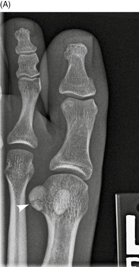
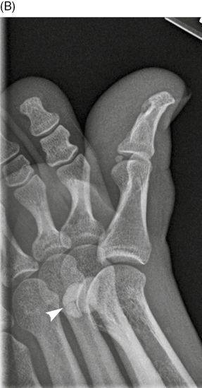
23-year-old woman with great toe pain the morning after marching many miles in a military reenactment. AP (A) and lateral (B) radiographs of the left great toe. There is a non-displaced transverse fracture of the lateral sesamoid of the great toe (arrows). A fracture may be distinguished from the more common bipartite sesamoid by the sharpness of the fracture line and the lack of cortical margins. In addition, the fracture fragments usually look as if they could fit back together perfectly, while the separate ossifications of a bipartite (or multipartite) bone typically do not. Most fractures of the sesamoids of the great toe are stress injuries [9–10].
Case 14–13
Medial sesamoid fracture
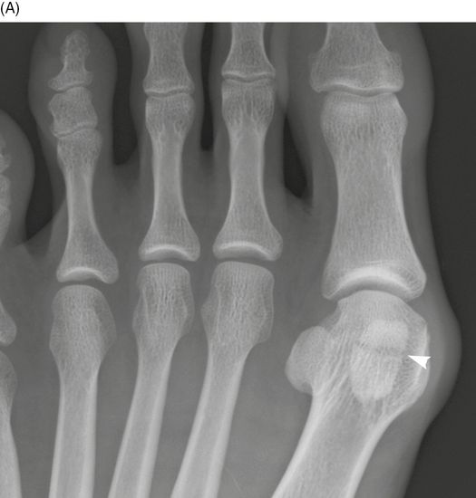
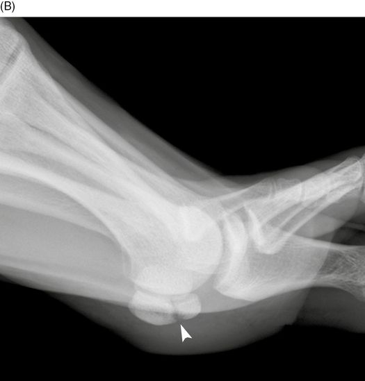
32-year-old woman who has had pain below the first metatarsophalangeal joint for 6 weeks. AP (A) and lateral (B) radiographs of the left great toe. There is a non-displaced transverse fracture of the medial sesamoid of the great toe (arrows). Bipartite or multipartite sesamoids are common findings, with one study reporting a prevalence of 14% and a preponderance of 82% on the medial side [11].
Case 14–14
First metatarsophalangeal dislocation
Related posts:


Stay updated, free articles. Join our Telegram channel

Full access? Get Clinical Tree


