Fig. 52.1
Anatomy. (1) Celiac plexus, (2) aorta, (3) inferior vena cava, (4) pancreas, (5) renal plexus, (6) abdominal aortic plexus, (7) inferior mesenteric ganglion, (8) inferior mesenteric plexus, (9) superior hypogastric plexus, (10) inferior hypogastric plexus (Reproduced with permission from Danilo Jankovic)
Fig 52.2
Transversal section through abdomen at the level of the first lumbar vertebra. (1) Celiac plexus with celiac ganglion, (2) spinal ganglion, (3) cauda equina, (4) medulla renalis, (5) aorta, (6) inferior vena cava, (7) psoas muscle, (8) quadratus lumborum muscle, (9) erector spinae muscle, (10) liver (Reproduced with permission from Danilo Jankovic)
Fig. 52.3
(1) Sympathetic trunk (underneath: greater and minor splanchnic nerves), (2) celiac plexus (underneath: superior mesenteric artery and renal artery), (3) vertebral body of the first lumbar vertebra with rami communicantes albi, (4) lumbar plexus (Reproduced with permission from Danilo Jankovic)
The celiac plexus is the largest of the three large sympathetic plexuses (cardiac plexus, thorax; celiac plexus, abdomen; hypogastric plexus, pelvis).
It receives its primary innervation from the preganglionic splanchnic nerves (greater splanchnic nerve T5–10, lesser splanchnic nerve T10–11, and least splanchnic nerve T11–12), the postganglionic fibers of which, after synapsing in the celiac ganglion, radiate to the associated plexus and innervate most of the abdominal organs. This large network, with a diameter of about 50 mm, surrounds the origins of the celiac artery and superior mesenteric artery and extends laterally as far as the adrenal glands, upward as far as the aortic hiatus, and downward as far as the root of the renal artery. It lies on the initial part of the abdominal aorta, at the level of the first lumbar vertebra, anterior to the medial crus of the diaphragm.
The most important roots of the celiac plexus are the splanchnic nerves, the abdominal branches of the vagus nerves, and several branches of the last thoracic ganglion and two highest lumbar ganglia. Cranially, the celiac plexus is connected to the thoracic aortic plexus, and caudally it continues into the abdominal aortic plexus. A paired celiac ganglion forms the basis for the celiac plexus although up to five ganglia may be involved.
The left ganglion lies closer to the midline and partly on the aorta, while the right one (ventrolateral to the vena cava) lies slightly more to the side in the area of the fissure between the medial and lateral crura of the diaphragm. The two ganglia are connected to one another. With closer approximation and fusion, the double ganglion takes on a ring shape, which is also known as the solar ganglion (solar plexus).
The smaller superior mesenteric ganglion and aorticorenal ganglion are associated with the celiac ganglion.
The lesser splanchnic nerve usually enters the latter ganglion, while the greater splanchnic nerve passes to the posterior surface of the lateral part of the celiac ganglion. The phrenic ganglion is a third, unpaired, ganglion. The following, sometimes paired and sometimes unpaired, secondary (associated) plexuses emerge from the celiac plexus:
Paired (phrenic plexus, suprarenal plexus, renal plexus, and spermatic plexus)
Unpaired (superior gastric plexus, hepatic plexus, splenic plexus, and superior mesenteric plexus)
Indications
Diagnostic
Differential diagnosis of pain in the abdominal region (visceral, epigastric, or cardiac pain).
Prognostic
In upper abdominal cancer pain, before a neurolytic block.
Therapeutic
1.
Relief of upper abdominal pain (pancreas, stomach)
2.
Treatment of the dumping syndrome
3.
Injection of neurolytics as palliation measure in malignant intra-abdominal disease-related pain (pancreas, stomach)
Contraindications
1.
Anticoagulant treatment
2.
Infections and skin diseases in the injection area
3.
Hypovolemia
4.
General debility
5.
Injection of a neurolytic without precise identification of the needle position (with guidance by CT or image intensifier)
Fluoroscopic Guided Procedure
This block should be carried out by very experienced anesthetists. Full information for the patient is mandatory.
Preparations
Check that the emergency equipment is complete and in working order: sterile precautions, intravenous access, ECG monitoring, pulse oximetry, intubation kit, ventilation facilities, and emergency medication.
Materials
Fine 26-G needle for local anesthesia. 20–22-G spinal needle, 0.7 (0.9) 120 mm (150 mm). Syringes: 2, 10 and 10 mL. Disinfectant, swabs, compresses, sterile gloves and drape, and flat, firm pillow.
Patient Positioning
1.
Prone position: supported with a pillow in the mid-abdomen (to relieve lumbar lordosis). The patient’s arms are dangling, with the head lying to the side. The patient breathes with the mouth open, to reduce tension in the back muscles. This position is preferable.
2.
Lateral decubitus position, with support under the flank. The side being blocked lies upward.
Location
1.
L4 iliac crest line (count the spinous processes cranially).
2.
L2: a parallel line is drawn about 7–8 cm lateral to the midline. The intersection between this line and the lower edge of the twelfth rib determines the level of the upper edge of L2.
3.
T12–L1 is located in the midline and joined with dots to the lower edge of the twelfth rib. This produces a triangle, the equal sides of which provide basic guidance for the needle direction (Fig. 52.4).
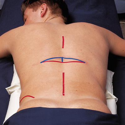
Fig. 52.4
Location (Reproduced with permission from Danilo Jankovic)
Skin prep, generous local anesthetic infiltration of the injection channel, covering with a sterile drape, drawing up the local anesthetic, checking the patency of the injection needle.
During the injection, the following points must be observed:
1.
Bilateral injection is usually unnecessary depending on the spread of the contrast agent.
2.
The operator performing the injection must stand on the side being blocked.
3.
In most patients, the distance between the skin and the celiac plexus is about 9–11 cm.
4.
Superficial bone contact (after 3–5 cm) indicates contact with the transverse process and requires cephalocaudad redirection.
5.
Paresthesias during the introduction of the needle arise due to stimulation of the lumbar somatic nerves.
6.


There is a risk of perforating the dural cuff (epidural or subarachnoid injection).
Related posts:


Stay updated, free articles. Join our Telegram channel
Full access? Get Clinical Tree
