Fig. 30.1
(a) The interscalene groove. Scalenus medius muscle (1) and scalenus anterior muscle (2). Injection of the local anesthetic into the proximal neurovascular sheath of the brachial plexus. The plexus is located in a kind of “sandwich” between the scalenus anterior muscle and scalenus medius muscle (With permission from Danilo Jankovic). (b) (1) Trunks of the brachial plexus with subclavian artery, (2) middle and anterior scalene muscles, (3) proximal supraclavicular plexus sheath, (4) clavicle, (5) strnocleidomastoid muscle (With permission from Danilo Jankovic)
Location of the Puncture Site
To locate the injection site, the patient’s arm is drawn in the direction of the knee (Fig. 30.2). The patient is asked to turn the head to the opposite side and to lift it slightly (ca. 20°), so that the posterior edge of the sternocleidomastoid muscle becomes evident (Fig. 30.3). The transverse process (C6) is palpated at the lateral edge of the sternocleidomastoid muscle. For confirmation (pleura) and guidance, the pulsation of the subclavian artery (at the lower end of the interscalene groove) and the upper edge of the clavicle can also be palpated and their distance from the injection site can be estimated (Fig. 30.4).
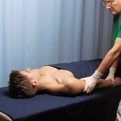
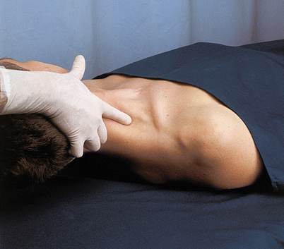
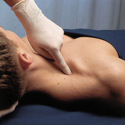
Fig. 30.2
Drawing the arm toward the knee (With permission from Danilo Jankovic)
Fig. 30.3
Turning the head to the opposite side and raising it slightly (With permission from Danilo Jankovic)
Fig. 30.4
Palpating the clavicle and subclavian artery (With permission from Danilo Jankovic)
Posterior to the sternocleidomastoid muscle, the scalenus anterior muscle is palpated. The interscalene groove between the scalenus anterior and scalenus medius muscles is felt with “rolling fingers” and located (Fig. 30.5).
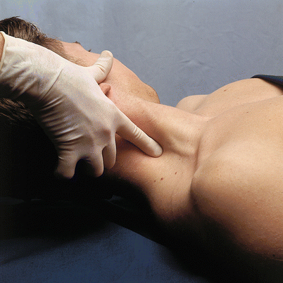
Fig. 30.5
Palpating the interscalene groove with “rolling” fingers (With permission from Danilo Jankovic)
The injection site in the interscalene groove lies at the level of the cricoid, opposite the transverse process of C6 (Chassaignac’s tubercle). The external jugular vein often crosses the level of the cricoid cartilage here (Fig. 30.6).
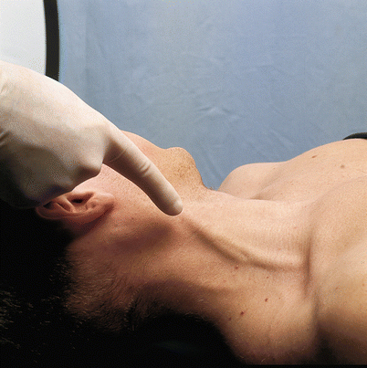
Fig. 30.6
Position of the external jugular vein (With permission from Danilo Jankovic)
When there are anatomical difficulties, it is helpful for the patient to inhale deeply or to try and blow out the cheeks. The scalene muscles then tense up, and the interscalene groove becomes more easily palpable.
Continuous Interscalene Block: Anterior Technique (Adapted from Meier)
Skin Prep
In all blocks.
Patient Positioning
See above the steps for locating the puncture site.
Landmarks (Fig. 30.7)
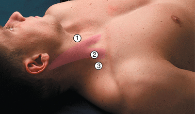
Fig. 30.7
Interscalene block (anterior access route). The posterior edge of the sternocleidomastoid muscle at the level of the superior thyroid notch. (1) Superior thyroid notch, (2) posterior edge of the sternocleidomastoid muscle, (3) external jugular vein and posterior scalene groove (With permission from Danilo Jankovic)
Superior thyroid notch
Posterior edge of the sternocleidomastoid muscle
Posterior scalene groove
External jugular vein
Transition from the middle to lateral third of the clavicle
Technique [14]
After identification of the posterior edge of the sternocleidomastoid muscle at the level of the superior thyroid notch, the block needle (55-mm short bevel insulated atraumatic needle or Tuohy needle, 38 or 52 mm) is introduced at an angle of 30° caudally and slightly laterally, in the direction of the transition from the middle to the lateral third of the clavicle (Fig. 30.8). A stimulation current of 1–2 mA and 2 Hz is selected with a stimulus duration of 0.1 ms. After a motor response from the relevant musculature (twitching in the biceps brachii muscle – musculocutaneous nerve and/or deltoid muscle – axillary nerve is regarded to be as reliable as twitching of the distal muscles [23, 27–29], the stimulation current is reduced to 0.2–0.3 mA. Slight twitching suggests that the stimulation needle is in the immediate vicinity of the nerve. The catheter is advanced approximately 3 cm beyond the end of the cannula or needle (Fig. 30.9). After removal of the cannula or needle, fixation of the catheter and placement of a bacterial filter, and after careful aspiration and injection of a test dose, the bolus administration of the local anesthetic follows.
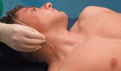
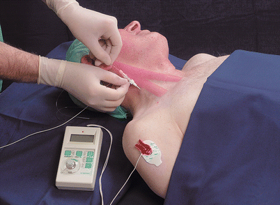
Fig. 30.8
Introducing the puncture needle at an angle of ca. 30° to the skin, caudally and laterally in the direction of the transition from the middle to the lateral third of the clavicle (With permission from Danilo Jankovic)
Fig. 30.9
Interscalene block (continuous technique). Introducing the catheter through a Tuohy needle (With permission from Danilo Jankovic)
Dosage
Surgical
“Single-shot” administration:
40 mL local anesthetic is sufficient for an adequate block of the brachial plexus and caudal part of the cervical plexus. In the literature [6, 11, 21, 24], the doses administered vary from 30 to 50 mL. A mixture of 20 mL 0.75 % ropivacaine or 0.5 % bupivacaine (0.5 % levobupivacaine) with 20 mL 1 % prilocaine (1 % mepivacaine) has proved its value very well in practice (in our own experience). This leads to a fast onset and long duration.
25 mL local anesthetic – e.g., 1 % prilocaine (1 % mepivacaine), in combination with 5–10 mg diazepam i.v. for reducing a dislocated shoulder.
20–25 mL local anesthetic – e.g., 0.75 % ropivacaine or 0.5 % bupivacaine (0.5 % levobupivacaine), in combination with basic general anesthesia for surgical interventions in the area of the shoulder and clavicle. This leads to very good postoperative pain control.
20 mL local anesthetic is sufficient to block the lower part of the cervical plexus and the upper part of the brachial plexus. The brachial plexus is only incompletely anesthetized with this amount and block of the ulnar nerve territory is often deficient.
Therapeutic
“Single-shot” administration (block series):
10 mL local anesthetic – e.g., 0.2 % ropivacaine or 0.125–0.25 % bupivacaine (0.125–0.25 % levobupivacaine) in shoulder and upper arm pain, shoulder arthritis, post-stroke pain, lymphedema after mastectomy.
10–20 mL local anesthetic – e.g., 0.2–0.375 % ropivacaine or 0.25 % bupivacaine (0.25 % levobupivacaine) in post-herpetic neuralgia, vascular diseases and injuries, complex regional pain syndrome (CRPS) types I and II, post-amputation pain.
25 mL local anesthetic – e.g., 1 % prilocaine or 1 % mepivacaine in combination with 5–10 mL diazepam i.v to mobilize the shoulder.
Continuous Interscalene Block: Posterior Technique (Pippa Technique)
The posterior cervical paravertebral block of the brachial plexus is an alternative to anterior route. This method was first described by Kappis in 1912 and was republished by Pippa in 1990 as a “loss of resistance” technique [15, 17, 20, 31]. The availability of electrical nerve stimulation has made this access route to the brachial plexus more important.
Indications and Contraindications
S. above.
Procedure
This block should only be carried out by experienced anesthetists or under their supervision. A detailed discussion with the patient is an absolute necessity.
Preparation
See anterior interscalene block.
Materials
See anterior interscalene block.
Skin Prep
In all blocks.
Patient Positioning
Sitting, with the neck flexed (to relax the cervical muscles) and supported by an assistant (the lateral recumbent position can be used as an alternative).
Landmarks
Spinous processes of the sixth (C6) and seventh (C7 – vertebra prominens) cervical vertebrae (Fig. 30.10).
Fig. 30.10
Interscalene block, posterior access route. Landmarks: spinous processes of C6 and C7 (vertebra prominens). (1) nuchal ligament, (2) vertebra prominens (With permission from Danilo Jankovic)Related posts:

Stay updated, free articles. Join our Telegram channel

Full access? Get Clinical Tree


