Fig. 8.1
Functional anatomy of the airways. (1) Trigeminal nerve (V) with the ophthalmic and maxillary nerves. Dotted red area: field of sensory olfactory innervation via the olfactory nerve. (2) The sensory innervation area of the glossopharyngeal nerve (IX), (3) the sensory innervation area of the vagus nerve (X), (4) mandible, (5) epiglottis, (6) trachea, (7) esophagus (With permission from Danilo Jankovic)
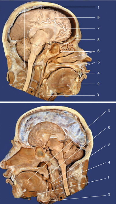
Fig. 8.2
(a) Functional anatomy of the airways. Median section through the head. (1) Posterior arch of the atlas, (2) cricoid cartilage, (3) geniohyoid, (4) genioglossus, (5) soft/hard palate, (6) nasal concha (superior, middle, inferior), (7) olfactory tract, (8) frontal sinus, (9) sphenoid sinus (With permission from Danilo Jankovic). (b) Functional anatomy of the airways. Median section through the head. (1) Hyoid bone, (2) epiglottis, (3) thyroid cartilage, (4) posterior pharyngeal wall, (5) genioglossus, (6) uvula (With permission from Danilo Jankovic)
The nasal cavity is divided by the nasal septum into right and left halves. The sensory innervation of the skin of the external nose is provided by the branches of the ophthalmic nerve and maxillary nerve (Fig. 8.1; cf. Chaps. 6 and 10), and the motor innervation of the facial muscles around the nose is from the facial nerve (cf. Chap. 7).
Each nasal cavity opens via a posterior choana into the upper level of the pharynx, the nasopharynx (or epipharynx). The nasopharynx connects with the choanae and is enclosed by the skull base superiorly and by the pharyngeal wall posteriorly. Inferiorly, the soft palate forms the boundary to the middle level of the pharynx (oropharynx). The larynx is an air-conducting organ that stretches from the lower pharyngeal space (laryngopharynx) as far as the trachea. In adult males, the larynx is located at the level of the C1–C3 vertebrae (Fig. 8.3), while in women and children, it lies further up. The laryngeal mucosa is innervated as far as the vocal folds by the purely sensory internal branch of the superior laryngeal nerve and below that by the recurrent laryngeal nerve, a branch of the vagus nerve. The inner laryngeal muscles are all supplied by the (inferior) recurrent laryngeal nerve. The only exterior laryngeal muscle, the cricothyroid, is innervated by the external branch of the superior laryngeal nerve (Fig. 8.4).
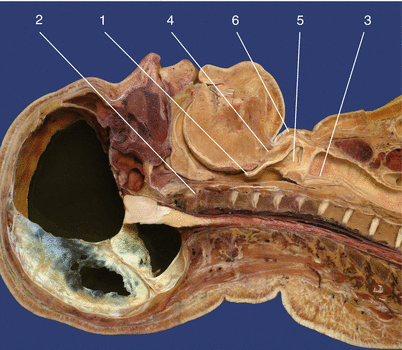
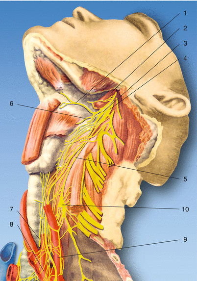

Fig. 8.3
Anatomic specimen. Paramedian sagittal section. In adults, the larynx is located over the C3–C6 vertebrae. (1) Epiglottis, (2) atlantodental joint, (3) trachea, (4) hyoid bone, (5) laryngeal ventricle, (6) thyroid cartilage (With permission from Danilo Jankovic)

Fig. 8.4
Anatomy. (1) Hypoglossal nerve, (2) internal carotid artery,(3) inferior ganglion of the vagus nerve, (4) inferior cervical ganglion, (5) vagus nerve, (6) superior laryngeal nerve, (7) recurrent laryngeal nerve, (8) aortic arch, (9) left subclavian artery, (10) cervicothoracic ganglion (With permission from Danilo Jankovic)
The tongue is basically a powerful muscle body that is covered with a mucosal layer, the mucosa of the tongue. The lingual mucosa in the anterior (presulcal) part of the tongue receives its sensory innervation from the lingual nerve (a branch of the mandibular nerve; cf. Chap. 10) and (with the exception of the vallate papillae) from the chorda tympani (branching from the intermediofacial nerve). The posterior (postsulcal) part of the tongue receives its sensory innervation from the glossopharyngeal nerve (with the exception of the epiglottic valleculae, which are supplied from the vagus nerve). The exterior lingual muscles (with the exception of the palatoglossus) are innervated from the hypoglossal nerve (Fig. 8.4).
The trachea is 10–12 cm long and stretches from the cricoid to the tracheal bifurcation. The smooth trachealis muscle is innervated from the recurrent laryngeal nerve, which is also responsible for the sensory and secretory innervation.
Anesthesia of the Larynx and Trachea
Various procedures can be used for anesthesia of the laryngeal and tracheal mucosa: spraying and advancement with the fiber endoscope, injection of local anesthetic through the cricothyroid membrane, bilateral blockade of the superior laryngeal nerve, and aerosol inhalation.
Fiberoptic Intubation [1–6, 8, 9]
The importance of fiberoptic devices for anesthesia was already recognized in the late 1960s by Murphy and Kronschwitz. Taylor and Towey were the first to report the use of the fiber bronchoscope, developed in 1968 by Ikeda, for intubation.
Indications
The principal indication for using flexible endoscopes is difficult intubation:
Limited mobility (jaw, head, cervical spine)
Malformations and acquired anomalies (face, jaw, oral cavity, neck)
Space-occupying lesions (oral cavity, pharynx, larynx, trachea)
Patient history (including failure with conventional techniques)
Plastic surgery in the facial region
Mobility disturbances in the head and nucha
Reintubation in high-risk patients
To avoid the need for contraindicated drugs (e.g., muscle relaxants)
When the patient has a full stomach as well as hindrances to intubation
Sedatives and Analgetics for Endoscopic Intubation
Benzodiazepines (Table 8.1) are used in combination with opioids (Table 8.2) for sedation and attenuation of the laryngeal reflexes. Benzodiazepines and opioids have synergistic effects, and the combination is highly effective at low dosage.
Dosage (mg/kg) | Clearance (mg/kg/min) | Elimination half-life (h) | |
|---|---|---|---|
Diazepam | 0.3–0.5 | 0.2–0.5 | 21–37 |
Midazolam | 0.15–0.3 | 6–8 | 1–4 |
Lorazepam | 0.05 | 0.7–1.0 | 10–20 |
Analgetic strength | Clearance (mg/kg/min) | Elimination half-life (h) | |
|---|---|---|---|
Morphine | 1 | 15–23 | 114 |
Fentanyl | 75–125 | 11–21 | 185–219 |
Sufentanil | 500–1,000 | 13 | 148–164 |
Alfentanil | 25 | 5.0–7.9 | 70–98 |
Local Anesthesia for Endoscopic Intubation
Adequate topical anesthesia of the mucosa in the upper respiratory tract is required for fiber-endoscopic intubation, in order to subdue coughing, swallowing movements, laryngospasm, and excessive secretion. Lidocaine (Xylocaine®) and cocaine are usually used for topical anesthesia in the respiratory tract for endoscopic intubation in conscious patients.
Lidocaine (Xylocaine®) is administered as a 4 % solution for mucosal anesthesia. A 10 % pump spray is also used for the oropharynx and nasopharynx. Gargling with a 2 % viscous lidocaine solution can also induce oropharyngeal anesthesia. Anesthesia of the nose can be achieved with a 2 % gel.
Bleeding in the region of the nasopharynx and mesopharynx can become a severe hindrance to endoscopic intubation. In the nasal procedure, an attempt therefore needs to be made to achieve not only good mucosal anesthesia (with lidocaine spraying) but also bleeding prophylaxis using vasoconstrictive agents. The local anesthetic and vasoconstrictive effects of cocaine are therefore advantageous during nasal endoscopic intubation. The synthetic analogs lack cocaine’s vasoconstrictive action. Cocaine nose drops (4–10 %, 0.5 mL into each nostril) have been found to be very useful in practice. The maximum dosage of cocaine is 100–200 mg. Alternatively, a mixture of lidocaine (4 %) and phenylephrine (1 %) at a proportion of 3:1 can be used in the nasal procedure. In the oral procedure, pretreatment of the mesopharynx with a lidocaine spray is necessary.
Possible alternatives to initial treatment with local anesthetics and mucosal decongestants in the nasal airways include:
10 % cocaine solution (1-mL ampullae)
10 % lidocaine spray + xylometazoline 0.1 % (Otrivin®)
4 % lidocaine + phenylephrine 1 % (Neo-Synephrine®)
Lidocaine spray + naphazoline nitrate 0.05 % (Privine®)
Before anesthesia in the oropharynx, an anticholinergic (atropine) should be injected to dry out the mucosa. Oropharyngeal anesthesia is administered with two or three spray shots of 10 % lidocaine or by gargling with 2–4 mL of a viscous solution of 2 % lidocaine for 30–40 s.
Practical Procedure for Nasal Endoscopic Intubation in a Conscious Patient
The anesthetist usually stands at the patient’s head, as this position provides easier orientation for conventional intubation. Steps in endoscopic intubation:
Adequate preoxygenation (5–10 min).
Secretory inhibition: atropine 0.5 mg i.m.
Inhibition of laryngeal reflexes: fentanyl 0.1 mg i.v. (alfentanil 1–2 mg in adults) combined with a sedative (e.g., midazolam)
Vasoconstriction, mucosal anesthesia: 10 % cocaine (0.5 mL into each nostril).
Preparation of the tube and flexible endoscope (Xylocaine® gel, antifogging agent) (Fig. 8.5a). The endotracheal tube is advanced over the flexible part of the endoscope and attached with adhesive plaster.
Introduction of the flexible endoscope via a nostril (under vision).
Adjustment of the laryngeal inlet (Fig. 8.5b)
Mucosal anesthesia via the biopsy channel (5-mL Xylocaine® 2 %). The onset of action should be awaited.
Introduction of the flexible part of the endoscope into the trachea.
Induction of anesthesia.
Advancement of the tube over the flexible endoscope, using it as a guide rail.
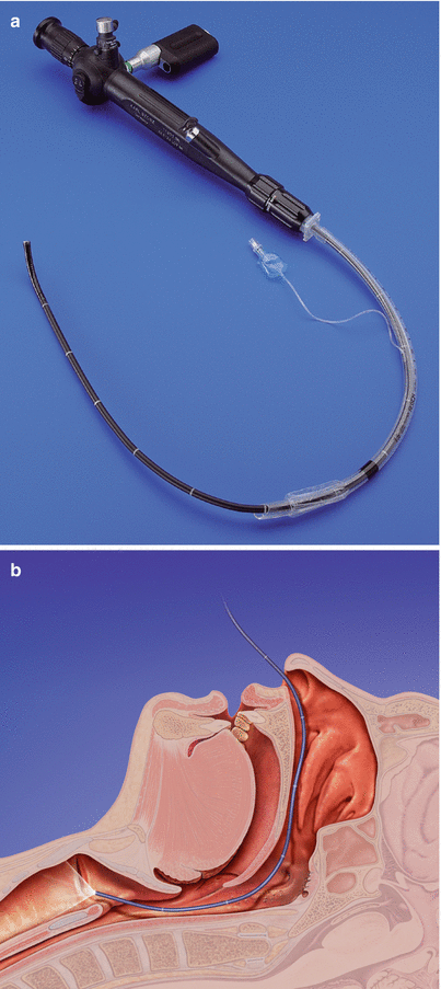
Fig. 8.5
(a) Flexible fiber endoscope for intubation (5.2 mm) with LED battery light source and tube switch attached (Karl Storz, Tuttlingen). (b) Introducing the flexible fiber endoscope into the trachea and positioning of the flexible endotracheal tube
Practical Procedure for Nasal Endoscopic Intubation in an Anesthetized Patient
Atropine for secretory inhibition
10 % cocaine nose drops (1 mL) (vasoconstriction local anesthesia)
Preoxygenation
Induction of anesthesia (etomidate, fentanyl)
Introduction of a spiral tube into the nasopharynx
Introduction of the endoscope, lubricated with gel, into the tube, after applying antifogging agent to the lens
Inspection of the laryngeal inlet and vocal folds
Introduction of the scope into the trachea with visual guidance, as far as the bifurcation
Advancement of the tube over the endoscope, using it as a guide rail
Checking of tube position
Continuation of anesthesia
Techniques for Blocking Individual Nerves of the Airway
The following nerve blockades are used in the region of the upper airways:
Superior laryngeal nerve blockade—above the vocal folds
Translaryngeal block—larynx and trachea below the vocal folds
Glossopharyngeal nerve blockade—oropharynx
Vagus Nerve, Superior Laryngeal Nerve, and Recurrent Laryngeal Nerve
Anatomy [11, 14–18, 21–23]
The vagus nerve (cranial nerve X) not only supplies areas of the head but also descends into the thoracic and abdominal space. It is the largest parasympathetic nerve and carries motor fibers (pharyngeal muscles), exteroceptive sensory fibers, visceromotor fibers, viscerosensory fibers, and gustatory fibers. The fibers emerge immediately behind the oliva, combine into a nerve trunk, and exit from the skull through the jugular foramen.
The nerve forms the superior ganglion of the vagus nerve (jugular ganglion) within the foramen and after passing through it forms the much larger inferior ganglion of the vagus nerve (nodose ganglion) (Fig. 8.6).


Fig. 8.6
Anatomy. (1) Glossopharyngeal nerve, (2) inferior ganglion of the vagus nerve, (3), superior cervical ganglion, (4) hypoglossal nerve, (5) superior laryngeal nerve, (6) vagus nerve, (7) hyoid bone, (8) recurrent laryngeal nerve, (9) vertebral artery, (10) left common carotid artery (With permission from Danilo Jankovic)
The superior ganglion of the vagus nerve is connected to the superior cervical ganglion and also to the facial nerve, glossopharyngeal nerve, and accessory nerve (Fig. 8.7a, b).
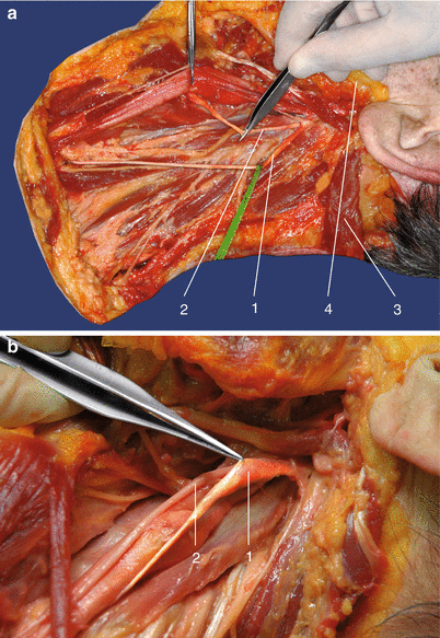
Fig. 8.7
(a) Anatomic specimen. (1) Division of the vagus nerve. (2) Branching of the superior laryngeal nerve, (3) sternocleidomastoid (reflected back), (4) mandible (With permission from Danilo Jankovic). (b) Anatomic specimen. (1) Superior cervical ganglion and (2) vagus nerve (With permission from Danilo Jankovic)
The inferior ganglion of the vagus nerve has connections with the cerebral part of the accessory nerve and with the superior cervical ganglion, the hypoglossal nerve, and the loop between the first and second spinal nerves.
Stay updated, free articles. Join our Telegram channel

Full access? Get Clinical Tree


