Probe placement and puncture site location for supraorbital nerve block. Ultrasound transverse view of disruption of the bone table at the foramina and vessel identification using color Doppler.
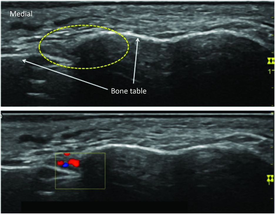
The infraorbital nerve block: ultrasound transverse view of disruption of the bone table (broken yellow circle) at the foramina and vessels identification using color Doppler.
The probe is located transversally above the orbital rim to localize the supraorbital foramen (frontal nerve). The infraorbital foramen image can be achieved by positioning the ultrasound probe horizontally or vertically in the sagittal plane (infraorbital nerve). Slight translation movements from medial to lateral along the lower orbital margin are performed to highlight the disruption of the bone table. Finally, the mental foramen is localized using a transverse or sagittal plane with dynamic scanning between the upper and lower borders of the mandible at the level of the second inferior premolar with scanning in the cephalic direction (mental nerve).
Suprazygomatic maxillary block
The maxillary nerve (V2 – second division of the trigeminal nerve) exits the skull through the rotundum foramen. The majority of its terminal branches (zygomatic branches, superior alveolar nerve, pterygopalatine and parasympathetic branches, palatine and pharyngeal branches) arise in the pterygopalatine fossa to supply sensory innervation of the cutaneous temporo-zygomatic area, the lower eyelid, the nasal septum, and of the lateral nasal wall, upper lip and teeth, the soft and bone palates, and maxillary sinus. At the upper part of the pterygopalatine fossa, the maxillary nerve is accessible for a complete maxillary block. In children, the ultrasound-guided suprazygomatic approach of maxillary nerve in pterygopalatine fossa seems to be safe and easily reproducible (Sola et al., 2012). The high frequency probe is placed in the infrazygomatic area, over the maxilla, with an inclination of 45 degrees in both the frontal and horizontal planes (Figure 21.3). The probe location allows visualization of the pterygopalatine fossa, limited anteriorly by the maxilla and posteriorly by the greater wing of the sphenoid (Figure 21.4). The needle is advanced using the out-of-plane approach. Real-time ultrasound guidance allows direct localization of the internal maxillary artery, needle-tip positioning –which is easily identified by movements applied to the needle and the spread of LA solution within the pterygopalatine fossa.
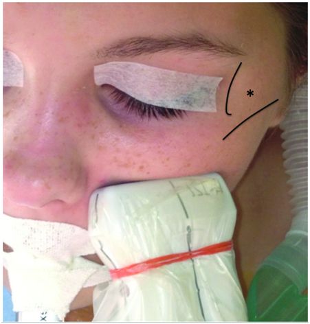
The suprazygomatic maxillary nerve block: probe placement and puncture site location (*) at the angle formed inferiorly by the superior edge of the zygomatic arch and anteriorly by the posterior margin of the lateral orbital rim.
Related posts:


Stay updated, free articles. Join our Telegram channel

Full access? Get Clinical Tree


