, Corinna Eleni Psomadakis2 and Bobby Buka3
(1)
Department of Family Medicine, Mount Sinai School of Medicine Attending Mount Sinai Doctors/Beth Israel Medical Group-Williamsburg, Brooklyn, NY, USA
(2)
School of Medicine Imperial College London, London, UK
(3)
Department of Dermatology, Mount Sinai School of Medicine, New York, NY, USA
Keywords
FungusFungalDermatophyteAthlete’s footOnychomycosisMacerationInterdigitalAntifungalClotrimazoleTerbinafine
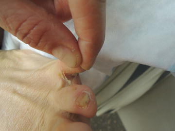
Fig. 15.1
Webspace maceration and itch are critical elements to making this diagnosis

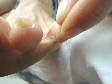
Fig. 15.2
Hyperkeratotic toenails present
Primary Care Visit Report
A 37-year-old female with no past medical history presented with left foot pain which started acutely 2 days prior. At the visit, the pain was so severe she had trouble putting her shoe on. The pain was in the toe webbing between her left fourth and fifth toes. She had applied over-the-counter Lotrimin for athlete’s foot but it did not help. She also had two thick yellow toenails on her left fourth and fifth toes, which had been like that for years.
Vitals were normal. On exam, there was a 5 × 5 mm yellow macular blister with some surrounding erythema in the toe webbing between the left 4th and 5th toes; there was trace edema and mild erythema on her 5th toe; there was some erythema on the dorsal aspect of the base of the 4th/5th toe webbing. The 4th and 5th toenails of the left foot were yellow, thickened, and ridged.
This was treated as an acute flare of tinea pedis, likely after longstanding onychomycosis . The patient was treated with 250 mg oral terbinafine for 2 weeks, as well as Burow’s solution topically three times daily, and foot powder to prevent maceration and to keep the area dry. The patient was advised to follow up with podiatry if the blister worsened over the following few days while on treatment. With respect to the toenail fungal infection, the patient wanted to try natural remedies (i.e., tea tree oil) as opposed to staying on the terbinafine for 12 weeks.
Discussion from Dermatology Clinic
Differential Dx
Tinea pedis
Dyshidrotic eczema
Pitted keratolysis
Candidiasis
Contact dermatitis
Psoriasis
Favored Dx
Clinical examination, maceration between toe spaces, and presence of onychomycosis are suggestive of a fungal infection.
Overview
Tinea pedis, commonly known as athlete’s foot , is a dermatophytic infection of the foot that causes scaling, flaking, itching, and maceration. It can sometimes lead to onychomycosis, or toenail fungus, which can be a source of reinfection in that the tinea pedis may be treated and cured, but the still existing onychomycosis can then lead to a recurrence of tinea pedis.
Tinea pedis is a condition that affects approximately 15 % of the global population [1]. Fungal infections generally affect men more than women. In the case of tinea pedis, the incidence is four times greater in men than women and increases with age [1]. Those who use communal pools, baths, and showers are more likely to develop an infection [2]. The dermatophytes that cause tinea pedis thrive in warm, humid environments, and are transmitted via direct contact, for example by retained skin in clothes, socks, or towels.
Stay updated, free articles. Join our Telegram channel

Full access? Get Clinical Tree


