, Corinna Eleni Psomadakis2 and Bobby Buka3
(1)
Department of Family Medicine, Mount Sinai School of Medicine Attending Mount Sinai Doctors/Beth Israel Medical Group-Williamsburg, Brooklyn, NY, USA
(2)
School of Medicine Imperial College London, London, UK
(3)
Department of Dermatology, Mount Sinai School of Medicine, New York, NY, USA
Keywords
MoleNevusNeviGrowthCongenitalHyperpigmentationBirthmarkLesionBenignMelanocyte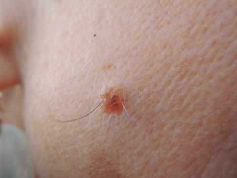
Fig. 49.1
Clear margined pink papule with central pigmentation. Hair growth has zero prognostic significance for the behavior of a mole
Primary Care Visit Report
A 47-year-old female presented with a mole on her right cheek. Per patient, this mole had been present throughout her life. Until 5 years prior, the mole had been more uniformly dark brown in color. In the past 5 years, the patient noted that the coloring was more varied, with the center remaining darker colored but the periphery becoming lighter. The mole grew a single black hair out of its center, which she regularly plucked.
Vitals were normal. On exam, there was a 0.5 cm brown, raised, round nevus with central dark brown pigmentation and surrounding lighter brown coloring. The borders were regular. There was a single dark-colored hair growing from the center of the nevus as well as several lighter colored hairs .
This patient was referred to dermatology for further evaluation.
Discussion from Dermatology Clinic
Differential Dx
Congenital nevus
Dysplastic nevus
Acquired melanocytic nevus
Favored Dx
Given its presence for the entirety of the patient’s life, the growth is most likely a congenital nevus.
Overview
Nevi, or moles, are a class of cutaneous pigmented growths. They are ideally well-circumscribed, can be macular or papular, and are typically round or oval in shape. They can be brown, pink, or flesh colored.
Moles present at birth are called congenital nevi . The American Academy of Dermatology estimates 1 in 100 people are born with one or more such lesions [1]. Men and women of all races are equally likely to have congenital nevi at, or within a few weeks, of birth, and the lesions grow proportionately with the child [2]. Congenital nevi are classified by size (small <1.5 cm; medium 1.5–19.9 cm, giant >20 cm) and they are most likely to occur on the trunk and extremities. They are often indistinguishable from common acquired nevi [3]. Most congenital nevi are benign and asymptomatic through life [2].
Presentation
Congenital nevi can exhibit great variance from patient to patient. They are generally brown in color but can feature shades of black, gray, red, yellow, or blue, and in some cases they may show variation in pigment [3]. Up to 75 % of cases present with an overlying dark, coarse terminal hair [4]. They may be elevated, verrucous, papular, pebbly, or cerebriform, and they can change with age [3]. Approximately one in six lighten with age [4].
Related posts:


Stay updated, free articles. Join our Telegram channel

Full access? Get Clinical Tree


