Fig. 63.1
(a) Dissection of front of thigh and adductor region. (1) Femoral nerve, (2) femoral artery and vein, (3) adductor longus muscle, (4) saphenous nerve, (5) adductor magnus muscle, (6) vastoadductor membrane and adductor canal with saphenous nerve and femoral artery and vein, (7) vastus medialis muscle, (8) vastus lateralis muscle, (9) sartorius muscle (With permission from Danilo Jankovic). (b) Cross-sectional depiction of the thigh. (1) Vastus lateralis muscle, (2) femur, (3) biceps femoris (short head), (4) biceps femoris (long head), (5) sciatic nerve, (6) adductor muscles, (7) femoral artery–nerve, (8) vastoadductor membrane, (9) saphenous nerve, (10) sartorius muscle, (11) vastus medialis muscle (With permission from Danilo Jankovic). (c) Depiction of medial thigh. (1) Lateral femoral cutaneous nerve, (2) femoral nerve, (3) saphenous nerve, (4) obturator nerve, (5) psoas major muscle, (6) vastus medialis muscle, (7) adductor longus muscle (With permission from Danilo Jankovic). (d) Medial view of cadaveric thigh. (1) Saphenous nerve (infrapatellar branch), (2) superior medial and inferior medial genicular arteries (With permission from Danilo Jankovic)
The adductor canal (canalis adductorius, Hunter’s canal, subsartorial canal) is a musculoaponeurotic tunnel in the middle third of the thigh, extending from the distal apex of the femoral triangle—defined by the crossing of the medial margin of the adductor longus muscle and the medial margin of the sartorius muscle—to the adductor hiatus, an opening in the adductor magnus [1, 2]. The muscular boundaries of the canal are the sartorius (anterior), vastus medialis (anterolateral), and adductor longus/magnus (posteromedial). The vastoadductor membrane, which varies in thickness, is a continuum of the aponeurotic roof of the canal that covers the distal part of the canal [3]. The canal contains the femoral artery and vein, the descending genicular artery and muscular branches of the femoral artery, the nerve to the vastus medialis, and the saphenous nerve.
The saphenous nerve is the largest and longest branch of the femoral nerve. It is a pure sensory nerve of the medial leg distal to the knee and arises from the posterior division of the femoral nerve. It courses lateral to the femoral artery in the proximal thigh, along the roof of the adductor canal, and then crosses anteriorly over the femoral artery to lie medial to it in the distal aspect of the canal [4]. The saphenous nerve enters the adductor canal after most of the motor branches of the femoral nerve have divided. The exception is the nerve to the vastus medialis, a motor nerve that travels with the saphenous nerve in the adductor canal under the sartorius muscle and anterolateral to the femoral artery [5, 6].
After entering the adductor canal, the saphenous nerve can divide either within the canal or distally in the subsartorial fat [7]. It gives off the infrapatellar branch and other cutaneous branches, which may anastomose with cutaneous branches from the obturator nerve and medial cutaneous nerve of the thigh to form a subsartorial plexus [7–9]. The infrapatellar branch can either run parallel to the saphenous nerve or pierce the distal part of the sartorius muscle. The saphenous nerve consistently emerges into the subcutaneous tissue between the tendons of the sartorius and gracilis muscles [10, 11]. The saphenous nerve travels with the saphenous branch of the descending genicular artery just beneath the sartorius muscle [12–14]. This occurs where the femoral artery descends through the adductor hiatus into the popliteal fossa.
The posterior articular branch of the obturator nerve either enters the distal part of the adductor canal or penetrates the adductor magnus muscle on its way to the posterior knee capsule in the popliteal fossa [7–9, 15]. This nerve branch may contribute to the analgesic effect of the adductor canal block for knee surgery. However, the course of the nerve in the distal thigh and its relation to the adductor canal varies, and the nerve may not have any meaningful clinical role following saphenous nerve blocks in the adductor canal.
Only a few of the nerves of the subsartorial plexus, specifically the saphenous nerve, infrapatellar nerve, and the nerve to the vastus medialis muscle, can be identified with ultrasound imaging. It is unclear to what extent all the plexus or just the saphenous nerve and its branches are involved in adductor canal block.
Landmarks
Based on the aforementioned anatomical definition of the adductor canal, identification by external surface landmarks can prove misleading. To assume that a position halfway between the base of the patella and the anterior superior iliac spine is within the canal can be erroneous and may mislead clinicians to choose a needle entry site proximal to the entrance of the canal within the femoral triangle [2]. Rather, the adductor canal may be reliably identified using sonographic landmarks (vastoadductor membrane, sartorius muscle, adductor longus muscle, vastus medialis muscle, and femoral vessels; Fig. 63.2).

Fig. 63.2
Serial sonograms demonstrating adductor canal block. Panel (a) baseline sonogram prior to injection (pre-scan). The saphenous nerve (long yellow arrow) lies deep to the sartorius muscle (SM) and hyperechoic vastoadductor membrane (white arrow) and is adjacent to the superficial femoral artery (SFA); adductor longus muscle (AL). Panel (b) in-plane approach to adductor canal block (the needle is shown with white arrowheads). Panel (c) distributions after injection tracked distally in the thigh demonstrating local anesthetic surrounding the saphenous (long yellow arrow) and infrapatellar (short yellow arrow) nerves. VM vastus medialis
A high-frequency linear ultrasound probe is placed on the medial aspect of the thigh using a transverse plane of imaging. The saphenous nerve can be identified in its short axis as it courses alongside the femoral artery in the adductor canal. As it travels distally in the thigh, the femoral artery, which appears immediately posterior to the sartorius muscle, eventually “dives” deep and moves away from the muscle plane of the sartorius toward the posterior aspect of the thigh where it becomes the popliteal artery [4]. This point is considered to be the adductor hiatus and the distal extent of the adductor canal.
Equipment
A standard regional anesthesia tray may include the following:
Ultrasound machine with linear transducer (8–14 MHz), sterile cover, and acoustic coupling gel
A 38-mm 25-gauge needle for skin infiltration
A syringe containing 5–15 mL of local anesthetic
A 50–70 mm, 22-gauge echogenic needle
Sterile gloves
A standard nerve catheter tray prepared with needle, catheter, and sterile drape (for continuous technique).
Suggested Block Technique
After placement of routine monitors, peripheral venous access, and sedation (if required), the patient is positioned supine with the operative leg slightly flexed at the knee and externally rotated; this will facilitate coverage of the saphenous nerve and superficial femoral artery by the sartorius muscle (Fig. 63.3). Sterile skin preparation should always occur prior to block.
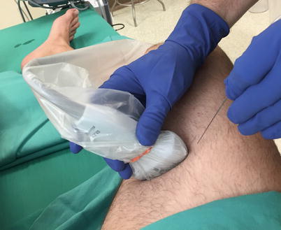
Fig. 63.3
External photography showing ultrasound-guided, in-plane approach to adductor canal block in the thigh (Photograph courtesy of Adam B. Collins, MD)
As previously mentioned, ultrasound can be used to guide the saphenous nerve block in the adductor canal and anywhere along its course. The choice of approach may be somewhat arbitrary—although too proximal, an approach can anesthetize motor branches of the femoral nerve, while too distal an approach may theoretically risk paresthesia, nerve puncture, or nerve entrapment [16–19]. Despite much debate regarding the anatomy and nomenclature of the adductor canal, present clinical studies have failed to show significant differences when comparing various locations and approaches to these regional blocks in the thigh [16, 20, 21].
A myriad of techniques have been proposed based on external vs. internal landmarks, nerve stimulation vs. ultrasound, and the amount of total local anesthetic used [2, 4, 16]. While debate and clinical research is ongoing, this section presents a safe and effective approach for blocking the saphenous nerve in the adductor canal.
Single Injection
Place the ultrasound machine on the opposite side of the bed so that the block site and display are both in front of the operator. Perform the ACB with the sartorius muscle viewed in short axis while advancing the needle in the plane of imaging. The needle approach is from the anterior side at a level sonographically determined within the adductor canal, typically with the superficial femoral artery directly in the midpoint and deep to the sartorius muscle (Figs. 63.2 and 63.4). Because of the relatively steep angle of insertion through the sartorius muscle, an echogenic needle is typically selected with this approach [3]. This steep approach may prove problematic in the morbidly obese patient, in which case the femoral nerve block may be a shallower alternative. However, as long as the echogenic vastoadductor membrane is visualized sonographically under the posterior border of the sartorius muscle, success can usually be achieved with this block.
Fig. 63.4
Serial sonograms demonstrating in-plane approach to adductor canal block deep to the sartorius muscle (SM). Panel (a) needle tip in plane for block injection (white arrowheads). Panel (b) long-axis view of the saphenous nerve (SN, long yellow arrows) after injection demonstrating local anesthetic tracking along the nerve course in the thigh
Once through skin and the subcutaneous tissues, the needle is placed through the sartorius muscle to enter the plane deep to the muscle (i.e., transsartorial approach; Fig. 63.4). There may be a loss of resistance as the needle tip crosses the vastoadductor membrane. Local anesthetic is injected within this plane, adjacent to the femoral artery. While the bevel of the needle initially faces the transducer to improve needle-tip visibility, rotating it 180° (bevel down) and advancing the needle just above the artery can extend distribution of local anesthetic under the sartorius muscle [3].
Another approach to the saphenous nerve involves advancing the needle in plane, with a short-axis view, through the superficial part of the vastus medialis muscle in a lateral-to-medial direction deep to the sartorius muscle and vastoadductor membrane. This may provide improved needle-tip visibility and minimize vascular puncture. The needle tip is then placed next to the saphenous nerve and local anesthetic injected [3].
While much of the clinical pharmacology of injection volume in relation to onset, duration, and degree of motor blockade is still under investigation, volumes of 5–15 mL are commonly used for the single-shot ACB.
Continuous Technique
The continuous technique is similar to the single-injection approach. After dilation of the adductor canal with sterile saline or local anesthetic, advance the catheter 1–3 cm beyond the needle tip posterior to the nerve under direct ultrasound visualization (if possible). Withdraw the needle carefully while gently advancing the catheter to prevent recoil of the catheter from the insertion site. Catheter tip position may be assessed by injecting 0.5 mL of air via the catheter under ultrasound imaging [22]. The catheter should then be secured either by tunneling or applying a biocompatible tissue adhesive and occlusive adhesive dressing.
If the catheter is placed preoperatively, there should be communication with the surgical team regarding the location of the catheter, as a more distal approach may lie within the planned sterile surgical field or underneath the thigh tourniquet. The presence of a continuous catheter under the pressure of a tourniquet may cause pressure damage to the nerves, although this notion has not yet been tested by formal research. There is no formal study to support the belief that a tourniquet increases the incidence of catheter dislodgement, and observations may actually show that a thigh tourniquet reinforces the security of the catheter [23]. There is also concern that, given intraoperative manipulation of the knee, catheters placed preoperatively may be at risk for migration or dislodgement out of the adductor canal. Neither catheter type (rigid stimulating vs. flexible) vs. nor subcutaneous tunneling has shown to have an effect on migration within the canal [24].
Related posts:


Stay updated, free articles. Join our Telegram channel

Full access? Get Clinical Tree


