, Corinna Eleni Psomadakis2 and Bobby Buka3
(1)
Department of Family Medicine, Mount Sinai School of Medicine Attending Mount Sinai Doctors/Beth Israel Medical Group-Williamsburg, Brooklyn, NY, USA
(2)
School of Medicine Imperial College London, London, UK
(3)
Department of Dermatology, Mount Sinai School of Medicine, New York, NY, USA
Keywords
Pyoderma gangrenosumViolaceous bordersInflammatory bowel diseaseCrohn’s diseaseInflammationInflammatory disorderNeutrophilic infiltrationUlcerUlcerationNecrosisNon-healing ulcer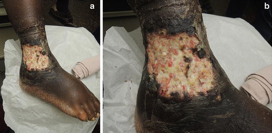
Fig. 16.1
(a and b) Painful ulceration with fresh granulation tissue and pinpoint bleeding
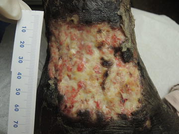
Fig. 16.2
Cribiform ulceration with “drop-off” or undermined edges
Primary Care Visit Report
A 69-year-old female with past medical history of hypertension and ulcerative colitis presented with right foot and ankle pain which she had been experiencing for about a week. She had no known trauma. The day prior to her visit, she had noticed some skin breakdown in the area. She had been applying over-the-counter antibiotic cream, Voltaren analgesic gel, and took some aspirin. About 8 months prior, the patient had started having crampy abdominal pain and blood in her stool. She was seeing a gastroenterologist for this.
Vitals were normal, except for elevated blood pressure of 150/80 mmHg. On exam, there was right ankle hyperemia, edema , and tenderness to palpation over the lateral malleolus with 1 cm of ulceration, and no lymphagitic streaking. Her complete blood count (CBC) revealed normal white blood cells, but significant anemia with a hemoglobin/hematocrit of 27.9/8.6.
This was initially treated as cellulitis with Bactrim DS BID for 10 days and topical Bactroban 2 % ointment TID.
Four days later, the patient returned with increased ankle pain and swelling, as well as a larger area of ulceration. At that point, her right foot and ankle had 2+ edema with a 2 cm pus-filled ulceration, which was exquisitely tender with surrounding erythema and warmth. The patient was sent to the Emergency Room for possible IV antibiotics given the lack of improvement on oral antibiotics.
She was admitted to the hospital and was treated with IV antibiotics for cellulitis without any improvement. The wound was surgically debrided and wound cultures showed no growth. She was then diagnosed with pyoderma gangrenosum and treated with oral prednisone and the wound started to improve.
These photos were taken 3 weeks after initially presenting at the Primary Care office.
Discussion from Dermatology Clinic
Differential Dx
Pyoderma gangrenosum
Cellulitis
Antiphospholipid antibody syndrome
Acute febrile neutrophilic dermatosis (Sweet syndrome)
Arterial or venous insufficiency
Verrucous carcinoma
Cutaneous anthrax
Blastomycosis
Favored Dx
A painful, deep ulcer with well-defined margins, with an associated history of ulcerative colitis is consistent with a diagnosis of pyoderma gangrenosum.
Overview
Pyoderma gangrenosum (PG) is an uncommon, chronic inflammatory skin condition characterized by neutrophilic infiltration of the skin and resulting in painful ulcers. The condition is immune-mediated; however, its exact cause is not known. While the name pyoderma suggests microbial etiology, PG is not infectious, though it is frequently misdiagnosed as such [1]. PG has recently been associated with mutations in the PSTPIP1, MTHFR, and JAK2 genes, and there are reports of familial cases without those mutations [2].
PG has a strong association with other autoimmune conditions, with underlying inflammatory bowel disease (IBD) , seropositive and seronegative polyarthritis, and hematological disorders present in about 50 % of cases [2–4]. The likelihood is that the development of PG is multifactorial and represents a complex interplay between dysfunctional neutrophils, dysregulated inflammatory cytokines, and genetic predisposition [5, 7].
PG can affect people of any age but it most commonly affects those between the ages of 40 and 60, and it has a slight female predominance [3, 6, 7]. The incidence among the general population is estimated to be between three and ten patients per million per year [7].
Related posts:


Stay updated, free articles. Join our Telegram channel

Full access? Get Clinical Tree


