Ophthalmologic Complications


Ophthalmologic Complications


Tariq M. Bhatti


What Is the Relevant Ophthalmic Anatomy and Physiology?

A basic knowledge and understanding of the anatomy and physiology of the human eye will help anesthesiologists in the delivery of ophthalmic anesthesia and in developing an appreciation of the potential ophthalmologic complications that may be associated with the administration of local or general anesthesia during ocular or nonocular surgery. Clinicians should be familiar with ocular and systemic risk factors that may place a patient at risk of a vision-threatening or life-threatening adverse event during ocular or nonocular surgery.

The anatomy and physiology of the eye and its associated structures is a very complex organ system with intricate anatomic structures, an elaborate physiologic system, and a wide array of pathologic processes. The visual system can be divided into three components: Optical, retinocortical and integrative.1 The following section is intended as a brief review of the pertinent anatomy and physiology of the eye for the practicing anesthesiologist. The interested reader is encouraged to refer to a more detailed text on the subject.2


▪ EYE OR GLOBE

The eye is composed of two modified spheres joined together, with the anterior sphere comprising the cornea, and the posterior sphere comprising the sclera. The junction of the cornea with the sclera is termed the limbus, which is clearly visible on external inspection of the eye. There is marked individual anatomic variation of the axial length of the globe, but on average it measures 24.0 mm. Each eye is located within the anterior eye socket and occupies only one fifth of the orbital volume (Fig. 29.2).

There are several internal chambers of the eye. The anterior chamber, filled with aqueous humor, is the space between the posterior cornea and iris-lens plane. The posterior chamber is bounded anteriorly by the iris, laterally by the ciliary body, and posteriorly by the anterior vitreous face. The volume of the posterior chamber is small compared with that of the anterior chamber, 60 µL and 250 µL, respectively. More than two thirds of the eye volume is formed by the vitreous cavity. In the nonvitrectomized (nonsurgical) eye the vitreous cavity is filled with vitreous humor, a gel composed of 98% water, hyaluronic acid, and collagenous fibrils.

The three layers of the eye, from outer to inner, are: (i) The corneosclera, (ii) the uveal tract, and (iii) the retina. The cornea is an elliptic-shaped structure that is
transparent and avascular. The sclera consists primarily of collagen, and is white and opaque. The several gaps found in the sclera are the locations for the optic nerve, and the various nerves, arteries, and veins of the eye. The conjunctiva, a nonkeratinized layer of epithelium, lines the ocular surface and extends over the eyelids to form the upper and lower eyelid fornices. Tenon capsule (fascia bulbi) covers the eye from the cornea to the optic nerve. The extraocular muscles travel from the orbital apex to the eye through openings within Tenon capsule.


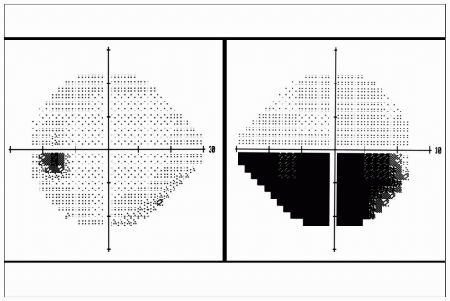
FIGURE 29.1 Automated visual field perimetry demonstrating an inferior visual field defect in the right eye.


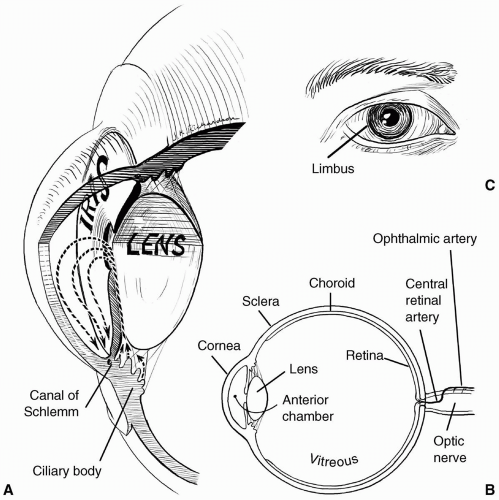
FIGURE 29.2 Illustration of the eye. A: Magnified view of the anterior segment of the eye. Arrows represent the flow of aqueous humor. B: Sagittal view of the eye and its major structures. C: External drawing of the eye and ocular adnexa.


The uveal tract is the middle layer of the eye and a highly vascularized structure. It can be divided from anterior to posterior into the iris, ciliary body, and choroid. The amount of light that enters the eye is controlled by the size of the central aperture of the iris or pupil. The ciliary body secretes aqueous humor and contains the ciliary muscle, which allows for accommodation. The choroidal circulation is the vascular supply of the outer two thirds of the retina.

The crystalline lens is suspended in the eye by fine glycoprotein fibrils, known as zonules. The lens is transparent; therefore, it allows light to enter the eye without any interference and, because of its elasticity, it can change shape, allowing for the ability to focus at different distances, a process known as accommodation.


▪ RETINA

Histologically, the retina is composed of 10 layers. There are two types of photoreceptors, rods and cones, which are in intimate contact with the retinal pigment epithelium cells. The photoreceptor cells synapse with the bipolar cells that, in turn, synapse with the retinal ganglion cells. The axons of the retinal ganglion cells make up the retinal NFL that go on to comprise the axons of the optic nerve. The outer two thirds of the vascular supply of the retina is derived from the choroidal circulation, with the remaining inner one third from the central retinal circulation.


▪ ANTERIOR AND POSTERIOR VISUAL PATHWAYS

The optic nerve is a white matter tract of the brain that stretches from the eye to the optic chiasm (see Fig. 29.3). The lamina cribrosa (LC) is a porous connective tissue structure that allows the transmission of the optic nerve from the eye through the scleral canal. The optic nerve axons are surrounded by myelin posterior to the LC. From the orbital apex, the optic canal allows the optic nerve to exit the orbit and enter the intracranial space. Each optic nerve is surrounded by pia, arachnoid, and dura matter, and contains approximately 1.2 million axons. The optic nerve can be divided into four sections: (i) Intraocular (optic nerve head), (ii) intraorbital, (iii) intracanalicular, and (iv) intracranial. The optic nerve head is the only portion of the optic nerve visible by clinical examination. Clinicians can assess several features of the optic nerve head, including the size of the disc (internal opening of the scleral canal), neuroretinal rim (nonmyelinated axons), and the optic cup (a slightly off-centered funnel-shaped depression) (see Fig. 29.4).


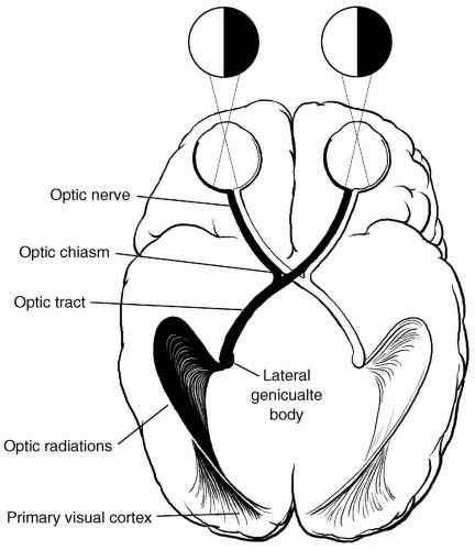
FIGURE 29.3 Drawing of the anterior and posterior visual pathways.

Intracranially the optic nerve joins the optic chiasm and is the crossing point of the nasal retinal fibers, which subserve the temporal visual field, to the contralateral optic tract. The temporal retinal fibers, which subserve the nasal visual field, extend to the ipsilateral optic tract. The
axons of each optic tract synapse at the lateral geniculate nucleus. The retrogeniculate pathway begins with the optic radiations located within the temporal and parietal lobes, traveling to and synapsing in the primary visual area of the occipital lobe or striate cortex. The integrative process of vision is accomplished by the transmission of visual impulses from the striate cortex to various vision-associated regions within the occipital, parietal, and temporal lobes.


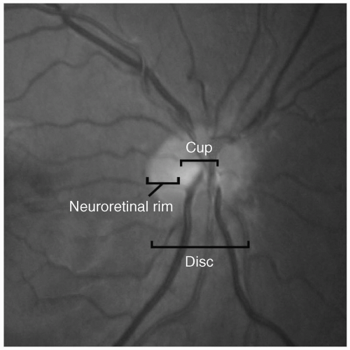
FIGURE 29.4 Photograph of normal-appearing optic nerve.


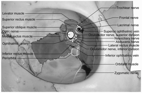
FIGURE 29.5 Anatomy of the bony orbit and structures within the orbital apex.


▪ ORBIT

The paired bony orbits are pyramidal in shape, covered by the orbital periosteum (periorbita), and contain the eye, extraocular muscles, orbital fat, connective tissue, lacrimal gland, nerves, veins, and arteries. The orbital apex comprises the optic foramen (opening to the optic canal), superior orbital fissure and inferior orbital fissure. It is the entry and exit point of the major neurovascular structures of the eye and orbit, and the site of the annulus of Zinn, the origin of the rectus muscles. Each orbit is composed of seven bones forming four walls (see Fig. 29.5).


▪ EXTRAOCULAR MUSCLES

There are six extraocular muscles that move the eye through the nine cardinal directions of gaze. The four recti muscles are the superior, inferior, medial, and lateral rectus muscles; and the two oblique muscles are the superior oblique and inferior oblique muscles. Aside from the inferior oblique muscle, which originates from the maxillary bone of the nasal orbit, all the extraocular muscles arise from the orbital apex. The extraocular muscles are connected to each other and various other orbital structures by a complex orbital connective tissue system. Conceptually, the four recti muscles form a cone-shaped compartment within the orbit between the orbital apex and the eye. On the basis of anatomic studies, the division of the orbit into an intraconal and extraconal space has not been validated, but remains a good practical concept to keep in mind. The extraocular muscles are innervated by three ocular motor cranial nerves: The third (oculomotor), fourth (trochlear) and sixth (abducens) nerves. Disturbances of the orbital facial system, dysfunction of the extraocular muscles, or neural interruption to the extraocular muscles can result in a misalignment of the two eyes (strabismus) and the subjective complaint of double vision.


▪ VASCULAR SUPPLY OF THE EYE, ORBIT, AND OPTIC NERVE

The main arterial supply of the eye and orbit is derived from the ophthalmic artery, the first intracranial branch of the internal carotid artery (see Fig. 29.6). The ophthalmic artery enters the orbit in partnership with the optic nerve through the optic canal in the orbital apex.

The blood supply to the proximal and distal segments of the optic nerve is quite variable from person to person, but is derived from the branches of the retinal vasculature, choroidal vasculature, and ophthalmic artery
(see Fig. 29.7).3 From anterior to posterior, the optic nerve head can be divided into four regions: The optic disc (surface of optic nerve head), prelaminar, LC, and postlaminar. The optic disc is supplied by the retinal arterioles. Segmental centripetal branches from the peripapillary choroid supply the prelaminar portion of the optic nerve head. The arterial supply of the LC region of the optic nerve head is derived from the centripetal branches of the short posterior ciliary arteries, which also contribute to the circle of Haller and Zinn (CHZ). Arterial branches from CHZ, the peripapillary choroid and, infrequently, the central retinal artery (CRA) supply the retrolaminar portion of the optic nerve head. The arterial supply of the intraorbital portion of the optic nerve is derived from pial branches of the ophthalmic artery.4 These vascular networks have watershed zones and are vulnerable to occlusion due to small vessel disease, hypoperfusion, and, rarely, embolic disease, all of which can result in ischemic disorders of the optic nerve, known as ischemic optic neuropathy. Depending on the vascular network affected, the clinical presentation can be either an AION or a posterior ischemic optic neuropathy (PION).


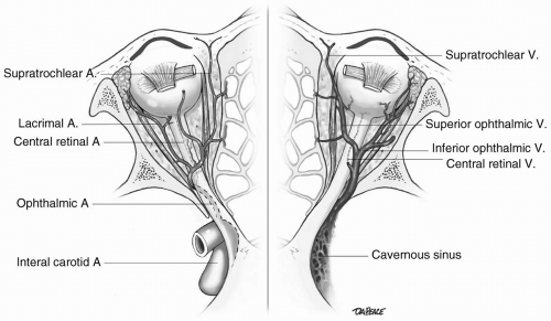
FIGURE 29.6 Vascular supply (arterial and venous) of the orbit. A, artery. V, vein.


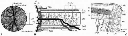
FIGURE 29.7 Schematic drawing of the blood supply to the optic nerve and optic nerve head. A, arachnoid; C, choroid; Col Br, collateral branches; CRV, central retinal vein; D, dura; LC, lamina cribrosa; NFL, surface nerve fiber layer of disc; OA, ophthalmic artery; OD, optic disc; ON, optic nerve; P, pia; PCA, posterior ciliary artery; PR and PLR, prelaminar region; R, retina; RA, retinal arteriole; S, sclera; SAS, subarachnoid space. (Reprinted with permission, Hayreh SS. The blood supply of the optic nerve head and the evaluation of it-myth and reality. Prog Retin Eye Res. 2001;20:563.)

Blood flow to the optic nerve head is determined by resistance to blood flow, BP and intraocular pressure
(IOP). Blood flow to the optic nerve head can be calculated by the formula:

Blood Flow = Perfusion Pressure/Resistance to Flow

The perfusion pressure (PP) of the optic nerve head is determined by the mean arterial pressure (MAP) within the optic nerve head and the IOP as stated by the following equation:


PP = MAP – IOP

In addition, the resistance of blood flow to the optic nerve head is influenced by the condition of the vessels (lumen size and integrity), autoregulation, endothelial-derived vasoactive substances, and “flowability” of blood.5,6

To a greater extent than the arterial system, the venous drainage system of the eye is anatomically highly variable (Fig. 29.6). The two sources that drain the eye are the central retinal vein (CRV) for the inner retina and the vortex veins for the uveal tract. The venous drainage of the optic nerve head is primarily into the CRV, with the prelaminar portion draining into the peripapillary choroidal veins.3 The multitude of venous orbital tributaries ultimately drain into the cavernous sinus through the superior and inferior ophthalmic veins. The central retinal venous pressure may have an influence on the perfusion pressure and ultimately the blood flow of the optic nerve head.5


▪ SENSORY INNERVATION OF THE EYE AND ORBIT

The primary sensory supply of the eye, the ocular adnexa, and orbit is from the first division (ophthalmic) of the trigeminal nerve (see Fig. 29.8). After entering the orbit through the superior orbital fissure, the ophthalmic nerve divides into the frontal, lacrimal, and nasociliary nerves.


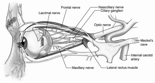
FIGURE 29.8 Sensory supply of the orbit and eye from the trigeminal nerve.


▪ INTRAOCULAR PRESSURE: AQUEOUS HUMOR PRODUCTION AND CIRCULATION

The aqueous humor is a clear fluid that provides nutrients and is a means of exchanging gases and metabolites to the anterior structures of the eye. Once secreted from the ciliary processes of the ciliary body, the aqueous humor circulates into the posterior chamber, traveling through the pupil, and then into the anterior chamber (Fig. 29.2). It is drained out of the eye through the structures within the anterior chamber angle, known as the trabecular meshwork and Schlemm canal, to eventually flow into the episcleral and scleral venous vessels. Approximately 15% of the aqueous humor drains through an alternative or unconventional pressure-independent system known as the uveoscleral drainage pathway.

Normal IOP ranges between 14 and 22 mm Hg (mean of 16 mm Hg) but are highly individually variable. The control and regulation of IOP is not fully understood, but there appear to be many factors involved, including diurnal variation, cardiorespiratory cycle, age, race, genetics, episcleral venous pressure, BP, and transmural pressure across ocular blood vessels. Any external pressure on the eye can raise the IOP. Furthermore, acute increases in central venous pressure (CVP), as can occur with Valsalva-type maneuvers (e.g., coughing, straining, vomiting, or endotracheal intubation) can result in a rapid rise in IOP.

The generation of IOP is based on the production and drainage rate of aqueous humor as described by the following equation:7


IOP = R(F – Fu) + Pv

where F = aqueous inflow, Fu = unconventional aqueous outflow, Pv = episcleral venous pressure and R = outflow
resistance (R = 1/C). C represents the outflow facility, which is influenced by F and IOP (C = ΔF/ΔIOP). An increase in IOP results in a collapse of Schlemm canal and thereby a decrease in outflow facility. Increases in IOP also influence the production of aqueous humor by decreasing the inflow rate.

IOP is also influenced by the hydrostatic pressure of the ocular blood vessels and can be summarized by the equation:


IOP = K([OpaqOPpl] + CP)

Where K = coefficient of outflow, Opaq = aqueous humor osmotic pressure, Oppl = plasma osmotic pressure and CP = capillary pressure. Clinically, IOP can be reduced effectively and rapidly by increasing the osmotic pressure of the plasma with the systemic administration of hypertonic solutions such as mannitol.

As mentioned earlier, the IOP contributes to the perfusion pressure of the optic nerve head. Elevations in IOP can lead to decreases in blood flow and ischemic disorders of the retina or optic nerve.


What Are the Anesthestic Considerations in Ophthalmic Surgery?


▪ PREOPERATIVE EVALUATION OF THE OPHTHALMIC PATIENT

Before eye surgery, or for that matter any surgical procedure, it is very important for the anesthesiologist to be familiar with the patient’s medical history and the ingestion of topical ocular or systemic medications. Routine laboratory testing has not been shown to increase the safety of cataract surgery;8 however, laboratory and ancillary testing (e.g., electrocardiogram, chest radiograph, etc.) should be performed on the basis of the ophthalmic procedure scheduled, the type of anesthesia to be used, and the patient’s current health status and past medical history. Most eye surgeries are elective cases; therefore, if the patient is found to be medically unstable, the surgery should be postponed so that the appropriate and necessary medical evaluation and treatment can be performed to optimize the patient’s health before proceeding with surgery. On the basis of several older studies, the perioperative mortality rate associated with eye surgery is low and has been quoted in the range of 0.06% to 0.18%, with the greatest risk factor of death occurring in patients with serious preexisting medical conditions.9 In patients undergoing cataract surgery, the incidence and prevalence of systemic illnesses is increased, in particular hypertension, diabetes mellitus and genitourinary diseases.10,11 Selfadministered health questionnaires are being developed to assist physicians in improving the cost-effectiveness of the preoperative evaluation of patients undergoing common elective eye surgeries and identifying patients at low risk of perioperative complications.12

Patients undergoing ocular surgery who are anticoagulated can pose a challenge to the ophthalmic surgeon and anesthesiologist. Because of the fear of a retrobulbar or intraocular hemorrhage during surgery, the discontinuation of anticoagulation or antiplatelet therapy is sometimes recommended.13 On the other hand, the risk of discontinuing treatment places the patient at risk of developing a stroke, myocardial infarction, or pulmonary embolism, depending on the reason for the therapy. In a prospective cohort study of more than 19,000 patients undergoing cataract surgery, the continued use of antiplatelet or anticoagulation therapy during surgery did not show an increase in the frequency of ocular hemorrhagic events nor an increased frequency of adverse medical events compared to those patients who discontinued medication before surgery.14 Although there does not appear to be a significant increase in the risk of hemorrhagic events with injectable orbital regional anesthesia in those patients on chronic antiplatelet or anticoagulation therapy,15 a careful evaluation of the risks and benefits of discontinuing antiplatelet or anticoagulation therapy should be made on a case-by-case basis and, if necessary, after consultation with the treating physician.

Because many diseases of the eye are manifestations of systemic genetic and metabolic disorders, especially in the pediatric age group, the anesthesiologist should be aware of these associations and proper steps taken to assure the successful delivery of anesthesia and performance of surgery (see Table 29.1).16,17,18

A wide variety of ophthalmic drugs (topical, intracameral, and systemic) are used in the medical and surgical treatment of ocular diseases that are relevant to the anesthetic management of the ophthalmic surgical patient. In particular, some of the glaucoma medications can have serious systemic adverse effects, as well as potentiate the effects of systemic medications, and therefore special attention should be made to note their use during the preoperative evaluation (see Table 29.2).19 Conversely, some of the systemic medications administered during surgery, in particular the general inhalants, can affect the IOP (see Table 29.3).20


▪ OPHTHALMIC SURGERY AND CHOICE OF ANESTHETIC TECHNIQUE

A general familiarity of the broad categories of ophthalmic surgeries is helpful in understanding and providing ophthalmic anesthesia. Ophthalmic surgery can be broadly divided into intraocular and extraocular surgery. Owing to the unique nature of many ophthalmic surgical procedures, anesthetic care should be custom-tailored for each patient. Some of the more common ophthalmic procedures performed include cataract extraction (often by phacoemulsification), penetrating keratoplasty (corneal
transplantation), strabismus surgery, glaucoma surgery (trabeculectomy or filtering surgery), oculoplastic surgery, orbital surgery, vitreoretinal surgery, and traumatic open globe repair.21


TABLE 29.1 Anesthetic Implications of Assorted Conditions with Ocular Pathology


Syndrome or Disease


Eye Findings


Features Affecting Anesthetic Management


Crouzon


Glaucoma Cataracts Strabismus Ectopia lentis Hypertelorism Proptosis


Upper airway obstruction Difficult intubation


Apert


See Crouzon


Difficult intubation Cardiac anomalies Anomalies of tracheobronchial tree


Goldenhar


Glaucoma Cataracts Strabismus


Difficult intubation Congenital heart disease


Down


Cataracts Strabismus Keratoconus


Retardation Airway obstruction Congenital heart disease Seizures Thyroid disorders


Homocystinuria


Ectopia lentis Pupillary block glaucoma Retinoschisis Retinal detachment Optic atrophy Central retinal artery occlusion Strabismus


Severe thromboembolic complications Hypoglycemic convulsions Hyphoscoliosis Osteoporosis


Lowe


Cataracts Glaucoma


Retardation Osteoporosis Decreased renal excretion of drugs Seizures Hyperchloremic acidosis


Marfan


Lens subluxation Anomalies of iris and iridocorneal angle Glaucoma Retinal detachment Myopia Cataracts Strabismus


Valvular heart disease Chest deformities Major vascular aneurysms Joint instability Difficult intubation


Myotonia congenital


Cataracts


Avoidance of depolarizing relaxants Avoidance of hypothermia Regional, if possible


Paramyotonia


Cataracts


Avoidance of hypothermia, exercise, potassium, all muscle relaxants, and neostigmine


Regional, if possible


Myotonia dystrophica


Cataracts Ptosis Strabismus


Avoidance of hypothermia, exercise, potassium, all muscle relaxants, neostigmine, digitalis, dilantin, barbiturates, cholinergics, and anticholinergics


Riley-Day


Corneal and other damage secondary to absence of lacrimation


Aspiration pneumonitis Abnormal respiratory control Postural hypotension Paroxysmal hypertension Temperature fluctuations


Rubella


Cataracts Microphthalmos Glaucoma Keratitis Iris atrophy Optic atrophy


Retardation and deafness Congenital heart disease Excretion of virus for several months


Sickle cell disease


Retinal detachment Vitreous hemorrhage Retinitis proliferans


Anemia Cardiopulmonary disease


Vulnerability to hypoxia, acidosis, dehydration, and hypothermia


Sturge-Weber


Vascular malformations Glaucoma Ectopia lentis


Retardation Seizures Airway angiomata


Von Recklinghausen


Ptosis Proptosis Optic glioma or meningioma Optic atrophy Glaucoma


Kyphoscoliosis Possible pheochromocytoma Abnormal response to muscle relaxants


Wagner-Stickler


Vitreous degeneration Chorioretinal degeneration Retinal holes and detachments Cataracts Glaucoma Strabismus


Difficult intubation Mitral valve prolapse Skeletal deformities


Zellweger


Glaucoma Cataract Corneal clouding Vitreous cellularity Retinal pigmentary disorders Optic atrophy Optic nerve hypoplasia


Seizures Hypoprothrombinemia Difficult intubation


Diabetes Mellitus


Cataracts Diabetic retinopathy Glaucoma Muscle palsy


“Silent” coronary artery disease Autonomic neuropathy Poor ventricular function Renal impairment Vulnerability to infection, sepsis, and aspiration Stiff joint syndrome


Reprinted with permission. McGoldrick KE, ed. Anesthesia for ophthalmic and otolaryngologic surgery. Philadelphia: WB Saunders; 1992:217-217.


Aside from the routine concern of the overall health and safety of the patient, specific operative and anesthetic conditions critical to the safety and successful outcome of patients who have undergone ophthalmic surgery are immobility of the eye and eyelids (akinesia), profound ocular analgesia, hemostasis, avoidance of the oculocardiac reflex, as well as uneventful induction, maintenance and emergence from general anesthesia. To maintain sterility of the eye during surgery, there may often not be direct access to the patient’s airway; therefore, it is vital that the anesthesiologist remain vigilant to any problems or concerns the surgeon or patient may raise during surgery.

The choice of anesthetic technique for ophthalmic surgery is dependent on physician preference and skill, as well as patient preference and cooperation. In some conditions, general anesthesia may be the anesthetic technique of choice, although, in most cases, ophthalmic surgery can be done under topical or orbital regional anesthesia (see Tables 29.4 and 29.5).22 In a select group of patients undergoing surgery by skilled cataract surgeons, the complete avoidance of topical or regional anesthesia has been demonstrated to be a feasible option.23

There is a myriad of local anesthetic solutions that are used for local ophthalmic anesthesia. The amide-linked agents (lidocaine, bupivacaine, and mepivacaine) are routinely used for ophthalmic surgery. The advantages of amide-linked agents over the ester-linked agents are increased stability, hypoallergenicity, and longer half-life.
In many cases, a combination of an agent with quicker onset, such as lidocaine, and a longer-acting agent, such as bupivacaine, is given in a 1:1 mixture. Also in many cases, a premixed local anesthetic solution with epinephrine is used to reduce bleeding, prolong the duration of the anesthesia, and improve the effectiveness of the anesthesia desired.24 Only a small amount of epinephrine is required, and the final concentration should not be greater than 5 µg/mL (1:200,000). The total dose should not exceed 0.1 mg.


TABLE 29.2 Summary of Nonocular Interactions Between Glaucoma Medications and Systemic Drugs


Glaucoma Medication


Systemic Drug


Interaction


Potential Result


Documentation Quality of References


Additive Antagonistic


Potential Result


β-Adrenergic antagonist


Anesthetic agents (inhalational)


X


Systemic hypotension


Poora


Hypoglycemic agents


X


A. Retard hypoglycemic rebound


None


B. Mask hypoglycemic symptoms


Poorb


C. Produce hypoglycemia


Poorb,c


β-Adrenergic


X


Increased toxic effects of β-antagonists


Poord,e,f


Calcium channel blockers


X


Cardiac depression


Poorg,h


Cholesterol-lowering medication


X


Decrease high-density lipoprotein cholesterol


Goodi,j,k


Cholinesterase inhibitors


X


Weakness of striated muscle


Fairl,m


Clonidine


X


Systemic hypertensive rebound after clonidine withdrawal


Poorn


Digitalis glycosides


X


Cardiac depression


Fairn,o,p,q,r,s


Fentanyl derivatives


X


Increased toxic effects of fentanyl


None


Phenothiazines


X


Increased serum levels of β-blocker and phenothiazine with potential toxide side effects


None


Prednisone


X


Increased serum potassium


Poort


Quinidine


X


Cardiac depression


Goodu,v


Reserpine


X


Cardiac depression


None


Sympathomimetic amines


1. Subcutaneous epi


X


Abrupt systemic hypertension


None


2. Xanthines


X


A. Bronchoconstriction


None


3. β-Adrenergic agonists for treatment of:


B. Reduced theophylline clearance Goodw,x,y,z Goodx,aa,bb,cc


a. Heart failure


X


Cardiac depression


b. Bronchoconstriction


X


Bronchoconstriction


Adrenergic agonist (nonselective)


Anesthetic agents (inhalational)


X


Cardiac arrhythmias


Poordd,ee


Digitalis glycoside


X


Cardiac arrhythmias


None


Monoamine oxidase inhibitors


X


Hypertensive crises


Goodff,gg,hh


Sympathomimetic amines


X


Systemic hypertension


None


Tri- and tetracyclic antidepressants


X


Cardiac arrhythmias


None


Adrenergic agonist (α2-selective)


Monoamine oxidase


X


Hypertensive crises


Goodff,gg,hh


Hypotension-producing medications


X


Systemic hypotension


Poorii


Cholinesterase inhibitors


Anesthetic agents (local: ester type)


X


Prolonged anesthetic action with cardiopulmonary depression


Fairjj


Cholinesterase inhibitors


X


Cholinergic toxicity


None


Succinylcholine


X


Prolonged neuromuscular blockade


Goodkk,ll,mm


Carbonic anhydrase inhibitors


Amphotericin-B


X


Increased hypokalemia


Poor


Anticholinergics


X


Enhanced anticholinergic action


Poor


β-Andrenergic blockers


X


Increased metabolic and respiratory acidosis


Fairnn


Corticosteroids


X


Potentiate hypokalemia


Poor


Cyclosporine


X


Increased cyclosporine toxicity


Pooroo


Digitalis glycosides


X


Digitalis toxicity increased by hypokalemia


Good


Ephedrine


X


Enhanced ephedrine action


Poor


Lithium


X


Increased lithium excretion


Fairpp


Mexiletine


X


Enhanced mexiletine effect


Poor


Phenytoin


X


Accelerated osteomalacia


Poor


Primidone


X


Decreased primidone effectiveness


Fairqq


Quinidine


X


Decreased quinidine excretion


Poor


Salicylates


X


Increased salicylate and carbonic anhydrase toxicity


Goodrr,ss,tt


a Mishra P, Calvey TN, Williams NE, et al. Intraoperative bradycardia and hypotension associated with timolol and pilocarpine eye drops. Br J Anaesth. 1983;55:897-9.

b Velde TM, Kaiser FE. Ophthalmic timolol treatment causing altered hypoglycemic response in a diabetic patient. Arch Intern Med. 1983;143:1627.

c Angelo-Nielsen K. Timolol topically and diabetes mellitus. JAMA. 1980;244:2263.

d Batchelor ED, O’Day DM, Shand DG, et al. Interaction of topical and oral timolol in glaucoma. Ophthalmology. 1979;86:60-5.

e Blondeau P, Cote M, Tetrault L. Effect of timolol eyedrops in subjects receiving systemic propanolol therapy. Can J Ophthalmol 1983;18:18-21.

f Chamberlain TJ. Myocardial infarction after ophthalmic betaxolol. N Engl J Med. 1989;321:1342-45.

g Pringle SD, McEwen CJ. Severe bradycardia due to interaction of timolol eye drops and verapamil. BMJ. 1987;294:155-6.

h Sinclair NI, Benzie JL. Timolol eye drops and verapamil—A dangerous combination. Med J. 1983;1:548.

i Sacks FM, Dzau VJ. Adrenergic effects on plasma lipoprotein metabolism. Speculation on mechanisms of action. Am J Med. 1986;80(Suppl 2A):71-81.

j Freedman SJ, Freedman NJ, Shields MB, et al. Effects of ocular carteolol and timolol on plasma high-density lipoprotein cholesterol level. Am J Ophthalmol. 1993;116:600-11.

k Coleman AL, Diehl DLC, Jampel HD, et al. Topical timolol decreases plasma high-density lipoprotein cholesterol level. Arch Ophthalmol. 1990;108:1260-63.

l Shavitz SA. Timolol and myasthenia gravis. JAMA. 1979;242:1611-2.

m Verkijk A. Worsening of myasthenia gravis with timolol maleate eye drops. Ann Neurol. 1985;17:211-12.

n Bailey RR, Neale TJ. Rapid clonidine withdrawal with blood pressure overshoot exaggerated by beta-blockade. BMJ. 1976;17:942-3.

o LeWinter MW, Crawford MH, O’Rourke RA, et al. The effects of oral propanolol, digoxin and combination therapy on the resting and exercise electrocardiogram. Am Heart J. 1977;93:202-9.

p Lowenthal DT, Porter S, Saris SD, et al. Clinical pharmacology, pharmacodynamics and interactions with esmolol. Am J Cardiol. 1985;56:14F-18F.

q Yamaoki K, Kakui M, Seko Y, et al. A case of digitalis poisoning after syncope due to beta-blocker eyedrops. Jpn Circ J. 1988;52:43.

r Rynne MV. Ophthalmic medication complicating systemic disease. J Maine Med Assoc. 1980;71:82.

s Ball S. Congestive heart failure from betaxolol. Arch Ophthalmol. 1987;105:320.

t Swanson ER. Severe hyperkalemia as a complication of timolol, a topically applied beta-adrenergic antagonist. Arch Intern Med. 1986;146:1220-21.

u Edeki TI, He II, Wood AJJ. Pharmacogenetic explanation for excessive β-blockade following timolol eye drops: Potential for oral-ophthalmic drug interaction. JAMA. 1995;274:1611-13.

v Dinai Y, Sharir M, Naveh N, et al. Bradycardia induced by interaction between quinidine and ophthalmic timolol. Ann Intern Med. 1985;103:890-1.

w Leier CV, Baker ND, Weber PA. Cardiovascular effects of ophthalmic timolol. Ann Intern Med. 1986;104:197-9.

x Munroe WP, Rindone JP, Kershner RM. Systemic side effects associated with the administration of timolol. Drug Intell Clin Pharm. 1985;19:85-9.

y Giudicelli JF, Chauvin M, Thuillez C, et al. β-Adrenoceptor blocking effects and pharmacokinetics of betaxol (SL 75212) in man. Br J Clin Pharmacol. 1980;10:41-9.

z Fraunfelder FT, Meyer SM. Systemic adverse reactions to glaucoma medications. Int Ophthalmol Clin 1989;29:143-6.

aa Guzman C. Exacerbation of bronchorrhea by topical timolol. Am Rev Respir Dis. 1980;121:899-900.

bb Schoene RB, Martin TR, Charan NB, et al. Timolol-induced broncho-spasm in asthmatic bronchitis. JAMA. 1981;245:1460-1.

cc Charan NB, Lakshminarayan S. Pulmonary effects of topical timolol. Arch Intern Med. 1980;140:843-4.

dd Francois J, Verbraeken H. Danger of collyrium with 2% levorenone (Abstract). Bull Soc Belge Ophtalmol. 1979;185:99-102.

ee Smith RB, Douglas H, Petruscak J, et al. Safety of intraocular adrenaline with halothane anesthesia. Br J Anaesth. 1972;44:1314-17.

ff Boakes AJ, Laurence DR, Teoh PC, et al. Interactions between sympathomimetic amines and antidepressant agents in man. BMJ. 1973;1:311-5.

gg Elis J, Lawrence DR, Mattie H. Modification by monoamine oxidase inhibitors of the effect of some sympathomimetics on blood pressure. BMJ. 1967;2:75-8.

hh Horwitz D, Goldberg LI, Sjoerdsma A. Increased blood pressure responses to dopamine and norepinephrine produced by monoamine oxidase inhibitors in man. J Lab Clin Med. 1960;56:747-53.

ii King MH, Richards DW. Near syncope and chest tightness after administration of apraclonidine before argon laser iridotomy. Am J Ophthalmol. 1990;110:308-9.

jj Zsigmoid EK, Eilderton TE. Abnormal reaction to procaine and succinylcholine in a patient with inherited atypical plasma cholinesterase. Can Anaesth Soc J. 1968;15:498-500.

kk Eilderton TE, Farmati O, Zsigmoid EK. Reduction in plasma cholinesterase levels after prolonged administration of echothiophate iodide eyedrops. Can Anaesth Soc J. 1968;15:291-6.

ll McGavi D. Depressed levels of pseudo-cholinesterase with echothiophate iodide eye drops. Lancet. 1975;2:272.

mm Cavallaro RJ, Krunperman LW, Kugle F. Effect of echothiophate therapy on metabolism of succinylcholine in man. Anesth Analg. 1968;47:570-4.

nn Boada JE. Severe mixed acidosis by combining therapy with acetazolamide and timolol eyedrops. Eur J Respir Dis. 1986;68:226-8.

oo Keogh A, Esmore D, Spratt P, et al. Acetazolamide and cyclosporine. Transplantation. 1988;46:478-9.

pp Thomsen K, Schor M. Renal lithium excretion in man. Am J Physiol. 1968;215:823-7.

qq Syversen GB, Morgan JP, Weintraub M, et al. Acetazolamide-induced interference with primidone absorption. Arch Neurol. 1977;34:80-4.

rr Anderson CJ, Kaufman PL, Sturm RJ, et al. Toxicity of combined therapy with carbonic anhydrase inhibitor and aspirin. Am J Ophthalmol. 1978;86:516-9.

ss Cowan RA, Hartnell GG, Lowdel CP, et al. Metabolic acidosis induced by carbonic anydrase inhibitors and salicylates in patients with normal renal function. BMJ. 1984;289:347.

tt Sweeney KR, Chapron DJ, Brandt JL, et al. Toxic interaction between acetazolamide and salicylate: Case reports and a pharmacokinetic explanation. Clin Pharmacol Ther. 1987;41:67.


Reproduced with permission. Albert DM, Jakobiec FA, Azar DT, eds. Principles and practice of ophthalmology, 2nd ed. Philadelphia: WB Saunders; 2000.


TABLE 29.3 Effects of Anesthetic Agents, Techniques and Adjuvant Drugs on Intraocular Pressure


Decreased IOP


Increased IOP


Inhalational agents


Ketamine (possibly)


Barbiturates, propofol, etomidate, narcotics


Hypoxemia


Neuroleptic agents


Hypercapnia


Nondepolarizing muscle relaxants (unless alveolar hypoventilation occurs)


Succinylcholine


Hyperventilation


Hypothermia


IOP, intraocular pressure.


The three types of adverse effects that may occur from the tissue infiltration of local anesthetic solution are an allergic reaction, tissue toxicity, and systemic toxicity.24 A true allergic reaction to local anesthetic solutions is rare, and the use of preservative-free preparations may avoid many problems. Neurotoxicity and myotoxicity of local anesthetic solutions may explain some of the cases of optic nerve dysfunction and strabismus following orbital regional anesthesia. Systemic toxicity may occur from the inadvertent intravascular injection or direct spread of the agent into the central nervous system, resulting in mental status changes, seizure activity, coma, or cardiovascular collapse. Theoretically, the use of epinephrine in local anesthetic solution for orbital regional anesthesia may promote vasconstriction of the ocular blood vessels, resulting in reduced blood flow and ocular ischemia.


TABLE 29.4 Advantages of Regional and General Anesthesia


Regional Anesthesia


General Anesthesia


Simple technique (minimal equipment)a


Less postoperative nausea and vomiting


Faster recovery


Postoperative analgesia superior


Blockade of oculocardiac reflex


Absence of respiratory depression


Less physiologic trespass


Full mental status retainedb


No loss of ‘control’ for patient


Potential for reduced stress


No risk of toxic hepatitis


Avoids trace gas exposure for staff


Less expensive


Not contraindicated with low serum K+


No risk of malignant hyperthermia


Easily applicable at high altitude


Complete control of patient


No risk of retrobulbar hemorrhage


No risk of globe perforation


No risk of myotoxicity


Applicable to all ages


a However, basic cardiopulmonary resuscitative equipment should be available.

b Enhanced ability to communicate intraoperatively and postoperatively.


Reprinted with permission. Smith GB, Hamilton RC, Carr CC, eds. Ophthalmic anaesthesia. A practical handbook, 2nd ed. London: Oxford University Press; 1996.


Hyaluronidase is very frequently added to the anesthetic solution to reduce the induction time and increase the effectiveness of anesthesia and akinesia. In addition, the depolymerization of hyaluronic acid (a major component of orbital connective tissue) allows a lower volume of anesthetic solution to be used and a more rapid distribution of the solution within the orbital tissue, thereby reducing the duration and degree of proptosis and elevated intraorbital pressure.25,26 Few complications have been reported with the use of hyaluronidase. An allergic reaction resulting in an orbital inflammatory syndrome has been described in five patients.27 During times of limited supply of hyaluronidase, clusters of strabismus cases have been reported and postulated to have occurred because of either the increased contact time of the anesthetic solution with the extraocular muscle, resulting in myotoxicity or focal increased intraorbital pressure, in turn leading to an extraocular muscle compartmental syndrome.28 Increasing the pH of the anesthetic solution by the addition of sodium bicarbonate improves the effectiveness of anesthesia and reduces injection pain. Its use in orbital regional anesthesia has been studied with mixed results and is not commonly used.29


Topical Anesthesia

Topical anesthesia refers to the administration of ophthalmic anesthetic drops directly onto the eye. With the
recent advances in intraocular lens design and microsurgical techniques, topical anesthesia (with or without intracameral application) is becoming a popular choice in the surgical management of patients requiring cataract extraction.30 In a 2003 survey of cataract and refractive surgeons, topical anesthesia was used in 61% of cases, an increase from 8% in 1995.31 Topical anesthetic agents used in modern day cataract surgery include 0.75% bupivacaine, 0.5% tetracaine, and 2% or 4% lidocaine. The main advantage of topical anesthesia is avoiding the risks associated with injectable orbital regional anesthesia (explained further in the text). Other benefits of performing surgery with topical anesthesia are:


TABLE 29.5 Contraindications to Regional and General Anesthesiaa


Regional Anesthesia (RA)


General Anesthesia (GA)


Reversible medical condition, uncorrected


Informed patient refusal of RA


Anesthetist inexperience


True allergy to RA (very rare)


Surgeon preference for GA


Emergency surgery (open eye wound)


Prolonged surgery (more than 2 h)


Children up to age of early teens


Unsuitable psychologic status:


▪ Behavioral or psychiatric disorder


▪ Agitated, or phobic patient


▪ Uncooperative patient


Mental retardation


Senile dementia


Head movements or tremors:


▪ Parkinson disease


▪ Tardive dyskinesia


Inability to lie flat (from cardiac or respiratory disease)


Intractable cough


Communication barrier:


▪ Language


▪ Deafness


Moderate to severe arthritis


Neurologic disease


Needle phobia


Claustrophobia


Complication from RA in same patient on an earlier occasion


Patients with high myopia


Caution with patients on anticoagulants


Reversible medical condition, uncorrected


Informed patient refusal of GA


History of serious adverse effect from GA


History of difficult airway


Known problems or disease states:


▪ Malignant hyperthermia history


▪ Muscle diseases—dystrophia myotonica, myasthenia gravis


▪ Hemoglobinopathies


▪ Chronic obstructive pulmonary disease


▪ Diabetes mellitus


Caution with:


▪ History of porphyria


▪ Atypical pseudocholinesterase


▪ Patients on MAO inhibitors


▪ Interactions with regular medications


▪ Patients on anticoagulants


aIn decreasing order of significance from absolute at top of table.


MAO, monoamine oxidase.


Reprinted with permission. Smith GB, Hamilton RC, Carr CC, eds. Ophthalmic anaesthesia. A practical handbook, 2nd ed. London: Oxford University Press; 1996.


  • Its safe use in anticoagulated patients


  • Decreased need for intravenous anxiolytic medications


  • Rapid onset of action


  • Intraoperative patient cooperation


  • Immediate return of vision


  • Lack of periorbital bruising


  • Absence of postoperative diplopia and


  • Avoidance of a postoperative eye patch

Although several studies have suggested that topical anesthesia is associated with an equivalent level of pain control during surgery compared with injectable orbital regional anesthesia,32 other studies have refuted this observation.33 A literature review of the randomized trials of orbital regional anesthesia found that injectable anesthesia provided better pain control than topical anesthesia during the surgical procedure but was more commonly reported to be painful on administration.34 The pain or discomfort experienced by a patient undergoing surgery with topical anesthesia is often due to manipulation of the iris and ciliary body. The intracameral application of unpreserved 1% lidocaine has been shown to improve patient comfort.35

Complications of topical anesthesia are confined to the eye and include corneal epithelial toxicity, difficult operative conditions due to lack of eyelid and globe akinesia, and intraoperative pain or discomfort. Intracameral lidocaine may result in temporary blindness due to retinal or optic nerve toxicity.36 The application of intracameral lidocaine does not result in any detectable systemic levels.37


Orbital Regional Anesthesia

The injectable orbital regional anesthesia techniques available for accomplishing ocular anesthesia and akinesia during opthalmic surgery are subconjunctival (perilimbal), parabulbar (sub-Tenon), peribulbar, and retrobulbar.38 Retrobulbar anesthesia was first performed by Knapp in 1884 and written about extensively in the literature by Atkinson starting in 1934.39 In contrast to peribulbar anesthesia, retrobulbar anesthesia is performed by injecting the anesthetic agent into the intraconal space of the orbit. To avoid the risk of complications associated with retrobulbar anesthesia, the perilimbal, parabulbar, and peribulbar techniques were developed (see Table 29.6). Although peribulbar anesthesia can result in ocular and systemic complications,40,41 several studies have suggested a lower complication rate and equal anesthesia effectiveness of peribulbar anesthesia compared with retrobulbar anesthesia.42,43,44


TABLE 29.6 Complications of Orbital Regional Anesthesia


Complication


Signs and Symptoms


Mechanism


Venous hemorrhage


Retrobulbar hematoma


Tearing or puncture of orbital vein


Arterial hemorrhage


Acute massive retrobulbar hematoma with ischemia


Tearing or puncture of orbital artery


Vascular occlusion


Occlusion of central retinal artery


Retrobulbar hematoma, intrasheath hematoma


Transient visual loss and visual field defects


Conduction block by anesthetic


Optic nerve penetration


Permanent visual loss and visual field defects, optic nerve head swelling, optic atrophy


Ischemic compression by hematoma, trauma to ciliary arteries, trauma to optic nerve


Penetration or perforation of the globe


Pain, loss of intraocular pressure, intraocular hemorrhage, retinal tear, retinal detachment


Needle penetrates or perforates globe with trauma to the choroids or to the retina


Needle penetration of optic nerve sheath


Increasing or decreasing cardiovascular vital signs, pulmonary edema, cardiac arrest, shivering, convulsions, hyperreflexia, hemiplegia, paraplegia, quadriplegia, contralateral amaurosis, contralateral oculomotor paralysis, facial palsy, deafness, vertigo, aphasia, loss of neck muscle power, loss of consciousness, vagolysis, respiratory depression, apnoea


Central spread of local anesthetics along submeningeal pathways


Intravenous injection


Cutaneous numbness, dizziness, confusion, drowsiness, twitching, unconsciousness, convulsions, coma, apnea, hypoxia, death, hypotension, bradycardia, cardiac standstill, ventricular fibrillation


Central nervous system and cardiovascular toxicity from increasing systemic levels of local anesthetics


Intra-arterial injection


Acute grand mal convulsive state


Acutely increased cerebral levels of local anesthetics


Slowing of pulse following strong stimulation


Bradycardia, nausea, increased blood pressure, loss of consciousness, cardiac arrest


Oculocardiac reflex elicited by dull or blunt needle


Reprinted with permission. Smith GB, Hamilton RC, Carr CC, eds. Ophthalmic anaesthesia. A practical handbook, 2nd ed. London: Oxford University Press; 1996.


Subconjunctival or Perilimbal Anesthesia

The subconjunctival injection of anesthesia has been advocated by some as an effective alternative to parabulbar, peribulbar, or retrobulbar anesthesia. In this technique, the conjunctiva is topically anesthetized and, bevel side up from the surface of the eye, a needle is inserted into the subconjunctival space, and 0.5 mL of an anesthetic agent, usually 2% lidocaine with or without epinephrine, is injected.45

Ocular side effects of this technique include lack of ocular akinesia, subconjunctival hemorrhage, chemosis, and postoperative discomfort. Scleral perforation has been reported, which resulted in a localized retinal
detachment following subconjunctival anesthesia.46 To date, no systemic complications have been reported with this technique.

Only gold members can continue reading. Log In or Register to continue

Stay updated, free articles. Join our Telegram channel

Jul 15, 2016 | Posted by in ANESTHESIA | Comments Off on Ophthalmologic Complications

Full access? Get Clinical Tree

Get Clinical Tree app for offline access