Fig. 12.1
Nerves supplying the surface of the back of the head: (1) great auricular nerve, (2) greater occipital nerve and occipital artery, and (3) lesser occipital nerve (With permission from Danilo Jankovic)
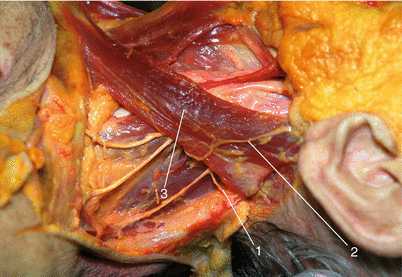
Fig. 12.2
(1) Lesser occipital nerve, (2) great auricular nerve, and (3) sternocleidomastoid muscle (With permission from Danilo Jankovic)
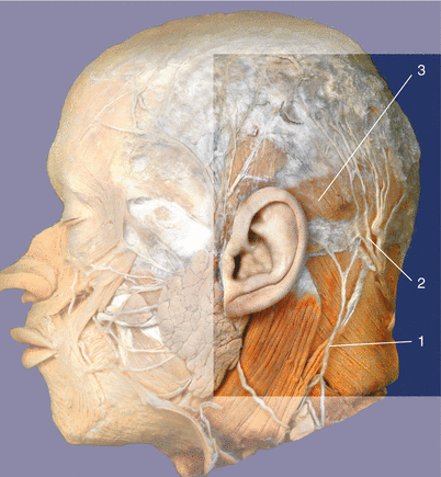
Fig. 12.3
Anatomy. (1) Lesser occipital nerve, (2) greater occipital nerve and occipital artery, and (3) occipital muscle (With permission from Danilo Jankovic)
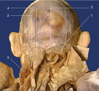
Fig. 12.4
Anatomy. (1) Greater occipital nerve, (2) the third occipital nerve, (3) inferior nuchal line, (4) semispinalis capitis muscle, and (5) splenius capitis muscle (With permission from Danilo Jankovic)
After exiting from the lower edge of the obliquus capitis inferior (or inferior oblique capitis) muscle, the second cervical spinal nerve divides into anterior and posterior branches. The posterior branch of the second cervical spinal nerve passes in a dorsal direction around the obliquus capitis inferior muscle and runs between the occipitovertebral muscles and the semispinalis capitis muscle. Here it divides into three branches: an ascending branch, which supplies the longissimus capitis muscle; a descending branch, which anastomoses with the posterior branch of C3 (the third occipital nerve); and the medial greater occipital nerve (posterior branch of C2).
The sensory greater occipital nerve passes in a cranial direction, goes through the semispinalis capitis muscle and trapezius muscle, and reaches the skin about 2–3 cm away from the midline in the area of the superior nuchal line. It gives off several branches toward the top of the head and extends laterally as far as the ear. The course of its branches follows the branches of the occipital artery.
Sonoanatomy
At the superior nuchal line, the greater occipital nerve is not always seen as it may already branch out into small nerves. The surrogate marker is the greater occipital artery. A high frequency linear ultrasound probe (8–12 MHz) is applied just lateral to the greater occipital protuberance. Because it is very superficial, one should apply minimal pressure at this site to prevent compression of the occipital artery. The ultrasound probe may slide caudally just off the nuchal line, and the greater occipital artery is seen deep to the trapezius muscle (Fig. 12.5).
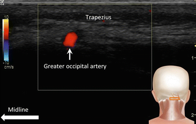
Fig. 12.5
Sonogram of occipital artery just caudal to the superior nuchal line. The artery indicates the plane where the greater occipital nerve lies. The probe position is indicated in the insert lower right corner (Reproduced with permission from Philip Peng Educational Series)
Other approach for greater occipital nerve is at the C2 level. At this level, the spinal process of C2 is well recognized for its bifid morphology (Fig. 12.6). The ultrasound probe is moved laterally to reveal the muscle layers from deep to superficial: obliquus capitis inferior, semispinalis capitis, splenius capitis, and trapezius muscles. To optimize the scan of the obliquus capitis inferior muscle, the lateral end of the probe is rotated toward the lateral mass of C1 to bring the probe along the long axis of the obliquus capitis inferior muscle. The greater occipital nerve is seen between the obliquus capitis inferior and semispinalis capitis muscles approximately 2–3 cm away from the midline (Fig. 12.7). The vertebral artery and dorsal root ganglion is usually revealed on the lateral side with Doppler scan (Fig. 12.8).
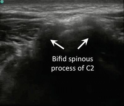
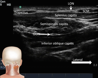
Fig. 12.6
Sonogram showing the bifid spinous process of C2 (Reproduced with permission from Philip Peng Educational Series)
Fig. 12.7


Sonogram at the same level as Fig. 12.6 but the probe is moved in the lateral direction with the lateral end of the probe tilted toward C1 lateral mass. The insert shows the probe position. GON greater occipital nerve, LON lesser occipital nerve, SCM sternocleidomastoid muscle (Reproduced with permission from Philip Peng Educational Series)
Related posts:


Stay updated, free articles. Join our Telegram channel
Full access? Get Clinical Tree
