Fig. 46.1
Dual neural supply of the facet joint. Oblique parasagittal view of overlapping segmental innervation of the facet joint. (1) Ventral branch of spinal nerve, (2) dorsal branch of spinal nerve, (3) ascendant branch of dorsal ramus, (4) medial branch of dorsal ramus, (5) distal branch of medial ramus to facet joint, (6) proximal branch of medial ramus to facet joint, (7) posterior ramus (sinuvertebral nerve of Luschka), (8) facet joint (Reproduced with permission of Danilo Jankovic)
Technique
The popular technique for both facet nerve and joint block is by fluoroscopic guidance, but ultrasound-guided techniques have been described and validated [7–10].
Fluoroscopic Guided Facet Nerve Block
The patient is placed in prone position with a pillow supporting the pelvis for comfort and to obliterate the lumbar lordosis. The techniques for the blockade of L1–L4 medial branches are the same [11]. The first step is to count the appropriate level. Once the level is counted, the target vertebral level is squared, that is, the anterior and posterior silhouettes of the lower border of vertebra are at the same level. The C-arm is then rotated ipsilaterally to the side of injection to obtain an oblique view. In this view, the outline of the “scotty dog” is clearly evident (Fig. 46.2). The target is the junction of the superior border of transverse process with the superior articular process. Alternatively, another end point has been described, which is the midpoint between the mamilloaccessory notch and the target point just described in Fig. 46.1 (Fig. 46.3). This target point has been shown to minimize the inadvertent spread of injectate into the intervertebral foramen or epidural space. For the L5 dorsal rami block, the target point is the ala of the sacrum at the base of the superior articular process of sacrum in an anteroposterior view (Fig. 46.4).
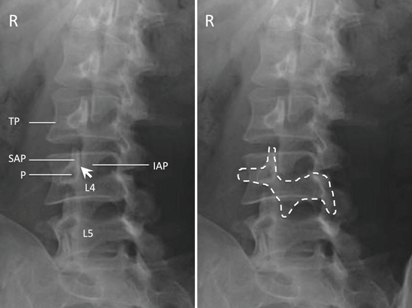
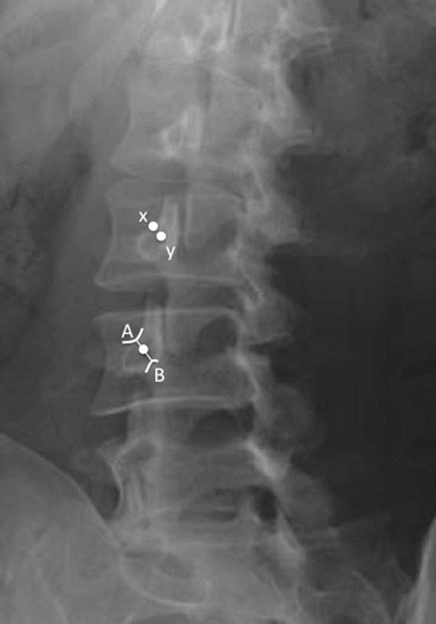
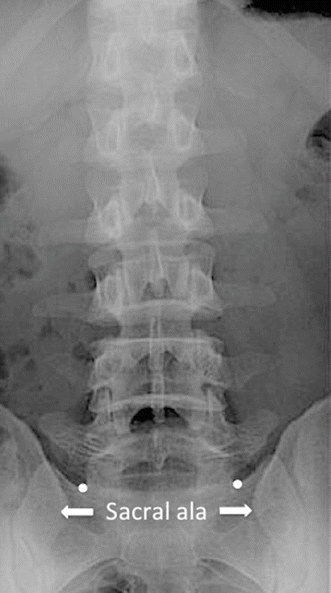
Fig. 46.2
Oblique radiograph of the lumbar spine. The left figure shows the individual structures: SAP superior articular process, IAP inferior articular process, P pedicle, TP transverse process; facet joint is indicated by the arrow. The right figure shows the “scotty dog” appearance of the same structure (Reproduced with permission from Philip Peng Educational Series)
Fig. 46.3
The radiograph shows the target points (white dots) for facet medial branch injection. A The junction of the superior border of transverse process with the superior articular process. B The mamilloaccessory notch. x The conventional target which is the junction of the superior border of transverse process with the superior articular process. Y The midpoint between A and B (Reproduced with permission from Philip Peng Educational Series)
Fig. 46.4
The target points for L5 dorsal rami was indicated by the white dots, which are the ala of the sacrum at the base of the superior articular process of sacrum (Reproduced with permission from Philip Peng Educational Series)
A 22-gauge spinal needle is inserted following local anesthetic infiltration of the skin. Once the bone is contacted at the target point, a small amount of contrast (0.2 mL) is injected to detect possible venous uptake. If there is no venous uptake, 0.5 mL of local anesthetic is injected.
Fluoroscopic Guided Facet Joint Injection
The patient is positioned as for the facet medial branch block. Oblique view is obtained until the joint space formed between the articular processes is optimally seen (Fig. 46.2). A 22-gauge spinal needle is inserted with coaxial technique. A small amount of contrast (0.2 mL) is injected to confirm the intra-articular placement. A volume of 0.5–1 mL injectate (20 mg methylprednisolone or triamcinolone) is sufficient. In patient with advanced disease, the joint space may be further obliterated. In that case, periarticular injection may be performed.
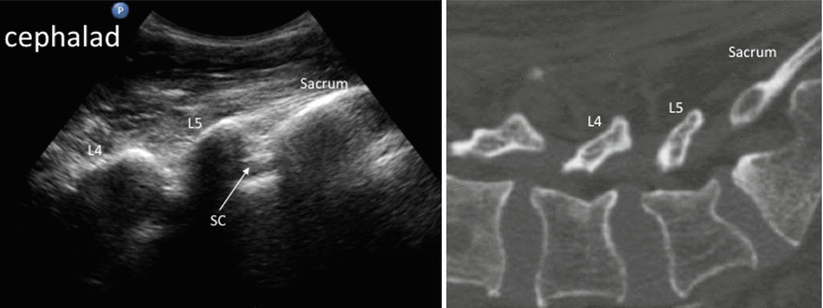
Ultrasound-Guided Facet Medial Branch Injection
The position of the patient is the same as the fluoroscopic guided technique. The target point is the junction between the superior articular process and transverse process. A curvilinear ultrasound probe (2–5MHz) is used to obtain a parasagittal oblique view. In this scan, both the sacrum and lamina can be seen and used for counting (Fig. 46.5). The probe is then moved laterally to visualize the transverse process (Fig. 46.6). Once an appropriate level is identified, the probe is rotated 90° to obtain a short axis view of the spine showing the transverse process and the corresponding superior articular process. The probe is then moved in cephalad direction until the cephalad aspect of the transverse process disappears. When this part of the transverse process reappears on sliding back the probe in caudal direction, the target is defined (Fig. 46.7). A 22-gauge spinal needle is inserted in-plane from lateral to medial to hit the bony target. Once this is achieved, the probe is turned 90° to demonstrate the needle is at the cephalic part of the transverse process (Fig. 46.8). The L5 dorsal ramus is usually difficult to assess because of the ilium.