Imaging
Angiography, Cerebral
(see Part 1, p. 7)
Angiography, Coronary
(see Part 2, p. 71)
Adenosine Stress Test
(see Stress Testing, p. 366)
Chest X-Ray
As an x-ray beam passes through a structure, different levels of absorption of the beam by different tissues occurs. The quality of the film, therefore, is dependent upon the amount of contrast between the tissues and different radiodensities. (The thickness of tissue through which the beam has to penetrate is a factor as well, i.e., the heart, a large structure, will appear more opaque than the much smaller right or left pulmonary artery.)
Remember:
The structure that absorbs the most x-rays will appear the lightest.
Thus:
Bony structures, lightest (absorb most x-rays)
Fat, somewhat darker
Body fluids (includes soft tissue, muscle, blood, heart), medium contrast
Gas or air, darkest (absorb minimal x-rays)
Accordingly, when a lung is referred to as being “whited out,” this means that air density of the lung has been replaced (or displaced) by a tissue or water-density disease process, and has resulted in increased opacity.
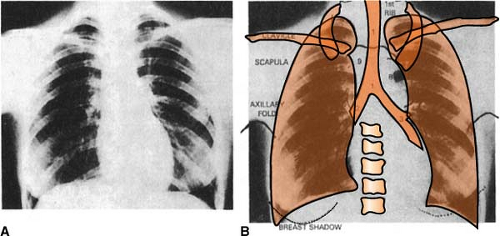 Figure. A, Normal posteroanterior chest radiograph. B, Outline of structures visible on normal posteroanterior chest radiograph. (Adapted from Fraser, R. G., Pare, J. A. P., et al. [1988]. Diagnosis of disease of the chest [pp. 288–290]. Philadelphia: WB Saunders. Reprinted with permission.) |
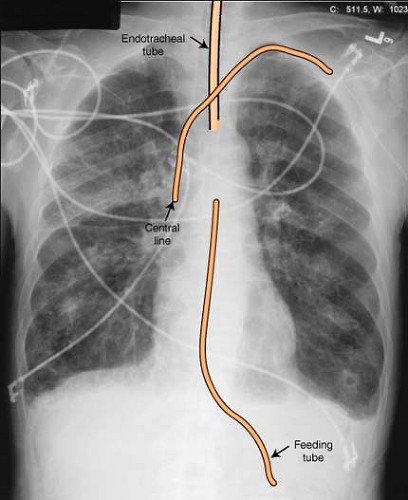 Figure. Radiograph depicting proper tube placements. Courtesy: Wausau Heart Institute, Wausau, WI. |
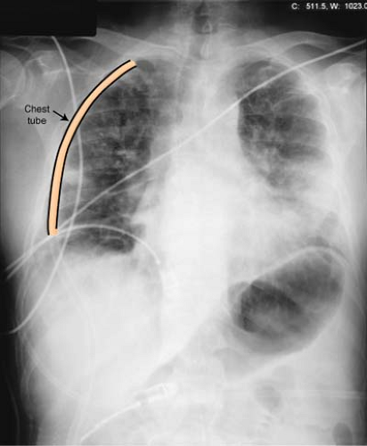 Figure. Radiograph denoting chest tube placement. Courtesy: Wausau Heart Institute, Wausau, WI. |
CT Scan (Computed Tomography)
Computed refers to the fact that the information obtained from the procedure is reconstructed by a computer, and tomography refers to the method of producing images on a plane. CT imaging is particularly useful because it can show several types of tissue with great clarity—lung, bone, soft tissue, and blood vessels. (Very fine soft tissue areas, such as the knee and shoulder, are better served by magnetic resonance imaging. See MRI.) As with conventional x-rays, dense objects (bone) will appear bright, while less dense material (tissue) will appear darker.
Inside the CT scanner is a rotating gantry that has an x-ray tube mounted on one side and an arc-shaped detector on the opposite side. An x-ray beam is emitted in a fan shape as the rotating frame spins the x-ray tube and detector around the patient. Each time the x-ray tube and detector make a 360-degree rotation and the x-ray passes through the body, an image is acquired. Think of the scan as a meat loaf cut into thin slices. When the “slices” are reassembled by the computer, a very detailed multi-dimensional view of the structure is obtained.
A relatively new technique, spiral CT, has improved CT scanning even more. It allows for faster scanning and higher resolution by advancing the examination table through the gantry at a constant rate while having the x-ray tube rotate continuously around the patient. Most common are 4 or 16 slice systems. With a 16 slice system, the radiologist can obtain 32 image slices per second, allowing for a complete scan of the chest or abdomen in 10 seconds or less.
Dobutamine Stress Test
(see Stress Testing, p. 365)
Doppler Echocardiography
(see also Echocardiography, p. 362)
The newest innovation in echocardiography is Doppler echocardiography. It is based on the principle that the frequency of sound reflected from a moving object is changed in a predictable way by the motion of that object. When the object moves toward the detector, the frequency increases; when it moves away from the detector, the frequency decreases. (Think of an approaching train: The whistle is loud, then recedes as the train passes). In a like manner, Doppler echocardiography consists of recording sound waves reflected from moving red blood cells, displayed as an audible signal or as a spectral image, graphed with frequency on the vertical axis and time on the horizontal axis.
Color Doppler is the most sophisticated form of echocardiography, making it possible to turn flow signals into different colors and then superimposing the image on the real-time 2-D image. Red denotes movement toward the transducer, and blue denotes movement away from it. The intensity of color denotes velocity: Faster signals are lighter, and slower signals are darker. Turbulence is indicated by a mosaic color pattern. Because an entire sector can be imaged, this is the preferred mode for detecting valvular regurgitation.
Ebus
(see Endobronchial Ultrasound, p. 363)
Echocardiography
(see also Doppler Echocardiography, p. 362; Transesophageal Echocardiography, p. 366)
Echocardiography is a study that uses ultrasound, or sound waves of frequencies higher than the human ear can detect, to examine the heart for motion, competency of cardiac valves, size of cardiac chambers, thickness and contractility of the myocardium, presence of structural abnormalities, and/or pericardial effusion or constriction.
M-Mode (Motion)
A hand-held transducer is applied to the front of the chest. High-frequency sound waves (2.5-5 mHz) are generated in short bursts (e.g., 1 microsecond), after which the transducer operates as a receiver (e.g., 1 millisecond). The returned echoes are then converted to impulses and transmitted to a machine for display and recording on videotape. The patient does not feel or hear the sound waves, and no tissue damage is produced.
Two-Dimensional (2-D) Mode
Two-dimensional (2-D) echocardiography is a further refinement of the M-mode. The transducer crystal rotates through an arc, or many crystals emit pulses, to provide a tomographic “slice” of cardiac structures, more closely resembling actual cardiac anatomy. The problem with 2-D echocardiography, however, is that incorrect positioning or beam angulation can significantly alter the appearance of the heart and can obscure diagnostic information. Thus, M-mode echocardiography continues to be used in addition to 2-D echocardiography, because the resolution is significantly better.
Related posts:


Stay updated, free articles. Join our Telegram channel

Full access? Get Clinical Tree


