 COMPLICATIONS ASSOCIATED WITH PROVOCATIVE DISCOGRAPHY
Because of its nonspecific features, the diagnosis of discogenic pain is not easily made by clinical means alone. Useful tools in the search for the origin of back pain are provocative discography and magnetic resonance imaging (MRI) studies. Although of questioned diagnostic value,6–8 provocative discography is the only test that may relate pathologic changes found on MRI to patient’s pain.8–11 Discography is performed in a procedure room under fluoroscopic guidance, which allows consistent visualization of the bony landmarks. The patient lies prone and the lumbar lordosis is corrected by placing a roll or soft wedge under the lower abdomen. Monitoring and light sedation are used and a fluoroscopy employing a lateral or extrapedicular approach using “tunnel vision” facilitates intradiscal needle placement.12
The overall rate of complications following provocative lumbar discography is low, with reports ranging from 0% to 2.5%. Discitis, epidural abscess, and bacterial meningitis have all been reported.5–11
More frequent procedural complications include acute paresthesias, muscle spasm, and minor bleeding. During provocative discography, excessive amounts of local anesthetic injected deep, closer to the disc/foramina just prior to intradiscal needle placement, may decrease the ability of the patient and proceduralist to detect spinal nerve contact during needle placement. To avoid potential neural injury, the needle should be directed into the region below the segmental spinal nerve, just lateral to the superior articular process, and above the superior vertebral endplate immediately below the targeted disc. The patient may experience a brief, sharp, painful sensation when the needle pierces the well-innervated outer annulus fibrosus. If the patient experiences any paresthesias during needle advancement, insertion of the needle must stop and needle redirected.
Provocative discography may lead to acute lumbar disc herniation. In five patients, postdiscography herniation was clinically manifest as an acute exacerbation of radicular leg pain and was accompanied by an acute foot drop in one of these patients. Acute disc pressurization caused either an increase in the size of preexisting herniation or formation of the new one, confirmed by the comparative lumbar MRI scans prior to and after completed discography. The authors concluded that annular weakness may be a predisposing factor to discography-related disc herniations.13
Chronic discitis is the most dreaded complication associated with discography, and its frequency is from 0% to 1.3% per disc injected,14,15 or more precisely 0.15% per patient and 0.08% per disc injected.14–17 It is believed to be due to inoculation of skin flora carried by the procedure needle into the intervertebral disc.14,15 Commonest skin flora microorganisms are Staphylococcus aureus and Staphylococcus epidermidis, although gram-negative bacteria also have been reported as the cause of discitis.15–19
Preprocedural intravenous and/or intradiscal antibiotics are frequently used as a prophylactic measure. The occurrence of infectious discitis may be decreased with the use of intradiscal antibiotics mixed with the water-soluble contrast. Iohexol lowers the MICs (minimal inhibitory concentrations) of the antibiotics if it is used together with cefazolin and gentamycin.20
Intravenous administration of some antibiotics may produce an unpredictable intradiscal concentrations. While clindamycin and vancomycin levels were detected in the rabbit discs after the intravenous administration, cephalothin and oxacillin were not.21 In humans, gentamicin and cefazolin do penetrate the intervertebral disc to a greater degree than other cephalosporins or oxacillin, indicating selective intradiscal availability of different antibiotics.22,23 The highest intradiscal concentration of cefazolins is achieved within the “golden period” of 15 to 81 minutes after 2 g are given intravenously.24
There were no cases of infectious discitis after the administration of antibiotics prior to discography in a 3-month follow-up study of 200 patients.15 Another large study reported no complications of 1,477 performed provocative discographies in 523 patients.25
The antibiotics can be mixed with the contrast agent into a stable, nonprecipitating solution before injecting them into the disc. None of the 127 patients, according to Osti et al.,26 developed clinical or radiological signs of chemical or bacterial discitis after discography, when 1 mg of cefazolin was added to 1 mL of contrast agent.26
It is difficult to determine if the incidence of discitis is more frequent following cervical, thoracic, or lumbar provocative discography. The incidence of discitis after cervical discography ranges from 0.16% to as high as 3%. One study reported cervical discitis in 7 out of 1,357 patients with the predominant presenting symptom being excessive neck pain soon after the discogram.27 Two smaller series reported acute discitis in 1 out of 31 and 2 out of 269 disc injections.28,29 There were no reported infectious complications after 89 thoracic discographies in 20 patients.30
In order to prevent discitis, both the North American Spine Society and the International Spinal Intervention Society guidelines recommend utilizing a two-needle approach, in which a larger needle is inserted through the skin first, and a smaller gauge needle is then passed through the larger needle to enter the disc.17,31 The theory behind the two-needle technique is that the smaller needle that enters the disc never passes through the skin, thereby reducing the chances of bacterial contamination of the needle tip. Prior to routine use of prophylactic antibiotics, Fraser et al.18 reported a rate of discitis with single nonstyletted versus double needles of 2.7% versus 0.7%.
The patient with discitis presents days or weeks after the procedure with severe, unremitting back or neck pain with or even without fever. It is needed to complete a physical exam, laboratory tests that include complete blood count with differential, C-reactive protein and blood cultures, and imaging. MRI is the preferred imaging modality32,33 and, within 3 to 4 days of symptoms, shows increased T2 signal in the disc and endplate hyperemia. It is recommended to do targeted biopsy during acute phase, before the endplate breach when sterile environment is created by the activation of immune system.34 Treatment of discitis is difficult and protracted, typically requiring long-term antibiotic. Discitis can lead to more complicated infections of the adjacent soft tissues, leading to abscess formation that may require surgical intervention.35,36 Epidural abscess formation is a rare complication of provocative discography. Frequently, decompressive lumbar laminectomy is needed to evacuate the abscess.37 There were reports of prevertebral abscess and a spinal subdural empyema as a unique complication of cervical discography.27,36
One case of urticaria was also reported in a study on 750 discographic injections performed in 250 patients.38
Prolonged pain after discography is often misinterpreted. The first study reporting prolonged low back pain after discography enrolled incarcerated subjects and an inappropriate contrast irritant, the ionic contrast media diatrizoate (Hypaque).39 More recently, a study conducted on six patients with a diagnosis of somatization disorder, abnormal psychometric testing, or worker’s compensation claims documented prolonged back pain after the discography.40 It is difficult to generalize such phenomena to a normal patient population, as persistent somatic complaints including chronic pain are common in this group.40,41
Finally, recent controversy has arisen surrounding the likelihood that discography itself can lead to accelerated disc degeneration after the diagnostic test is performed. Carragee et al.42 compared 52 control patient with 50 patients who underwent lumbar discography and analyzed their subsequent imaging studies 10 years later, looking for signs of progressive disc degeneration. They concluded that modern discography techniques using small gauge needles and limited pressurization resulted in accelerated disc degeneration, disc herniation, loss of disc height, and signal and the development of reactive endplate changes compared to match controls. The authors recommended that careful consideration of risk and benefit should be used in recommending procedures involving disc injection. This case-control study has significant limitations, but has raised significant controversy and concern about the role for discography in the diagnosis of discogenic pain.
It is important that lumbar discography be performed by a well-experienced physician, under sterile conditions using fluoroscopic imaging for proper needle placement, in order to minimize the risk of complications.5,9
 COMPLICATIONS ASSOCIATED WITH ANALGESIC DISCOGRAPHY
Analgesic discography is an alternative to traditional provocative discography and was designed in efforts to increase the diagnostic precision associated with tests aimed at identifying symptomatic disc degeneration. This test employs the intradiscal placement of small-bore infusion catheters, through which local anesthetic can be infused after placement. The catheters are placed, and then each disc is successively anesthetized in efforts to locate the symptomatic level. Little has appeared with regard to complications associated with this new procedure. Nonetheless, during analgesic discography, the diameter of the needle inserted is significantly larger than the needle size used during provocative discography (Fig. 29-1). This is necessary in order to thread the balloon catheter, used later on for postinsertion testing43 (Fig. 29-1B). As we have already discussed, puncturing an intervertebral disc with a needle may lead to progressive disc disruption.42 Greater progression of degenerative disc disease has been suggested in postdiscography discs, and it seems to be more rapid and severe in patients who had larger diameter size needles inserted into their intervertebral discs, suggesting that the large cannulae used during analgesic discography may be problematic.
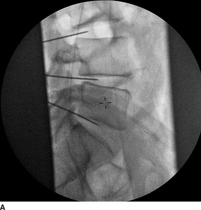
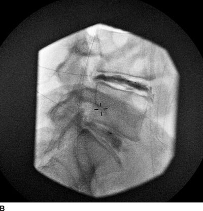
FIGURE 29-1. Intradiscal placement of the 18G introducers (A) used for the analgesic discography followed by insertion of intradiscal balloon (B) used during analgesic discography. Note a large diameter of the introducer that is required to be positioned within the intervertebral disc nucleus.
 COMPLICATIONS ASSOCIATED WITH INTRADISCAL INJECTIONS
Chymopapain
Chymopapain has been used for years as a substance for intradiscal nuclear ablation (chemonucleolysis) in patients with contained disc herniations and ongoing back and leg pain. Chemonucleolysis uses chymopapain B, an injectable proteolytic enzyme. Chymopapain produces hydrolysis of noncollagenous protein that interconnects long-chain mucopolysaccharides; chymopapain also has a neurolytic effects on free nerve endings within the disc. This series of biochemical reactions leads to depolymerization of the nucleus pulposus, thereby lowering intradiscal pressure. Most patients who received such therapy were those suffering from contained disc protrusion and predominantly leg pain, but it has been used also for the treatment of discogenic lower back pain. An overall success rate of 72% was reported in one analysis of 17,000 patients treated with chymopapain for acute disc herniation and leg pain.44,45
There were 2.4% of patients with various side effects.46 In 20% to 40% of cases, postinjection back pain and muscle spasm occurred as the most common side effects.44–46 Chymopapain has not used in the United States since 1999 largely due to a series of patients with severe anaphylactic reactions after injection; nonetheless, this treatment is still common in other parts of the world.44 The incidence of allergic reaction to chymopapain can be lowered from 1% to 0.3% by using IgE serum sensitivity testing prior to procedure.47 It also appears that chemonucleolysis performed under local anesthesia lowers the risk of anaphylactic reaction.48 Serious complications, like transverse myelitis, hemorrhage, and discitis, are rare. The overall morbidity rate related to this procedure is 0.399%, and the mortality rate is 0.02%.46
Intradiscal Steroids
It is questionable if there is any clinical benefit from repeated intradiscal steroid injections for the treatment of discogenic lower back pain; nonetheless, this treatment is still used by some practitioners. Observational studies suggest that patients with uncomplicated axial back pain presumed to be of discogenic origin had no long-term improvement in either functional capacity or pain scores.49,50 However, when intradiscal steroid injections were used in patients with inflammatory end-plate changes, significant short-term improvements in pain scores and functional capacity have been reported.38,39
The most frequent and serious complication of such intradiscal steroid injections is formation of epidural calcifications, mainly when triamcinolone hexacetonide is used.51 On occasion, this complication can first appear many years after an intradiscal injection of this agent.52 Expanding calcifications become symptomatic in 14% to 68% of patients.51,52 While this treatment is seldom used, it is important to keep in mind what previous study has revealed about the problems with this seemingly innocuous treatment.
 COMPLICATIONS ASSOCIATED WITH ANNULOPLASTY
The term annuloplasty is meant to refer to a group of minimally invasive procedures that are aimed at treating symptomatic degenerative disc disease. The dense innervation of the outer portion of the annulus fibrosus has been implicated as a cause of ongoing back pain associated with disruption of the outer annulus that appears with progressive disc degeneration. There are a number of different devices used to perform annuloplasty, but all employ some form of thermal energy in efforts to destroy free nerve endings. The incidence of complications during and after various annuloplasty procedures for discogenic pain range from 0% to as high as 10%53,54 in different reports. These complications can be divided into early (appearing immediately after or within days of the procedure) and late (appearing weeks to months after the procedure) complications. Nerve injuries related to needle placement and/or thermal injuries together with infection, bleeding, and burns are early complications. Late complications include accelerated disc degeneration, vertebral avascular necrosis, and postprocedural disc herniation. Instrument malfunction or breakage during the procedure may also lead to significant injury of neural or vascular structures (Table 29-1). In the sections that follow, we will discuss the complications that have been associated with each specific method for performing percutaneous annuloplasty.
Intradiscal Electrothermal Therapy
Intradiscal electrothermal therapy (IDET) is a minimally invasive procedure in which controlled thermal energy is gradually delivered to the posterior the annulus fibrosus via an intradiscaly placed resistive coil.55 IDET is performed under fluoroscopic guidance, while the patient is lightly sedated. A 17-gauge introducer needle is first inserted into the anterolateral part of the disc to be treated. The catheter is then threaded along the central aspect of the annulus. The distal part of the catheter is approximately 5 cm in length; once in position, the catheter is gradually heated to 90°C over 16 to 17 minutes. During this procedure, the intradiscal temperatures may range anywhere from 37°C to 65°C.56 Inappropriate catheter placement and high temperatures delivered to the disc and the surrounding neural structures may cause nerve injury or osteonecrosis of endplate, which would manifest as a radicular pain, axial pain, or transient palsy. These complications, however, may also happen during other percutaneous invasive procedures and are not specific only to IDET. Catheter breakage57(Figs.s 29-2 and 29-3), vertebral osteonecrosis,58 and thermal injury to the cauda equina59 have all been reported as serious complications of IDET. The proposed mechanism leading to catheter breakage and thermal injury to the cauda equine are shown in Figure 29-4. Some of the commonly listed risk factors for postsurgical spinal complications, such as obesity, history of leg pain, smoking, diabetes mellitus, and duration of the back pain, are unlikely to predict higher risk for complications after the IDET.53 When compared to the nucleoplasty (see further discussion below) alone, frequency of complications was not higher in patients who received IDET combined with the nucleoplasty.60 A study by Freeman et al.54 reported no major complications in 38 patients with chronic discogenic back pain following the IDET. However, four patients had transient radiculopathy that resolved in a period of <6 weeks. In another study, temporary (<6 weeks) radicular signs and symptoms were reported by several patients, one patient complained of a new burning sensation in one leg and another patient had a foot drop. In two patients, nondermatomal paresthesias were present.57 The paresthesias will occur in a small percentage of patients undergoing the IDET procedure, even when IDET is performed by the most skilled practitioners. Recently, Orr and Thomas published a case report in which an IDET catheter tip was broken off after an intradiscal kink, leaving a fragment of the catheter retained within the disc. The fragment subsequently migrated out of the disc and into the dural sac. The patient presented weeks later complaining of increased back pain, leg paresthesias, and dysesthesias, and the catheter fragment had to be surgically removed.61 If the catheter is sheared, it should be left within the disc, as the current experience, aside from this one report, suggests a low likelihood for migration leading to need for surgical removal.
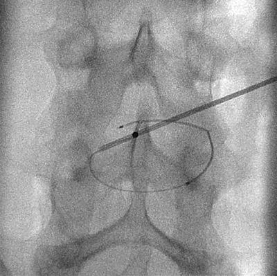
FIGURE 29-2. Repeated attempts to advance the IDET catheter can lead to kinking of the catheter and potential catheter breakage. Fluoroscopic anterior-posterior view combined with cranial tilt of the fluoroscope illustrates the extent of the IDET resistive coil kink.


