Fig. 43.1
Identifying the epidural space using the loss-of-resistance technique (With permission from Danilo Jankovic)
After identification of the epidural space and injection of a test dose (Fig. 43.2), a thin 27-G pencil-point spinal needle is carefully advanced through the epidural needle in the direction of the subarachnoid space, until dural perforation is confirmed by a click (Fig. 43.3a).
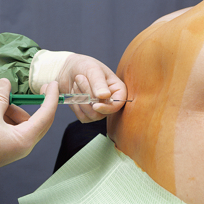
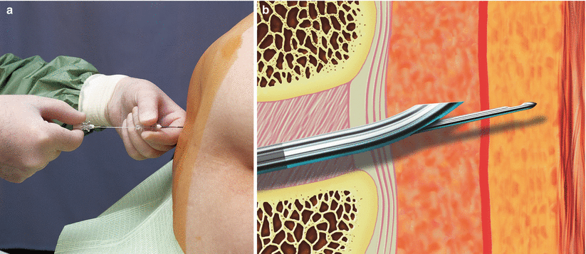

Fig. 43.2
Injecting a test dose (With permission from Danilo Jankovic)

Fig. 43.3
(a) 27-G pencil-point spinal needle is introduced through the positioned epidural needle. (b) Identification of the subarachnoid space with the dural click (With permission from Danilo Jankovic)
The adapted form of the Tuohy needle tip, which has a central opening positioned in the needle axis (“back eye”), allows the needle to take a direct path, so that the spinal needle does not need to bend. The plastic coating of the spinal needle expands its outer diameter, so that it fits the epidural needle precisely, maintains its central position as it is advanced, and easily passes through the axial opening (“back eye”) (Fig. 43.3b). After careful aspiration of CSF, subarachnoid injection of a local anesthetic, opioid, or a combination of the two is carried out (Fig. 43.4). As this is done, the hub of the spinal needle should be secured with the thumb and index finger of the left hand, which rests on the patient’s back. This is the critical phase of the puncture procedure.
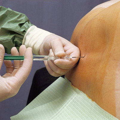

Fig. 43.4
Subarachnoid injection. The spinal needle is then withdrawn (With permission from Danilo Jankovic)
The spinal needle is then withdrawn, and the epidural catheter is introduced up to a maximum of 3–4 cm (Fig. 43.5a, b).
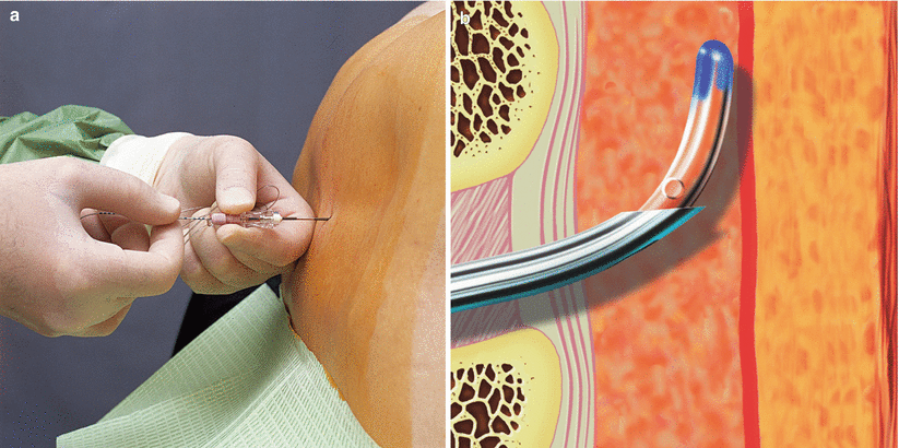

Fig. 43.5
(a) Introducing the epidural catheter. (b) The epidural catheter is advanced by a maximum of 3–4 cm cranially (With permission from Danilo Jankovic)
After aspiration, the open end of the catheter is laid on a sterile surface below the puncture site and any escaping fluid (CSF or blood) is noted (Fig. 43.6).
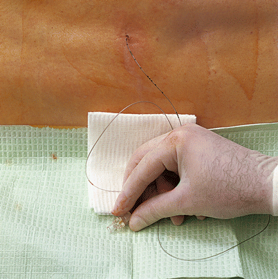

Fig. 43.6
The open end of the catheter is placed below the puncture site (With permission from Danilo Jankovic))
To test the patency of the catheter, 1–2 mL saline is then injected. The catheter is secured and a bacterial filter is attached (Fig. 43.7).
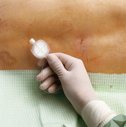

Fig. 43.7
The catheter is secured and a bacterial filter is attached (With permission from Danilo Jankovic)
Repeated aspiration.
As low a dose as possible.
Always use incremental injections (several test doses).
Maintain verbal contact.
Check the spread of anesthesia carefully.
Careful monitoring.
Problem Situations
Specific problems during CSE occur in connection with the administration of a test dose to exclude subarachnoid positioning of the catheter.
As spinal anesthesia is being given, it is not possible to test for incorrect intrathecal positioning of the catheter, and incorrect positioning usually becomes evident through high or total spinal anesthesia.
This technique should therefore only be carried out by experienced anesthetists.
Local anesthetics must only be injected in small incremental amounts (test doses), the spread of the anesthesia must be carefully checked and verbal contact with the patient must be maintained.
Two-Segment Technique
Puncture is carried out in the midline at the level of L2/3 or L3/4. An 18-G Tuohy needle is used. After identification of the epidural space, the epidural catheter is advanced to a maximum of 3–4 cm. A test dose is then administered. One or two segments lower, conventional spinal anesthesia is then carried out using a 27-G pencil-point needle. The remainder of the procedure is the same as in the “needle-through-needle” technique.
Dosages in the “Needle-Through-Needle” and Two-Segment Techniques [7–11, 17]
Subarachnoid
Opioid: 10 |jg sufentanil + 1 mL 0.9 % saline
Local anesthetic:
0.5 % ropivacaine 1–1.5 mL (5–7.5 mg) ± 0.2 mL
0.5 % hyperbaric bupivacaine 1–1.5 mL (5–7.5 mg) ± 0.2 mL
Local anesthetic + opioid:
0.5 % ropivacaine + sufentanil 7.5–10 pg or
Stay updated, free articles. Join our Telegram channel

Full access? Get Clinical Tree


