Bleeding Complications


Bleeding Complications


John C. Drummond

Charise T. Petrovitch


INTRODUCTION

Abnormalities of hemostasis that result in clinical coagulopathies are common in the operating room and the intensive care unit (ICU). Understanding the etiology and treatment of these abnormalities requires a knowledge of the normal coagulation mechanism, which is discussed in greater detail in Chapter 38, but a brief review is presented here (see Section “How Does the Normal Hemostatic Mechanism Function?). Abnormalities of hemostasis may be either congenital (see Section What Are the Important Congenital Abnormalities of Hemostasis?) or acquired (see Section What Are The Important Acquired Disorders of Hemostasis?). The acquired disorders may be the result of disease states or the administration of pharmacologic agents. The Section “How Should the Clinician Approach the Diagnosis and Treatment of Bleeding Diatheses?” presents an approach to the diagnosis and treatment of bleeding disorders. Disorders of the hemostatic mechanism may also result in hypercoagulable states (see Chapter 38).


How Does the Normal Hemostatic Mechanism Function?

In this chapter, only an abbreviated discussion follows. A detailed description of the coagulation mechanism can be found in another publication.1 The nomenclature (numerals and common names) and the half-lives of the clotting factors are presented in Table 36.1.


▪ THE COAGULATION MECHANISM

Although the classical, dual cascade model of coagulation with its intrinsic and extrinsic pathways (see Fig. 36.1) probably provides a reasonable model of coagulation, as it is evaluated in vitro by the activated partial thromboplastin time (aPTT) and prothrombin time (PT) determinations respectively, that model provides an inadequate representation of in vivo coagulation. It suggests important roles for factors XII and XI, congenital deficiencies of which cause relatively little clinical disturbance of coagulation. In addition, it fails to explain why a patient with hemophilia who lacks an intrinsic pathway factor (hemophilia A, factor VIII; hemophilia B, factor IX) cannot achieve hemostasis through the unaffected extrinsic pathway. A description of the current understanding of the three stages of the hemostatic process, which has
been thoroughly defined and described by Hoffman, are summarized in the following text and in Figure 36.2.2


TABLE 36.1 Factor Nomenclature and Half-Lives


Factor


Common Names and Synonyms


Half-life (Hours)


I


Fibrinogen


100-150


II


Prothrombin


50-80


III


Tissue factor, tissue thromboplastin


IV


Calcium ion


V


Proaccelerin, labile factor


24


VII


Serum prothrombin conversion accelerator, stable factor


6


VIII


Antihemophilic factor


12


vWF


von Willebrand factor


24


IX


Christmas factor


24


X


Stuart-Power factor, Stuart factor, autoprothrombin


25-60


XI


Plasma thromboplastin antecedent


40-80


XII


Hageman factor


50-70


XIII


Fibrin stabilizing factor


150


Prekallikrein


Fletcher factor


35


HMW kininogen


Fitzgerald, Flaujeac, Williams factor; contact activation cofactor


150


HMW, high molecular weight.


Activation

Activation of the coagulation process begins when disruption of the vascular endothelium exposes blood to tissue factor (TF) (Fig. 36.2A). TF activates factor VII to yield a complex of TF and activated (indicated by a lower case “a”) factor VII (Fig. 36.2B). The TF-VIIa complex then activates factors IX and X (Fig. 36.2C). Activated factor X (Xa) then activates factor V (Fig. 36.2D), leading to the formation of the “prothrombinase complex” (Xa and Va) on the phospholipid surface provided by TF. The prothrombinase complex catalyzes the conversion of prothrombin (factor II to thrombin (FIIa) (Fig. 36.2E). This initial formation of thrombin serves to advance the coagulation process to the more efficient “amplification” phase that follows. Note that the TF-VIIa complex-mediated activation of Xa is a self-limited one that generates only small amounts of thrombin. It is regulated in a negative feedback manner by the factor Xamediated generation of tissue factor pathway inhibitor (TFPI).3 Teleologically, this pathway probably serves to prevent extensive and spontaneous clot formation in the extravascular space. It provides the explanation for why patients with hemophilia (A or B) cannot simply rely on the extrinsic pathway (TF-VIIa mediated formation of Xa) to generate thrombin. The thrombin yield of that pathway is too modest to provide the thrombin burst necessary for the eventual consolidation of the platelet plug by fibrin (Fig. 36.2I).


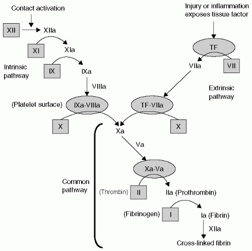
FIGURE 36.1 The intrinsic and extrinsic pathways of coagulation. Factors in the inactive form are within shaded squares. The shaded ovals represent phospholipid surfaces (tissue factor [TF] or platelets).


Amplification

Whereas it was the phospholipid surface provided by membrane-bound TF that initiated the coagulation process, it is now the phospholipid surface provided by activated platelets that serves to perpetuate it. The breach in the vascular tree that began the activation process also exposed platelets to collagen, to which they become bound by von Willebrand factor (vWF) through the glycoprotein (GP) Ib-IX-V receptor complex on the platelet surface (see Fig. 36.3). The thrombin just generated by the TF-bound
prothrombinase complex supports the amplification of the coagulation process in four ways, which are as follows:


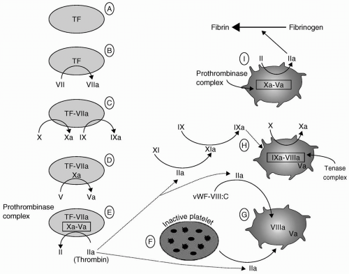
FIGURE 36.2 The coagulation mechanism. See text for a detailed explanation. TF, extravascular membrane-bound tissue factor; vWF-VIII:C, circulating factor VIII bound to its carrier protein, the von Willebrand factor. (Reproduced with permission from Drummond JC, Petrovitch CT. Hemotherapy and hemostasis. In: Barash PG, Cullen BF, Stoelting RK, eds. Clinical anesthesia, 5th ed. Philadelphia: Lippincott Williams & Wilkins; 2006:208.)


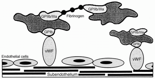
FIGURE 36.3 Platelet adhesion and aggregation. When the endothelium is denuded, von Willebrand factor (vWF) binds to collagen in the subendothelial layer. Platelets adhere through their glycoprotein 1b (GPIb)-IX-V receptors to vWF. Platelets aggregate to one another by cross-linking through fibrinogen (or vWF, not shown) between GPIIb-IIIa receptors expressed on the platelet surface during the process of platelet activation. (Reproduced with permission from Drummond JC, Petrovitch CT. Hemotherapy and hemostasis. In: Barash PG, Cullen BF, Stoelting RK, eds. Clinical anesthesia, 5th ed. Philadelphia: Lippincott Williams & Wilkins; 2006:208.)


  • First, it further activates the adjacent platelets (Fig. 36.2F). That activation results in platelet surface changes, most notably the appearance of the GPIIb-IIIa receptor, and the release of the contents of platelet granules. Among those contents are adenosine diphosphate (ADP), a powerful platelet activator and proaggregant that rapidly recruits other platelets to the growing platelet mass, and FV.


  • Thrombin’s second effect promotes the activation of FV to FVa (Fig. 36.2G).


  • Third, thrombin releases circulating FVIII from its carrier molecule (vWF) and activates it (Fig. 36.2G).


  • Fourth, thrombin activates factor XI (see Fig. 36.2H). Factor XIa in turn activates factor IX (Fig. 36.2H), further adding to the pool of factor IXa that first formed during the activation phase above (Fig. 36.2C). The net result of this amplification stage is the availability of activated platelets and activated factors V, VIII, and IX.


Propagation

The platelet then provides the phospholipid surface on which two coagulation factor complexes form to lead to the explosive generation of thrombin. First, factors VIIIa and IXa form the “tenase complex”, which activates factor X (Fig. 36.2H). The resultant Xa forms an additional prothrombinase complex (Xa-Va), and large amounts of thrombin are elaborated (Fig. 36.2I). Thrombin (IIa) catalyzes the formation of fibrin, which acts to cross-link the platelets to stabilize the friable platelet plug (Fig. 36.3). Thrombin also activates the thrombin activatable fibrinolysis inhibitor and factor XIII (fibrin stabilizing factor), both of which serve to stabilize the fibrin clot. Fibrin monomers initially aggregate relatively loosely to form clot composed of soluble fibrin (fibrin S), held together loosely by hydrogen bonds. Fibrin stabilizing factor (not shown in figure) mediates the formation of covalent peptide bonds between the fibrin monomers to produce a stable (insoluble) fibrin clot.


Fibrinolysis

Fibrinolysis leads to the dissolution of fibrin clots. Fibrinolysis, which requires the production of plasmin from plasminogen, serves to eventually remodel fibrin clots and “recanalize” vessels that have been occluded by thrombosis. The degradation products yielded by plasmin action on fibrin are called fibrin degradation products (FDPs)—or fibrin split products. Under normal circumstances, FDPs are removed from the blood by the liver, kidney, and reticuloendothelial system. However, if FDPs are produced at a rate that exceeds their normal clearance capacity, they will accumulate. This occurs most often when the fibrinolytic system is excessively active (e.g., disseminated intravascular coagulation [DIC]), or when liver function is impaired. In high concentrations, FDPs impair platelet function, inhibit thrombin, and prevent the cross-linking of fibrin strands,4 and thereby lead to bleeding. Excess plasmin can also lead to a bleeding diathesis. This is in large part because plasmin degrades fibrin. However, because it is a serine protease, plasmin can also degrade other coagulation factors, including fibrinogen, factors V, VIII, XIII, and vWF.5 In addition, plasmin can also digest the platelet receptor, GPIb. Accordingly, circulating plasmin inhibits platelet function and disrupts coagulation in several ways. Circulating plasmin is normally inhibited immediately by various antiplasmins, the most important of which is α2-antiplasmin. Antiplasmin capacity may be exceeded in conditions in which the fibrinolytic system produces large quantities of plasmin (primary fibrinolysis, DIC) and may contribute to the bleeding diathesis that occurs in these conditions.


▪ NATIVE CONTROL OF COAGULATION

There are numerous anticoagulant mechanisms that hold the normal coagulation mechanism in check. These are mentioned only briefly here because abnormalities of these mechanisms are more likely to manifest themselves as hypercoagulable states rather than bleeding diatheses. The first line of defense is composed of substances secreted by normal endothelium that inhibit platelet aggregation (prostacyclin, nitric oxide) and coagulation (heparan sulphate, ADPases, thrombomodulin) and promote fibrinolysis (tissue plasminogen activator [tPA]). Circulating inhibitors include protein C and antithrombin III (ATIII). Protein C (with protein S as a cofactor), which is activated at sites of coagulation by a complex of thrombomodulin and thrombin, inhibits factors Va and VIIIa. ATIII is a circulating serine protease inhibitor that binds to thrombin and thereby inactivates it. It also binds and inactivates, although less avidly, each of the activated clotting factors of the classical “intrinsic” coagulation cascade: Factors XIIa, XIa, IXa, and Xa.6


What Are the Important Congenital Abnormalities of Hemostasis?

Patient history is invaluable in the identification of disorders of hemostasis. Abnormalities of primary hemostasis, which are usually the result of reduced platelet number or function, will be revealed by evidence of skin and mucosal bleeding, including easy bruising, petechiae, prolonged bleeding from minor skin lacerations, recurrent epistaxis, and menorrhagia. Coagulation abnormalities are associated with “deep” bleeding events including hemarthroses or hematomas after blunt trauma.


▪ CONGENITAL PRIMARY HEMOSTASIS (PLATELET) DISORDERS

There are a substantial number of rare congenital disorders involving platelet receptor morphology or intracellular signaling processes.7,8 The most frequent (although still uncommon) among them are the Bernard-Soulier syndrome and Glanzmann thrombasthenia.7,8 vWD also commonly presents with mucocutaneous bleeding because of the role of vWF in primary hemostasis. It is discussed in the following section with the other congenital disorders that affect coagulation.


Bernard-Soulier Syndrome

This syndrome is the result of an autosomal recessive inheritance of one of several abnormalities of the GPIb-IX-V receptor complex by which the vWF binds platelets to the subendothelium (Fig. 36.3). Patients present with mucocutaneous bleeding. Evaluation reveals thrombocytopenia with enlarged platelets.


Glanzmann Thrombasthenia

The syndrome is caused by spontaneous or autosomal, recessively inherited mutations of the various genes that encode components of the GPIIb-IIIa receptor complex. The IIb-IIIa complex is required for cross-linking of platelets by fibrin and vWF. Although there is some variation with the specific mutation, patients generally present with mucocutaneous bleeding. Laboratory evaluation reveals a normal platelet count, a prolonged bleeding time (BT), and an impaired aggregation response to most agonists, for example, ADP, epinephrine, and collagen.


▪ CONGENITAL COAGULATION (CLOTTING FACTOR) DISORDERS


von Willebrand Disease

vWD is the most common hereditary bleeding disorder. It occurs in approximately 1% of the general population, although only a minority of those with the disorder are symptomatic.9 vWD is the result of abnormal synthesis of the vWF, a protein synthesized by endothelial cells, megakaryoctyes, and platelets. The vWF is important for both primary hemostasis and coagulation and essential for platelet plug formation. It mediates platelet adhesion to the subendothelial surface of blood vessels. After binding to the exposed subendothelium, the vWF undergoes a conformational change that allows platelets to adhere through GPIb receptors on the platelet surface. The antibiotic, ristocetin, can induce the platelet GPIb-vWF interaction and, accordingly, is the basis for one laboratory test of platelet function. The vWF also participates in platelet-to-platelet aggregation, which is accomplished by crossbridging of vWF molecules between GPIIb-IIIa receptors on the surface of several platelets. The vWF’s role in coagulation occurs through its function as a carrier protein/stabilizer for the coagulant activity of factor VIII (VIII:C).

The vWF has distinct binding sites that are responsible for its individual hemostatic functions.10 These include sites that are specific for collagen (for adherence to the subendothelium), the platelet GPIb receptor (for collagen to platelet bridging), the platelet GPIIb-IIIa receptor (for platelet aggregation), and factor VIII:C (for its carrier protein function). There are more than 50 recognized genetic variations of vWD, which explains the wide variation in clinical presentation and severity. There are three principal subtypes, which are as follows:


  • Type I (70% to 80% of vWD) is a quantitative defect. A functional vWF protein is secreted in reduced amount. Type I vWD presents with the mucocutaneous bleeding that is characteristic of abnormalities of primary hemostasis, that is, appears clinically as a platelet defect.


  • Type II vWD (20% to 30% of vWD) includes numerous qualitative defects of vWF. Some mutations affect the vWF’s interaction with platelets and others affect the interaction with factor VIII. Type II is subdivided into four subtypes. IIB is characterized by vWF molecule that causes abnormal aggregation of platelets and thrombocytopenia. The abnormal vWF in type IIB has a high affinity for the platelet GPIb receptor. The bleeding diathesis is probably the result of formation and clearance of vWF-platelet complexes and the resultant thrombocytopenia. 1-Deamino-8-D-arginine vasopressin (DDAVP), which promotes the release of vWF from the endothelium, will aggravate this variant of vWD. In the subtype IIN (Normandy), the vWF has a markedly reduced affinity for factor VIII. These patients demonstrate normal platelet function, but bleed because of decreased factor VIII coagulant activity. These patients are readily misdiagnosed as having mild hemophilia A.


  • Type III (very rare) is characterized by the complete absence of vWF, resulting in a severe abnormality of both primary hemostasis and coagulation.


Diagnosis and Treatment of von Willebrand Disease

History will commonly reveal abnormal bleeding from mucosal and other superficial surfaces, including, in order of frequency, epistaxis, menorrhagia, gingival bleeding, easy bruising, and hematomas. A history of unexplained postoperative bleeding, particularly following tonsillectomy or dental extraction, should prompt the consideration of vWD. Although vWD is an inherited disease, a clear family history is not always evident because of the variability of disease severity.

Specialized laboratory tests may be required to confirm the diagnosis and type of vWD. One or more vWF markers—including vWF antigen (vWF:Ag), vWF ristocetin cofactor activity (vWF:RCo), vWF collagen binding activity (vWF:CB) will be diminished or absent. Because vWD is a carrier protein/stabilizer of factor VIII, its half-life is diminished, and factor VIII levels are also characteristically decreased.11 It should be appreciated
that the results of common coagulation tests (platelet count, aPTT, PT) may be normal in the patient with vWD. Although the half-life of VIII:C is diminished in vWD, there is usually sufficient factor VIII:C to yield a normal aPTT in basal conditions.

The established treatments for vWD are DDAVP and factor concentrates.9 DDAVP, which promotes release of vWF, is effective first therapy for the majority (approximately 80%) of patients with vWD, including those with type I and type IIA disease. However, the identification of subtype IIB (described previously) is important because DDAVP will cause thrombocytopenia in these patients.9 DDAVP (also discussed in Section “How Should the Clinician Approach the Diagnosis and Treatment of Bleeding Diatheses?”), given intravenously in a dose of 0.3 µg per kg, increases factor VIII:C and vWF two-to fivefold in most patients. Its effect is maximal after 30 minutes, and increased levels persist for 6 to 8 hours.12 For patients in whom the response to DDAVP is inadequate, factor concentrates containing vWF and factor VIII will be appropriate;9 virally inactivated concentrates are available. Antifibrinolytic agents, epsilon-aminocaproic acid (EACA) and tranexamic acid (TXA), are sometimes used in combination with DDAVP to manage these patients during the perioperative period,13 and may be given intravenously, or orally. They have also been administered topically as mouthwashes in patients with vWD undergoing dental extractions. Oral contraceptives (estrogens) have been used to treat patients with vWD and menorrhagia or who are undergoing elective surgery.13 The mechanism of action of the estrogens is not understood, although an effect on vWF synthesis is suspected. Antiplatelet drugs should be avoided.


The Hemophilias

Hemophilia A and B are sex-linked recessive disorders, which occur almost exclusively in males. Hemophilia A is the result of a deficient or functionally defective factor VIII:C. Hemophilia B (Christmas disease) and C are caused by a deficiency or abnormality of factors IX and XI, respectively.14 Hemophilia C is an autosomal recessive disorder that occurs almost exclusively in Ashkenazi Jews.14 The relative frequencies of the three hemophilias are: Factor VIII:C (85%); factor IX (14%); and factor XI (1%). Inherited deficiencies of factors II, VII, V, and XI also occur but are rare.14 Patients with hemophilia most commonly experience deep tissue bleeding, hemarthroses, and hematuria. Approximately 50% of the operations performed in patients with hemophilia are orthopedic procedures required for treatment of the arthritic consequences of hemarthroses.


Hemophilia A

Factor VIII is a large macromolecule with two components: Coagulant factor VIII (VIII:C) and vWF. The VIII:C molecule circulates bound to and protected by vWF. Patients with hemophilia A have normal levels of vWF but reduced or defective factor VIII:C. Hemophilia A occurs in approximately 1 in 10,000 males. Hemophilia A is classified as mild, moderate, and severe. With mild disease, factor levels are 5% to 30% of normal, and abnormal bleeding usually occurs only following trauma. With moderate disease, factor levels are 1% to 5% of normal, and spontaneous bleeding occasionally occurs. Most patients with hemophilia have the severe form of the disease. Factor VIII:C levels are <1% of normal, and spontaneous bleeding episodes are frequent. Disease severity typically correlates with the level of clotting factor activity. As with vWD, patients with hemophilia should avoid other agents that interfere with hemostasis, for example, heparins, aspirin, and other platelet-inhibiting agents.


Diagnosis and Treatment of Hemophilia A

History will typically reveal the x-linked recessive pattern of disease inheritance. The diagnosis is confirmed by a prolonged aPTT (with a normal PT and BT) and factor assays demonstrating a deficiency of factor VIII coagulant activity with normal levels of vWF, factor IX, and factor XI. Hemophilia A is treated with plasma-derived concentrates that have been treated by viral attenuation procedures or with recombinant factor VIII (rFVIII).14 Before elective surgery, factor supplementation should be managed by a hematologist. Factor replacement is typically chosen to achieve a target procoagulant activity. A procoagulant level of 25% is a common target for achieving control of a spontaneous bleeding episode. The necessary replacement must be calculated on the basis of the patient’s plasma volume (˜40 mL of plasma per kilogram of body weight). One unit of procoagulant activity is defined as the amount of procoagulant activity present in 1 mL of plasma with 100% of the normal level. For an 80-kg patient (plasma volume 3,200 mL), 800 units of factor VIII:C (25% × 3,200 mL) would be required. For elective surgery, the target level of factor VIII:C activity is typically 50% to 100% of normal. Many patients with hemophilia develop inhibitors to factor VIII:C. The presence of the inhibitor increases the amount of factor VIII:C that must be administered to manage a given hemostatic challenge. Recombinant activated factor VIIa (see the following text) may be required for the patient with inhibitors.

DDAVP may also be effective in mild hemophilia, and is thought to cause the release of factor VIII:C from liver endothelial cells. There is a large variation in patient response to DDAVP. It is most effective in patients with factor VIII:C levels >5%.15,16 As with vWD (above), DDAVP is administered intravenously in a dose of 0.3 µg per kg, in 50 mL of saline, over 15 to 30 minutes. It causes a prompt increase in factor VIII:C. Tachyphylaxis, however, limits its usefulness.

The antifibrinolytics, EACA and TXA, have been widely used before dental procedures. However, they are contraindicated in bleeding episodes involving joints or the urinary tract because they inhibit the clearance of clots from those spaces.


Hemophilia B

Like hemophilia A, factor IX deficiency is also an x-linked recessive disorder. It occurs in approximately 1 in 25,000 males14 and produces a bleeding diathesis
that is clinically indistinguishable from hemophilia A. Typically, minor spontaneous hemorrhage is managed by achieving factor IX levels of 20% to 30% of normal. Levels of 50% to 100% are targeted in the event of more severe hemorrhage and in anticipation of elective surgery. Factor IX complex concentrates, also known as prothrombin complex concentrates (II, VII, IX, X), have been used in the face of resistance to factor IX concentrates. However, they convey an infectious hazard and may entail the risk of thrombosis and DIC because of the presence of activated factors. An rFIX concentrate is now available and is the preferred therapy.


What Are the Important Acquired Disorders of Hemostasis?

For organizational purposes, bleeding disorders can be classified according to which of the three hemostatic processes are involved: Primary hemostasis (platelet disorders); coagulation (clotting factor disorders); and fibrinolysis (production of inhibitors, e.g., FDPs). Some disorders involve more than one process. Coagulation tests may also focus on determining whether the clinical problem involves primary hemostasis (decreased platelet count, increased BT, etc.), coagulation (prolonged PT and aPTT, decreased factor levels, etc.), fibrinolysis (increased FDPs, increased D-dimer), or a combination of the three.


▪ ACQUIRED DISORDERS OF PLATELETS

The clinical conditions that cause an isolated disorder of primary hemostasis typically involve abnormalities of either platelet number or function.8


Thrombocytopenia

Platelets are derived from megakaryocytes in the bone marrow in response to thrombopoietin, which is synthesized by the liver. The causes of thrombocytopenia may be categorized (see Table 36.2) as inadequate production by the bone marrow, increased peripheral consumption or destruction (nonimmune mediated), increased peripheral destruction (immune mediated), dilution, and sequestration.


Decreased Bone Marrow Production

Platelets are derived from megakaryocytes in response to thrombopoietin, which is synthesized by the liver. Physical and chemical agents (radiation and chemotherapy), various drugs (thiazide diuretics, sulfonamides, diphenylhydantoin, alcohol), infectious agents (hepatitis B, tuberculosis [TB], overwhelming sepsis), and chronic disease states (uremia, liver disease) can all cause bone marrow suppression. Infiltration of the bone marrow by cancer cells or replacement by fibrosis will also result in inadequate platelet production.


TABLE 36.2 Causes of Thrombocytopenia


Decreased bone marrow production


Chronic disease (uremia, infection, hepatic failure)


Radiation, chemotherapy


Drugs (thiazides and others)


Nonimmunologically mediated consumption


Disseminated intravascular coagulation


Vasculitis, e.g., toxemia of pregnancy


Extensive tissue injury (burns, trauma)


Immunologically mediated consumption


Drugs


Autoimmune disorders


Alloimmunization


Dilution


Sequestration


Splenomegaly (any cause)


Nonimmunologically Mediated Consumption

This type of consumption occurs with the extensive activation of coagulation with or without the occurrence of DIC. After extensive tissue damage, for example, burns or massive crush injuries, with the associated denuding of the vascular endothelium, the normal process of hemostasis activates platelets, leading to their consumption and to thrombocytopenia. Similarly, the interaction of platelets with nonendothelialized structures, such as large vascular grafts or with native vessels during any extensive vasculitis (e.g., toxemia of pregnancy), can also lead to a transient thrombocytopenia. The many conditions that cause DIC (discussed in the following text) will also cause platelets to be consumed or destroyed faster than they can be produced.


Immunologically Mediated Consumption

This pattern of consumption can be caused by various drugs (heparin, quinidine, cephalosporins), autoimmune disorders (thrombotic thrombocytopenic purpura, systemic lupus erythematosis, rheumatoid arthritis), and alloimmunization resulting from previous blood transfusions or pregnancy. Heparin-induced thrombocytopenia is discussed in the subsequent text.


Dilution of Platelets

Massive transfusion may be associated with dilutional thrombocytopenia (see subsection Massive Transfusion).


Sequestration

Normally, approximately one third of platelets are sequestered in the spleen. With splenic enlargement, more are sequestered, and thrombocytopenia may result. This may occur with splenomegaly of any cause, including cirrhosis of the liver, although in that condition, decreased production also contributes to thrombocytopenia.


Disorders of Platelet Function


Uremia

Platelet dysfunction occurs commonly in patients with uremia. It is attributed to the accumulation of acids that are thought to interfere with the platelet’s ability to expose the PF3 phospholipid surface. These compounds can be dialyzed, and dialysis frequently improves the hemostatic defect. An abnormality in the interaction of vWF with platelet receptors is also suspected. DDAVP rapidly improves platelet adhesiveness in patients with uremia;17 the mechanism is not known with certainty. However, DDAVP has been shown to cause increased expression of GPIb-IX in platelets.18 Erythropoietin and conjugated estrogens have also been observed to cause gradual improvement of the hemostatic defect associated with uremia. The mechanisms of these effects are similarly unidentified. Cryoprecipitate will also improve the platelet dysfunction of uremia but, given the efficacy of DDAVP, the associated risks are not justified. Life-threatening bleeding in the patient with uremia should be managed by the administration of platelet concentrates.


Antiplatelet Agents

Numerous platelet-inhibiting medications are administered to reduce the risk of myocardial infarction (MI), stroke, and other thromboembolic complications. They induce platelet dysfunction by several mechanisms, including inhibition of cyclooxygenase (COX), inhibition of phosphodiesterase, ADP receptor antagonism, and blockade of the GPIIb-IIIa receptor.


Cyclooxygenase Inhibitors.

Aspirin is the prototype. Aspirin produces irreversible inhibition of platelet COX, and therefore prevents the synthesis of thromboxane A2, a potent platelet proaggregant and vasoconstrictor. In moderate doses, there is selective sparing of the synthesis of prostacyclin (antiaggregant, vasodilator), which results in shifting the balance substantially in favor of platelet inhibition. All of the nonsteroidal anti-inflammatory agents (e.g., ibuprofen, indomethacin, phenylbutazone) similarly inhibit COX. However, their inhibition is promptly reversible upon drug clearance. The recently introduced COX-2 inhibitors selectively inhibit the COX-2 isoform (responsible for generating the mediators of pain and inflammation) while sparing the COX-1 isoform (responsible for many of the adverse effects of COX inhibitors including gastric damage, decreased renal blood flow and inhibition of platelet thromboxane A2). Accordingly, platelet function should not be impaired. However, probably because COX-2 inhibitors reduce prostacyclin generation by vascular endothelial cells, they appear to tilt the natural balance toward platelet aggregation,19 which may explain in part the increased rate of myocardial ischemic events in patients taking specific COX-2 inhibitors.


Phosphodiesterase Inhibitors.

Cyclic adenosine monophosphate (AMP) inhibits platelet aggregation, and levels of cyclic AMP are increased by the inhibition of phosphodiesterase. Dipyridamole, used for stroke, and cilostazol appear to act primarily by this mechanism. Caffeine, aminophylline, and theophylline are also inhibitors of phosphodiesterase and similarly produce mild, reversible platelet inhibition.


Adenosine Diphosphate Receptor Antagonists.

Activation of the ADP receptor leads to expression of the IIb-IIIa receptor on the platelet surface. Ticlopidine and clopidogrel, both used primarily for stroke prophylaxis, block the ADP receptor noncompetitively and irreversibly, and thereby inhibit ADP-induced platelet aggregation. Ticlopidine has been withdrawn from the market because of the occurrence of neutropenia and thrombotic thrombocytopenic purpura.


Glycoprotein IIb-IIIa Receptor Antagonists.

The GPIIb-IIIa site, by which fibrinogen crosslinks platelets, is the final common pathway for platelet aggregation. The IIb-IIIa antagonists (abciximab, tirofiban, eptifibatide), which cause reversible inhibition of this cross-linking, have been used principally in the management of acute coronary syndromes. These agents all require intravenous administration. The half-lives are approximately 12 hours for abciximab and 2.5 hours for tirofiban and eptifibatide.20 However, abciximab has a relatively high affinity for the IIb-IIIa receptor, and platelet dysfunction may be longer (approximately 48 hours) than would be inferred from the half-life. All these agents may cause thrombocytopenia (incidence: Abciximab 2.5%; tirofiban and eptifibatide 0.5%).21,22 These agents also cause prolongation of the activated clotting time (ACT).20


Herbal Medications and Vitamins

Several herbal medications, including ginseng, gingko biloba, garlic, and ginger (for mnemonic purposes, “the Gs”) may cause inhibition of platelet function. The actual risks are not well defined. Nonetheless, they should be discontinued before surgery, and in particular, before neurologic, cardiac, and cosmetic surgical procedures. Vitamin E is also a platelet inhibitor and should similarly be withheld.23,24 (See also Chapter 65.)


Other Conditions

Myeloproliferative and myelodysplastic syndromes are associated with intrinsic defects of both platelet morphology and function. The platelet dysfunction that occurs in conjunction with other complex hemostatic disorders (liver disease, fibrinolytic states including DIC) is discussed in the following text.


▪ ACQUIRED DISORDERS OF CLOTTING FACTORS (INCLUDING ANTICOAGULANT THERAPY)


Vitamin K Deficiency

Synthesis of clotting factors II, VII, IX and X, as well as protein C and protein S by the liver, requires the presence of vitamin K. Vitamin K is required for the carboxylation of these factors. Without the carboxyl group, these factors
are unable to adhere to phospholipid surfaces during the coagulation process. When vitamin K deficiency occurs, the K-dependent factors are depleted in an order determined by their half-lives. Factor VII has the shortest half-life and is depleted first, followed by factors IX and X, and finally factor II.

Vitamin K refers to a group of vitamins.24 Vitamin K1 (phylloquinone) is found in leafy green vegetables. Vitamin K2 (menaquinone) is synthesized by the normal intestinal flora, and it is therefore uncommon for patients to develop vitamin K deficiency solely because of dietary deficiency. However, it may occur commonly in patients who are receiving parenteral nutrition without vitamin K supplementation and who are being treated concurrently with broad-spectrum antibiotics that alter or destroy the gut flora. Vitamin K deficiency can develop in as little as 1 week. Newborns, who have a sterile gut at birth, have been noted to develop vitamin K deficiency. Because vitamin K is a fat-soluble vitamin, it requires bile salts for absorption from the jejunum. Patients with biliary obstruction, pancreatic insufficiency, malabsorption syndromes, gastrointestinal (GI) obstruction, or rapid GI transit can develop vitamin K deficiency because of inadequate absorption.


Diagnosis and Treatment of Vitamin K Deficiency

Vitamin K deficiency will cause prolongation of the PT. This occurs because factor VII is depleted first. With more severe deficiency, as levels of factors IX and X decrease, the aPTT will also be increased. Platelet count will remain normal. Vitamin K may be administered orally, intramuscularly, or intravenously. Urgent treatment of vitamin K deficiency is best accomplished by the intramuscular or intravenous administration of vitamin K (Aquamephyton), usually in doses of 1 to 5 mg. Vitamin K should be administered slowly to avoid hypotension. Improvement of the coagulation abnormality will begin to be apparent within 6 to 8 hours.


Warfarin Therapy.

Warfarin produces its anticoagulant effect by competing with vitamin K for the carboxylation-binding sites (see the preceding text), and thereby causing depletion of factors II, VII, IX, X, protein C, and protein S. As with vitamin K deficiency, factor VII is the first factor to be depleted. Thereafter, factors IX and X are depleted, and then factor II. As a result, initially only the PT will be prolonged. With greater doses, the aPTT will become prolonged as well.

Warfarin therapy (most commonly for deep vein thrombosis [DVT], pulmonary embolus [PE], atrial fibrillation, prosthetic cardiac valves, and protein S or protein C deficiency) is adjusted according to the International Normalized Ratio (INR) (see tests of the hemostatic mechanism). Bleeding is the principal untoward effect. Rapid reversal (12 to 24 hours) of warfarin effect25 can be accomplished by the intravenous, slow administration of 5 mg of vitamin K. Smaller doses, 0.5 to 3 mg, should be used in situations of lesser urgency or when the objective is to reduce rather than normalize INR. The INR should be rechecked at 6-hour intervals. Vitamin K administration may have to be repeated at 12-hour intervals. In situations of greater urgency, fresh frozen plasma (FFP) is commonly administered. However, prothrombin complex concentrate, which contains factors II, VII, IX, X, has been reported to be more effective than FFP26,27 because FFP administration frequently fails to achieve adequate levels of factor IX26 and furthermore, some patients cannot tolerate the requisite volume, that is, approximately 15 mL per kg of FFP. If FFP or concentrates are administered, and sustained reversal is desired, vitamin K should also be administered because of the short (6 hours) half-life of factor VII. rFVIIa (discussed in the following text) has also been reported to achieve rapid normalization of INR.28


Heparin Therapy.

Heparin inhibits coagulation principally through its interaction with ATIII. Heparin, in binding to ATIII, causes a conformational change that greatly increases ATIII’s thrombin inhibitory activity. ATIII also inhibits several activated factors including, in addition to IIa (thrombin), Xa, IXa, XIa, and XIIa. It is most active against thrombin and Xa. Heparin similarly increases the activity of a second circulating antithrombin, heparin cofactor II, which inhibits thrombin but not the other activated factors. Its contribution to the clinical effects of heparin is uncertain. Heparin resistance can occur in patients who are deficient in ATIII on either a hereditary or an acquired basis. The latter may occur in patients on sustained heparin therapy or in the presence of depletion by a consumptive coagulopathy. Heparin responsiveness can be restored by the administration of ATIII concentrates29,30 or FFP.


  • LOW MOLECULAR WEIGHT HEPARIN (LMWH): Low molecular weight fractions of heparin are supplanting subcutaneous unfractionated heparin and coumadin for DVT prophylaxis and treatment.31 There are several available agents, including certoparin, dalteparin, danaparoid, enoxaparin, reviparin, and tinzaparin. These agents do not appear to differ in terms of efficacy.32 Enoxaparin is used most widely in the United States. LMWHs also act through ATIII but have greater activity against factor Xa than thrombin (IIa). The ratio of Xa/IIa activity varies among the agents (enoxaparin 3.8:1; tinzaparin 1.9:1).33 As a consequence, the effect of these agents on standard coagulation tests will vary (minimal for enoxaparin34), as will the effect of protamine neutralization, which is very incomplete for enoxaparin. Coagulation testing is usually not required or performed. If laboratory testing is deemed necessary, the anti-Xa level is the appropriate test. LMWH causes less platelet inhibition and is associated with a lesser incidence of heparin-induced thrombocytopenia.35 There has been considerable variation in the dosage regimens employed. In Europe, it has been common to begin prophylactic administration 12 to 24 hours preoperatively. Postprocedure administration is more common in North America, but there is little evidence to suggest the superiority of either practice.32 Twice daily dosing with enoxaparin has been common in North America. However, once-daily regimens are usually sufficient and may actually be safer in some circumstances. Because of the relatively long half-life of enoxaparin, twice daily dosing poses a problem with
    respect to removal of epidural catheters because there is no anticoagulant nadir.


  • HEPARIN-INDUCED THROMBOCYTOPENIA/THROMBOSIS (HITT): The clinical manifestations of heparin-induced thrombocytopenia/thrombosis (HITT) are most commonly thrombotic and thromboembolic events (DVT, PE, limb or acral ischemia, MI, stroke), rather than a bleeding diathesis.36 Accordingly, this chapter will not provide a detailed description. As many as 5% of patients who receive heparin therapy for 5 days will develop thrombocytopenia that results from the development of antibodies (usually immunoglobulin G [IgG]) directed against platelet factor 4-heparin complexes. HITT appears to be dose-related and is more common with bovine than porcine heparin. Onset usually occurs after several days in the heparin naive patient but can occur much more quickly (10 to 12 hours) in those exposed within the preceding 100 days.37 LMWH-associated HITT occurs at a much lesser frequency and requires longer periods of exposure.38 Treatment requires withdrawal of heparin and institution of an alternate anticoagulant (e.g., a direct thrombin inhibitor [DTI] such as hirudin, argatroban, lepirudin, bivalirudin or a heparinoid such as danaparoid), but not a LMWH. Warfarin is contraindicated because the inhibition of proteins C and S by warfarin in the face of ongoing platelet clumping may aggravate thrombosis. Platelets may similarly contribute to thrombosis and should not be administered unless thrombocytopenia is extreme.


  • HEPARIN IN CARDIOPULMONARY BYPASS: A comprehensive review of the use and monitoring of heparin therapy in cardiopulmonary bypass (CPB) is beyond the scope of this chapter, and extensive reviews are available elsewhere.39 In brief, the common practice is to administer sufficient heparin to maintain ACT >480 to 500 seconds for the duration of bypass. Although there is no universal agreement, it appears that there is greater hazard in allowing ACT to be on the “low side” than in maintaining more complete heparinization.40 Platelet activation is less apparent when longer ACTs are maintained. Whether this is a function of direct inhibition of platelets, which are subject to contact activation by the CPB circuit, binding of vWF or the result of reduced formation of thrombin and inhibition of platelets by fibrin breakdown products (FDPs) is not apparent to the authors of this chapter. Protamine is used to reverse heparin’s effect. Many clinicians employ a “milliliter for a milliliter” technique. However, titration of protamine against ACT is ideal to avoid the excessive administration of protamine, which has inherent anticoagulant effects including platelet inhibition, stimulation of tPA release from endothelium, and inhibition of fibrinogen cleavage by thrombin.41 Various alternatives have been used for the patient with HIT who requires CPB. Plasmapheresis before surgery with subsequent use of heparin has been reported.42 Nonheparin anticoagulants, including the defibrinogenating agent Ancrod (from the venom of the Malaysian pit viper) and, more commonly DTIs, have been employed.43 The contact activation of platelets is not inhibited, and it may be appropriate to administer platelet inhibitors simultaneously.


Direct Thrombin Inhibitors.

The DTIs are now used most commonly for CPB when there are contraindications to heparin. The available DTIs include hirudin, argatroban, lepirudin, and bivalirudin. Hirudin occurs naturally (in the saliva of the medicinal leech), and the others are synthetic. Bivalirudin has been used most frequently during CPB, and effective suppression of hemostatic activation has been reported.44 There are disadvantages, however. The first is that there is no antidote, and termination of effect is therefore largely dependent on renal elimination. The exception is bivalirudin, which is in part cleared by proteolysis by thrombin. In the patient with renal failure or in urgent situations, elimination can be accomplished by dialysis or hemofiltration.45 Accurate monitoring of the anticoagulant cannot be accomplished with the common coagulation tests. Although the DTIs will prolong the ACT, as well as the TT, aPTT, and PT, they do not do so in a reliable dose-related manner. The ecarin clotting time (ECT) (see the following text) is probably the preferred method of monitoring.46 However, the ECT is not widely available in North America, and the use of DTIs during CPB has been reported using either protocol-driven administration43,47 or dosing to maintain kaolin-ACT values not less than 450 seconds.48

Only gold members can continue reading. Log In or Register to continue

Stay updated, free articles. Join our Telegram channel

Jul 15, 2016 | Posted by in ANESTHESIA | Comments Off on Bleeding Complications

Full access? Get Clinical Tree

Get Clinical Tree app for offline access