Popliteal Sciatic Block
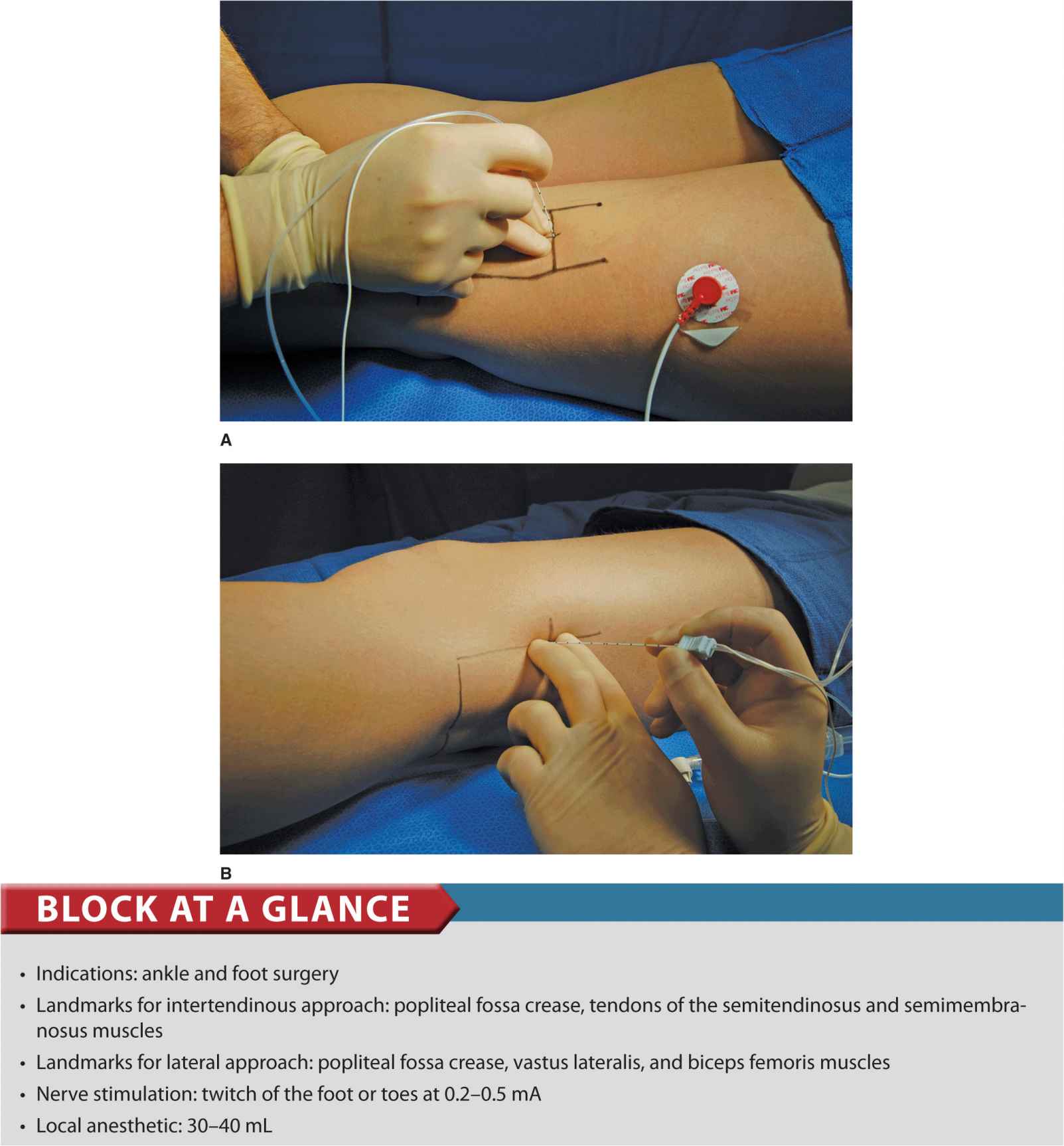
FIGURE 20.1-1. (A) Needle insertion for the popliteal intertendinous approach. (B) Needle insertion for lateral approach to popliteal block.
PART 1: INTERTENDINOUS APPROACH
General Considerations
The popliteal block is a block of the sciatic nerve at the level of the popliteal fossa. This block is one of the most useful blocks in our practice. Common indications include corrective foot surgery, foot debridement, and Achilles tendon repair. A sound knowledge of the principles of nerve stimulation and the anatomic characteristics of the connective tissue sheaths of the sciatic nerve in the popliteal fossa are essential for its successful implementation.
Functional Anatomy
The sciatic nerve is a nerve bundle consisting of two separate nerve trunks, the tibial and the common peroneal nerves (Figure 20.1-2). A common epineural sheath envelops these two nerves at their outset in pelvis. As the sciatic nerve descends toward the knee, the two components eventually diverge in the popliteal fossa to continue their paths separately as the tibial and the common peroneal nerves. This division of the sciatic nerve usually occurs between 4 and 10 cm proximal to the popliteal fossa crease. From its divergence from the sciatic nerve, the common peroneal nerve continues its path downward and laterally, descending along the head and neck of the fibula, Figure 20.1-2. Its major branches in this region are branches to the knee joint and cutaneous branches to the sural nerve. Its terminal branches are the superficial and deep peroneal nerves. The tibial nerve is the larger of the two divisions of the sciatic nerve. It continues its path vertically through the popliteal fossa, and its terminal branches are the medial and lateral plantar nerves, Figure 20.1-2. Its collateral branches give rise to the medial cutaneous sural nerve, muscular branches to the muscles of the calf, and articular branches to the ankle joint. It is important to note that the sciatic nerve in the popliteal fossa is lateral and superficial to the popliteal artery and vein, and it is contained in its own tissue (epineural) sheath rather than in a common neurovascular tissue sheath. This anatomic characteristic explains the relatively low risk of systemic toxicity and vascular punctures with a popliteal block (Figure 20.1-3). However, the proximity of the large vessels, popliteal artery, and vein still makes it imperative to carefully rule out an intravascular needle placement by careful aspiration and meticulously slow injection (e.g., ≤20 mL/min).
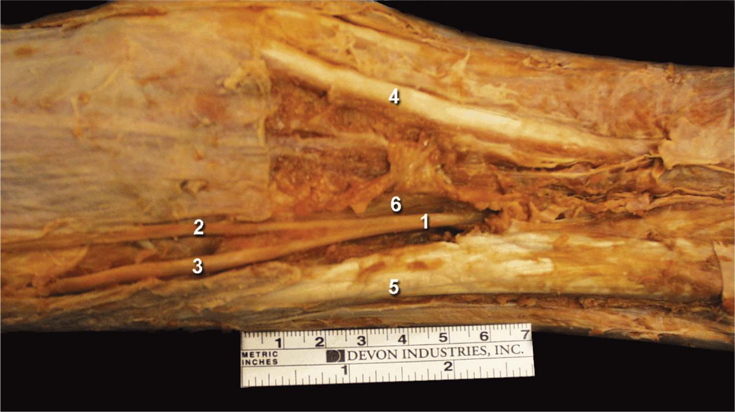
FIGURE 20.1-2. Anatomy of the sciatic nerve in the popliteal fossa. The sciatic nerve ![]() is shown with its two divisions, tibial
is shown with its two divisions, tibial ![]() and common peroneal
and common peroneal ![]() nerves. The common sciatic nerve (1) is is seen between semitendinosus (
nerves. The common sciatic nerve (1) is is seen between semitendinosus (![]() , medially) and biceps (
, medially) and biceps (![]() , laterally) muscles enveloped by the thick epineural sheath
, laterally) muscles enveloped by the thick epineural sheath ![]() .
.
FIGURE 20.1-3. The spread of the contrast solution after injection into the common epineural sheath of the sciatic nerve (SN). The sciatic nerve is positioned between the biceps femoris (BF) and semimembranosus (SM) muscles. An extensive spread within the epineural sheath is seen.
Distribution of Blockade
A popliteal block results in anesthesia of the entire distal two thirds of the lower leg, with the exception of the skin on the medial aspect. Cutaneous innervation of the medial leg below the knee is provided by the saphenous nerve, a cutaneous terminal extension of the femoral nerve. When the surgery is on the medial aspect of the leg, the addition of a saphenous nerve block or local anesthetic infiltration at the incision site may be required for complete anesthesia. Popliteal block alone is usually sufficient for tourniquet on the calf because the tourniquet discomfort is the result of pressure and ischemia of the deep muscle beds and not of the skin and subcutaneous tissues.
Technique
Equipment
A standard regional anesthesia tray is prepared with the following equipment:
• Sterile towels and gauze packs
• Two 20-mL syringes containing local anesthetic
• A 3- to 5-mL syringe plus 25-gauge needle with local anesthetic for skin infiltration
• A 50 cm, 22-gauge short-bevel insulated stimulating needle
• Peripheral nerve stimulator
• Sterile gloves; marking pen
Landmarks and Patient Positioning
The patient is in the prone position. The foot on the side to be blocked should be positioned so that even the slightest movements of the foot or toes can be easily observed. This is best achieved by allowing the foot to extend beyond the operating room bed.
Landmarks for the intertendinous approach to a popliteal block are easily recognizable even in obese patients (Figure 20.1-4):
FIGURE 20.1-4. Landmarks for the popliteal block. ![]() popliteal fossa crease.
popliteal fossa crease. ![]() biceps femoris tendon.
biceps femoris tendon. ![]() semitendinosus semimembranous muscles.
semitendinosus semimembranous muscles.
1. Popliteal fossa crease
2. Tendon of the biceps femoris muscle (laterally)
3. Tendons of the semitendinosus and semimembranosus muscles (medially)
Maneuvers to Facilitate Landmark Identification
The anatomic structures are best accentuated by asking the patient to elevate the foot while palpating muscles against resistance. This allows for easier and more reliable identification of the hamstring tendons. All three landmarks should be outlined with a marking pen.
The needle insertion point is marked at 7 cm above the popliteal fossa crease at the midpoint between the two tendons (Figure 20.1-5).
FIGURE 20.1-5. The point of needle insertion (circle) is marked at 7 cm above the popliteal crease between tendons of semitendinosus and semimembranosus muscles.
Technique
After a thorough cleaning of the injection site with an antiseptic solution, local anesthetic is infiltrated subcutaneously. The anesthesiologist stands at the side of the patient with the palpating hand on the biceps femoris muscle. The needle is introduced at the midpoint between the tendons (Figure 20.1-6). This position allows the anesthesiologist to both observe the responses to nerve stimulation and monitor the patient. The nerve stimulator should be initially set to deliver a current of 1.5 mA (2 Hz, 0.1 ms) because this higher current allows the detection of inadvertent needle placement into the hamstring muscles (local twitches). When the needle is inserted in the correct plane, its advancement should not result in any local muscle twitches; the first response to nerve stimulation is typically that of the sciatic nerve (a foot twitch).
FIGURE 20.1-6. Needle insertion for the popliteal intertendinous approach. Needle is inserted at the midpoint between the biceps femoris laterally and semitendinosus muscles medially.
FIGURE 20.1-7. The needle is re-oriented laterally when the contractions of the local twitches of the semitendinosus and semimembranous muscles are elicited.
FIGURE 20.1-8. The needle is re-oriented medially when local twitches of the biceps femoris muscle are elicited.
After initial stimulation of the sciatic nerve is obtained, the stimulating current is gradually decreased until twitches are still seen or felt at 0.2 to 0.5 mA. This typically occurs at a depth of 3 to 5 cm. After obtaining negative results from an aspiration test for blood, 30 to 40 mL of local anesthetic is slowly injected.
Troubleshooting
When insertion of the needle does not result in stimulation of the sciatic nerve (foot twitches), implement the following maneuvers:
1. Keep the palpating hand in the same position.
2. Withdraw the needle to skin level, redirect it 15° laterally, and reinsert it.
3. When step 2 fails to result in sciatic nerve stimulation, withdraw the needle to skin level, reinsert it 1 cm laterally, and repeat the procedure first with perpendicular needle insertion.
4. When the step 3 fails, reinsert the needle 15° laterally. These maneuvers should facilitate localization of the sciatic nerve when it proves to be challenging.
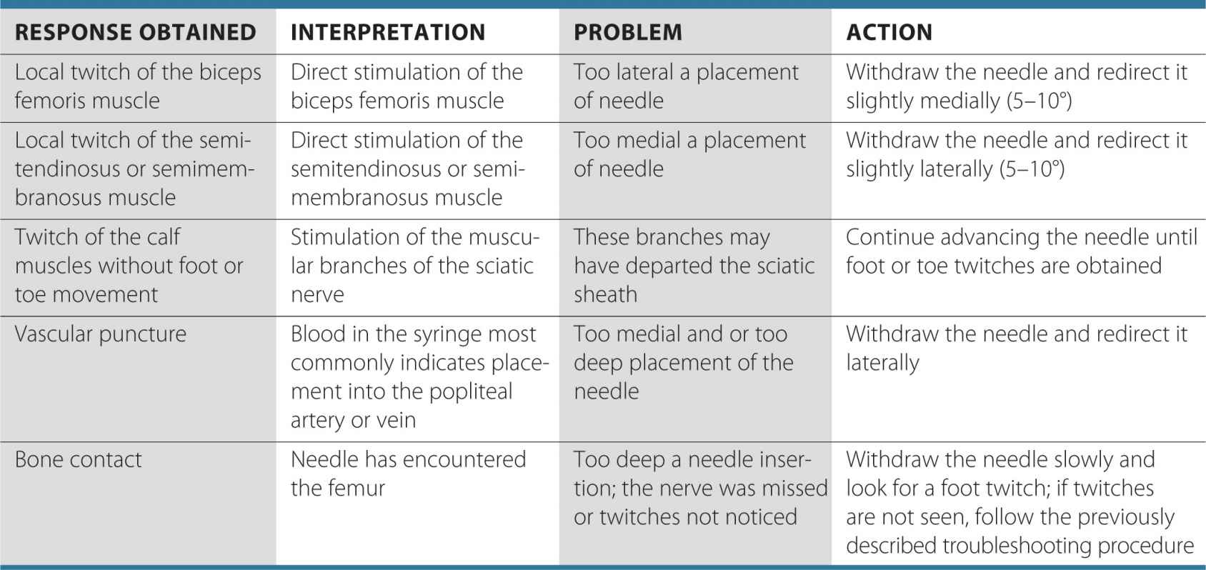
Table 20.1-1 lists some common responses to nerve stimulation and the course of action to take to obtain the proper response.
TABLE 20.1-1 Common Responses to Nerve Stimulation and the Course of Action to Obtain the Proper Response
Block Dynamics and Perioperative Management
This technique is associated with minor patient discomfort because the needle passes only through the adipose tissue of the popliteal fossa. Administration of midazolam 1 to 2 mg after the patient is positioned and alfentanil 250 to 500 μg just before block placement suffice as premedication for most patients. A typical onset time for this block is 15 to 30 minutes, depending on the type, concentration, and volume of local anesthetic used. The first signs of the onset of blockade are usually reports by the patient’s of inability to move their toes or that the foot “feels different.” With this block, sensory anesthesia of the skin is often the last to develop. Inadequate skin anesthesia despite the apparently timely onset of the blockade is common and it may take up to 30 minutes to develop. Thus local infiltration by the surgeon at the site of the incision is often all that is needed to allow the surgery to proceed.
Continuous Popliteal Block
The technique is similar to a single-injection procedure; however, slight angulation of the needle cephalad is necessary to facilitate threading the catheter. Securing and maintaining the catheter is easy and convenient. This technique can be used for surgery and postoperative pain management in patients undergoing a wide variety of lower leg, foot, and ankle surgeries.
Equipment
A standard regional anesthesia tray is prepared with the following equipment:
• Sterile towels and gauze packs
• Two 20-mL syringes containing local anesthetic
• Sterile gloves, marking pen, and surface electrode
• A 3- to 5-mL syringe plus 25-gauge needle with local anesthetic for skin infiltration
• Peripheral nerve stimulator
• Catheter kit (including an 8- to 10-cm large-gauge stimulating needle and catheter).
Kits come in two varieties based on catheter construction: nonstimulating (conventional) and stimulating catheters. During the placement of a conventional nonstimulating catheter, the stimulating needle is first advanced until appropriate twitches are obtained. Then 5–10 mL of local anesthetic or other injectate (e.g., D5W) is then injected to “open up” a space for the catheter to advance freely without resistance. The catheter is then threaded through the needle until approximately 3 to 5 cm is protruding beyond the tip of the needle. The needle is then withdrawn, the catheter secured, and the remaining local anesthetic injected via the catheter. Stimulating catheters are insulated and have a filament or core that transmits current to a bare metal tip. After obtaining twitches with the needle, the catheter is advanced with the nerve stimulator connected until the sought motor response is obtained. This method requires that no conducting solution (i.e., local anesthetic, saline) be injected through the needle prior to catheter advancement, or difficulty obtaining a motor response will result.
Landmarks and Patient Positioning
The patient is positioned in the prone position with the feet extending beyond the table to facilitate monitoring of foot or toe responses to nerve stimulation.
The landmarks for a continuous popliteal block are essentially the same as those for the single-injection technique (Figure 20.1-4). These include the following:
1. Popliteal fossa crease
2. Tendon of the biceps femoris muscle (laterally)
3. Tendons of the semitendinosus and semimembranosus muscles (medially)
The needle insertion site is marked 7 cm proximal to the popliteal fossa crease and between the tendons of the biceps femoris and semitendinosus muscles (Figure 20.1-5).
Technique
The continuous popliteal block technique is similar to the single-injection technique. With the patient in the prone position, infiltrate the skin with local anesthetic using a 25-gauge needle at an injection site 7 cm above the popliteal fossa crease and between the tendons of biceps femoris and semitendinosus muscles. An 8- to 10-cm needle connected to the nerve stimulator (1.5 mA current) is inserted at the midpoint between the tendons of the biceps femoris and semitendinosus muscles. Advance the block needle slowly in a slightly cranial direction while observing the patient for rhythmic plantar or dorsiflexion of the foot or toes. After appropriate twitches are noted, continue manipulating the needle until the desired response is seen or felt using a current ≤0.5 mA. The catheter should be advanced no more than 5 cm beyond the needle tip (Figure 20.1-9). The needle is then withdrawn back to skin level while advancing the catheter simultaneously to prevent inadvertent removal of the catheter.
FIGURE 20.1-9. Needle direction and insertion of the catheter for continuous popliteal sciatic block. The catheter is inserted 3–5 cm beyond the needle tip.
The catheter is checked for inadvertent intravascular placement and secured using an adhesive skin preparation, followed by application of a clear dressing. The infusion port should be clearly marked “continuous nerve block.”
Related posts:


Stay updated, free articles. Join our Telegram channel

Full access? Get Clinical Tree


