Ultrasound-Guided Ankle Block
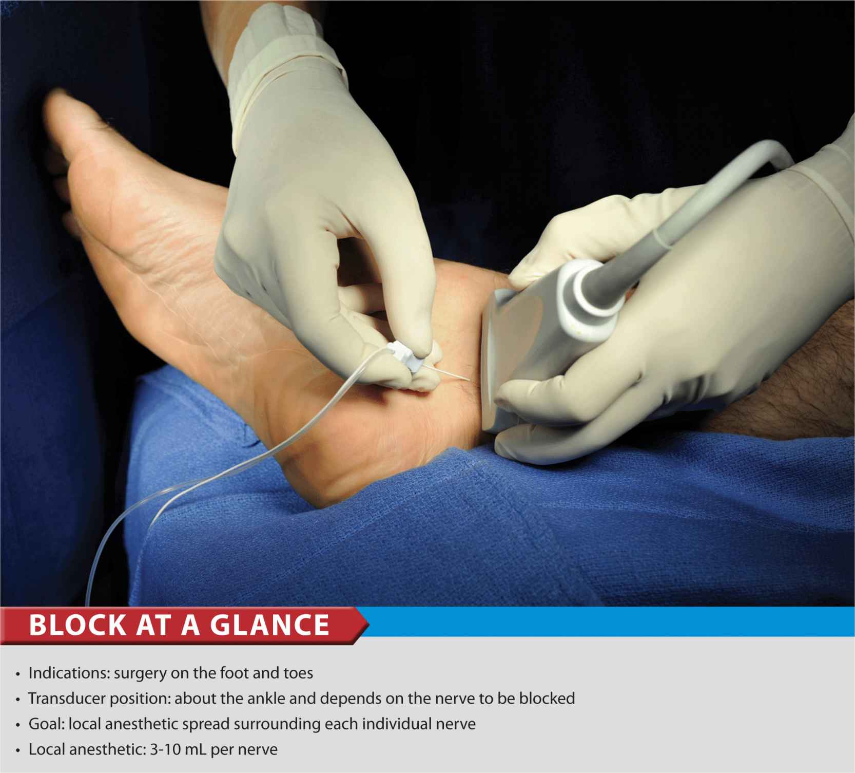
FIGURE 41-1. Block of the posterior tibial nerve using an out-of-plane technique.
General Considerations
Using an ultrasound-guided technique affords a practitioner the ability to reduce the volume of local anesthetic required for ankle blockade. Because the nerves involved are located relatively close to the surface, ankle blocks are easy to perform technically; however, knowledge of the anatomy of the ankle is essential to ensure success.
Ultrasound Anatomy
Ankle block involves anesthetizing five separate nerves: 2 deep nerves and 3 superficial nerves. The 2 deep nerves are tibial (TN) and deep peroneal nerve (DPN). The superficial nerves are superficial peroneal, sural and saphenous. All nerves except saphenous nerve are terminal branches of the sciatic nerve; saphenous nerve is a cutaneous extension of the femoral nerve.
Tibial Nerve
The tibial nerve is the largest of the five nerves at the ankle level and provides innervation to the heel and sole of the foot. With a linear transducer placed transversely at (or just proximal to) the level of the medial malleolus, the nerve can be seen immediately posterior to the posterior tibial artery (Figures 41-1, 41-2, and 41-3A and B). Color Doppler can be very useful in depicting the posterior tibial artery when it is not readily apparent. The nerve typically appears hyperechoic with dark stippling. A useful mnemonic for the relevant structures in the vicinity is Tom, Dick ANd Harry, which refers to, from anterior to posterior, the tibialis anterior tendon, flexor digitorum longus tendon, artery/nerve/vein, and flexor hallucis longus tendon. These tendons can resemble the nerve in appearance, which can be confusing. The nerve’s intimate relationship with the artery should be kept in mind to avoid misidentification.
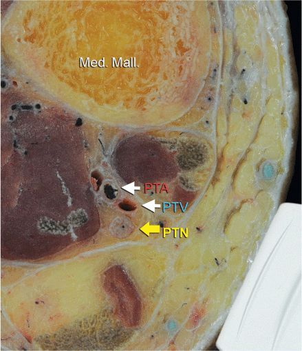
FIGURE 41-2. Cross-sectional anatomy of the posterior tibial nerve at the level of the ankle. Shown are posterior tibial artery (PTA) and vein (PTV) behind the medial malleolus (Med. Mall.) The posterior tibial nerve (PTN) is just posterior and superficial to the posterior tibial vessels.
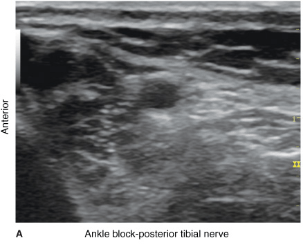
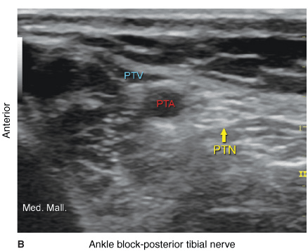
FIGURE 41-3. (A) Ultrasound image of the posterior tibial nerve. (B) Posterior tibial nerve (PTN) is seen posterior to the posterior tibial artery (PTA). Med. Mall., medial malleolus; PTV, posterior tibial vein.
Deep Peroneal Nerve
This branch of the common peroneal nerve innervates the web space between the first and second toes. As it approaches the ankle, the nerve crosses the anterior tibial artery from a medial to lateral position. A transducer placed in the transverse orientation at the level of the extensor retinaculum will show the nerve lying immediately lateral to the artery, on the surface of the tibia (Figures 41-4, 41-5, and 41-6A, B). The nerve usually appears hyperechoic, but it is small and often difficult to distinguish from the surrounding tissue.
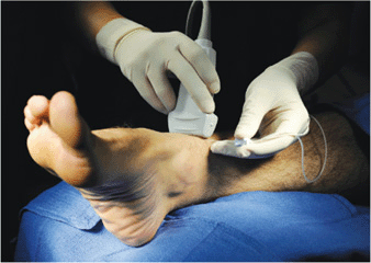
FIGURE 41-4. Deep peroneal nerve block: the transducer position and needle insertion to block the deep peroneal nerve at the level of the ankle.
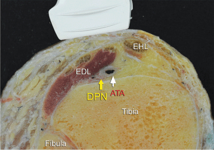
FIGURE 41-5. Cross-sectional anatomy of the deep peroneal nerve at the level of the ankle. The deep peroneal nerve is located just lateral to anterior tibial artery (ATA) and between the extensor digitorum longus (EDL) and tibia. Note the proximity of the extensor hallucis longus (EHL) that can serve as an important landmark. DPN, deep peroneal nerve.
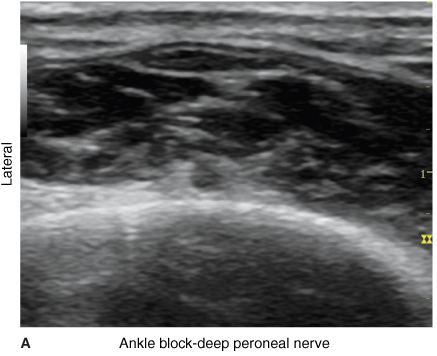
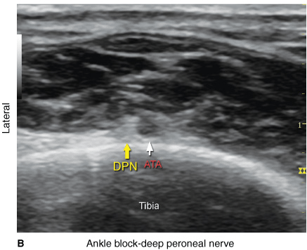
FIGURE 41-6. (A) Ultrasound image of the deep peroneal nerve (DPN) is seen at the surface of the tibia just lateral to the anterior tibial artery (ATA). (B) Ultrasound anatomy of the DPN at the level of the ankle with the structures labeled.
Related posts:


Stay updated, free articles. Join our Telegram channel

Full access? Get Clinical Tree


