Ultrasound of the Lumbar Paravertebral Space and Considerations for Lumbar Plexus Block
Manoj Karmakar and Catherine Vandepitte
Introduction
Lumbar plexus block (LPB) traditionally is performed using surface anatomic landmarks to identify the site for needle insertion and eliciting quadriceps muscle contraction in response to nerve electro-localization, as described in the nerve stimulator–guided chapter. The main challenges in accomplishing LPB relate to the depth at which the lumbar plexus is located and the size of the plexus, which requires a large volume of local anesthetic for success. Due to the deep anatomic location of the lumbar plexus, small errors in landmark estimation or angle miscalculations during needle advancement can result in needle placement away from the plexus or at unwanted locations. Therefore, monitoring of the needle path and final needle tip placement should increase the precision of the needle placement and the delivery of the local anesthetic. Although computed tomography and fluoroscopy can be used to increase the precision during LPB, these technologies are impractical in the busy operating room environment, costly, and associated with radiation exposure. It is only logical, then, that ultrasound-guided LPB is of interest because of the ever-increasing availability of portable machines and the improvement in the quality of the images obtained.1,2
Anatomy and Sonoanatomy
Lumbar plexus block, also known as psoas compartment block, comprises an injection of local anesthetic in the fascial plane within the posterior aspect of the psoas major muscle. Because the roots of the lumbar plexus are located in this plane, an injection of a sufficient volume of local anesthetic in the postero-medial compartment of the psoas muscle results in block of the majority of the plexus (femoral nerve, lateral femoral cutaneous nerve, and the obturator nerve). The anterior boundary of the fascial plane that contains the lumbar plexus is formed by the fascia between the anterior two thirds of the compartment of the psoas muscle that originates from the anterolateral aspect of the vertebral body and the posterior one third of the muscle that originates from the anterior aspect of the transverse processes. This arrangement explains why the transverse processes are closely related to the plexus and therefore are used as the main landmark during LPB.
A scan for the LPB can be performed in the transverse or longitudinal axes. The ultrasound transducer is positioned 3 to 4 cm lateral to the lumbar spine for either orientation. The following settings usually are used to start the scanning:
• Abdominal preset
• Depth: 11–12 cm
• Frequency: 4–8 MHz
• Tissue harmonic imaging and compound imaging functions engaged where available
• Adjustment of the overall gain and time-gain compensation
Longitudinal Scan Anatomy
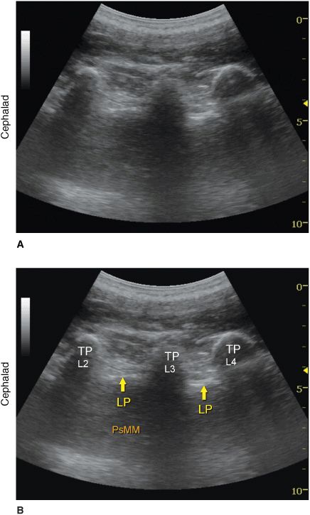
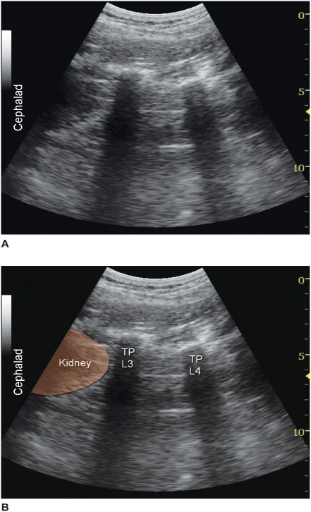
Regardless of the technique, the operator first should identify the transverse processes on a longitudinal sonogram (Figure 46-1). One technique is to first identify the flat surface of the sacrum and then scan proximally until the intervertebral space between L5 and S1 is recognized as an interruption of the sacral line continuity. Once the operator identifies the transverse process of L5, the transverse process of the other lumbar vertebrae are easily identified by a dynamic cephalad scan in ascending order. The acoustic shadow of the transverse process has a characteristic appearance, often referred to as a “trident sign” (Figure 46-2A). Once the transverse processes are recognized, the psoas muscle is imaged through the acoustic window of the transverse processes. The psoas muscle appears as a combination of longitudinal hyperechoic striations within a typical hypoechoic muscle appearance just deep to the transverse processes (Figure 46-2B). Although some of the hyperechoic striations may appear particularly intense and mislead the operator to interpret them as roots of lumbar plexus, the identification of the roots in a longitudinal scan is not reliable without nerve stimulation. This unreliability is partly due to the fact that intramuscular connective tissue (e.g., septa, tendons) within the psoas muscle are thick and may be indistinguishable from the nerve roots at such a deep location. As the transducer is moved progressively cephalad, the lower pole of the kidney often comes into view as low as L2-L4 in some patients (Figure 46-3A and B).

FIGURE 46-1. Transducer position (curved transducer, longitudinal view) to image the central neuroaxis, transverse processes, and estimate the needle and depth to the lumbar plexus using a longitudinal view.

FIGURE 46-2. (A) Ultrasound anatomy of the lumbar paravertebral space demonstrating transverse processes at a depth of approximately 3 cm. Lower frequency, curved transducer is used optimize imaging at the deep location and obtain a greater angular view, respectively. (B) Labeled ultrasound anatomy of the lumbar paravertebral space with structures labeled. TP, transverse process; LP, lumbar plexus roots (most likely); PsMM, psoas major muscle.

FIGURE 46-3
Stay updated, free articles. Join our Telegram channel

Full access? Get Clinical Tree


