Ultrasound-Guided Forearm Blocks
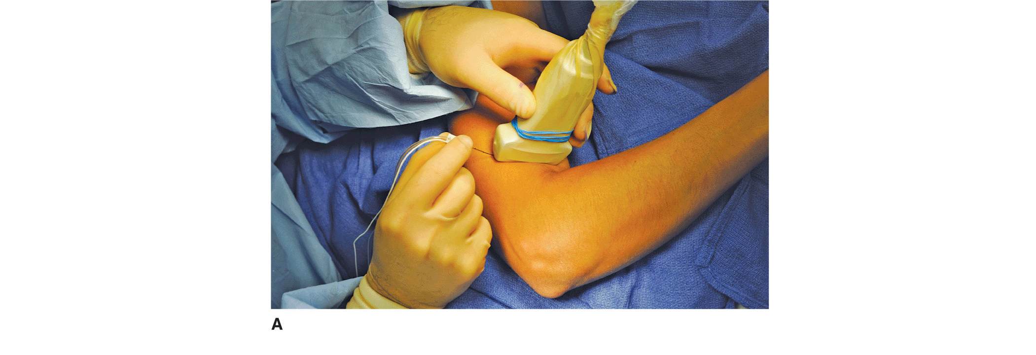
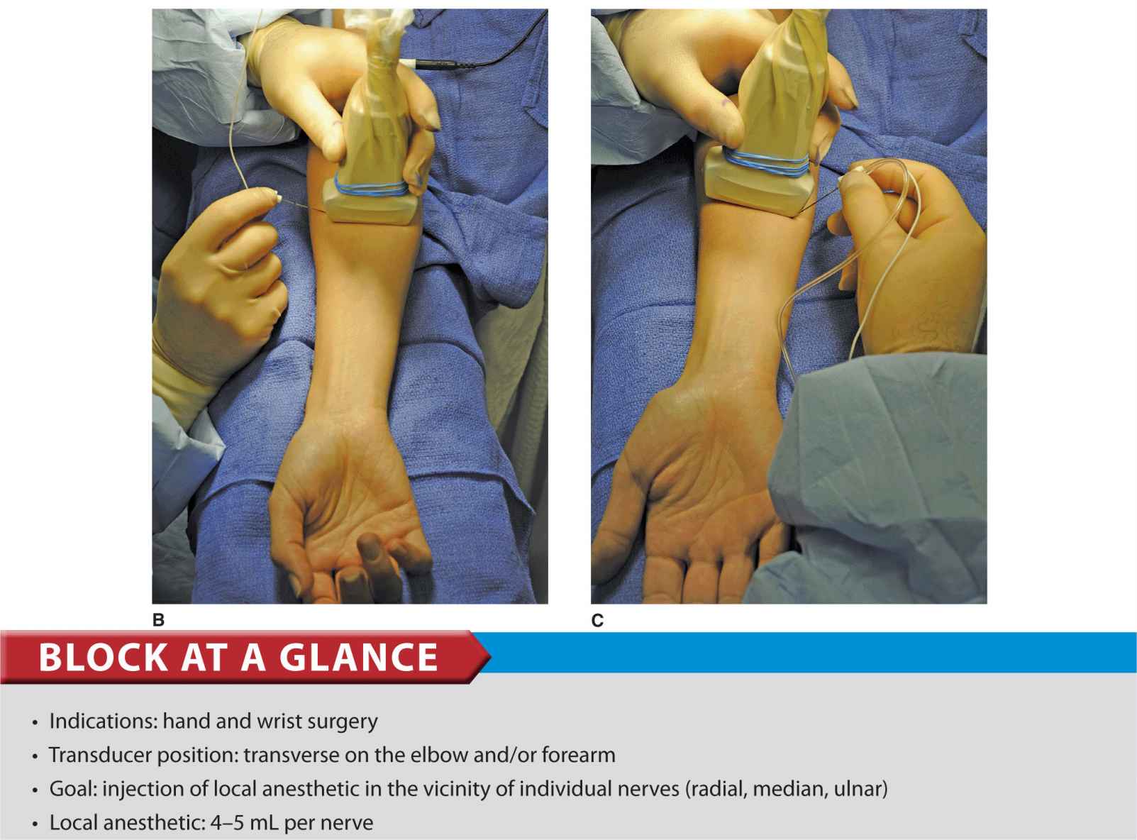
FIGURE 33-1. (A) Radial nerve block above the elbow. The needle is inserted in-plane from lateral to medial direction. (B) Median nerve block at the level of the midforearm. (C) Ulnar nerve block at the level of the midforearm.
General Considerations
Ultrasound imaging of individual nerves in the distal upper limb allows for reliable nerve blockade. The two main indications for these blocks are a stand-alone technique for hand and/or wrist surgery and as a means of rescuing or supplementing a patchy or failed proximal brachial plexus block. The main advantages of the ultrasound-guided technique over the surface-based or nerve stimulator–based techniques are the avoidance of unnecessary proximal motor and sensory blockade, that is, greater specificity. Additional advantages are avoidance of the risk of vascular puncture and a reduction in the overall volume of local anesthetic used. There are a variety of locations where a practitioner could approach each of these nerves, most of which are similar in efficacy. In this chapter, we present the approach for each nerve that we favor in our practice.
Ultrasound Anatomy
Radial Nerve
The radial nerve is best visualized above the lateral aspect of the elbow, lying in the fascia between the brachioradialis and the brachialis muscles (Figure 33-2). The transducer is placed transversely on the anterolateral aspect of the distal arm, 3–4 cm above the elbow crease (Figure 33-1A). The nerve appears as a hyperechoic, triangular, or oval structure with the characteristic stippled appearance of a distal peripheral nerve. The nerve divides just above the elbow crease into superficial (sensory) and deep (motor) branches. These smaller divisions of the radial nerve are more challenging to identify in the forearm; therefore, a single injection above the elbow is favored because it ensures blockade of both. The transducer can be slid up and down the axis of the arm to better appreciate the nerve within the musculature surrounding it. As the transducer is moved proximally, the nerve will be seen to travel posteriorly and closer to the humerus, to lie deep to the triceps muscles in the spiral groove (Figure 33-3).
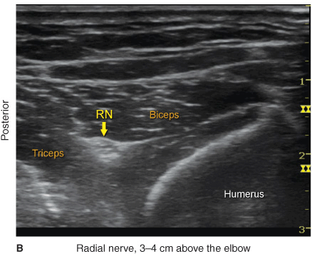
FIGURE 33-2. (A) Radial nerve anatomy at the distal third of the humerus. (B) Sonoanatomy of the radial nerve at the distal humerus. Radial nerve (RN) is shown between the biceps and triceps muscles at a depth of approximately 2 cm.
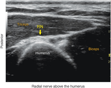
FIGURE 33-3. Sonoanatomy of the radial nerve in the spiral groove of the humerus. RN, radial nerve.
Median Nerve
The median nerve is easily imaged in the midforearm, between the flexor digitorum superficialis and flexor digitorum profundus, where the nerve typically appears as a round or oval hyperechoic structure (Figure 33-4A and B). The transducer is placed on the volar aspect of the arm in the transverse orientation and tilted back and forth until the nerve is identified (Figure 33-1B). The nerve is located in the midline of the forearm, 1–2 cm medial and deep to the pulsating radial artery. The course of the median nerve can be traced with the transducer up and down the forearm, but as it approaches the elbow or the wrist, its differentiation from adjacent tendons and connective tissue becomes more challenging.
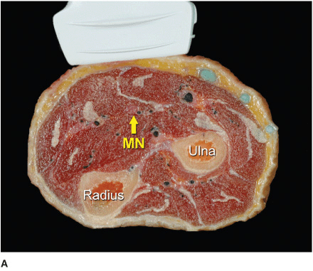
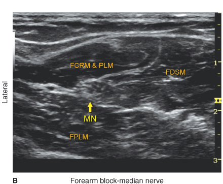
FIGURE 33-4. (A) Anatomy of the medianus nerve (MN) of the midforearm. (B) Sonoanatomy of the MN at the midforearm. FDSM, Flexor Digitorum Superficialis Muscle; FCRM, Flexor Carpi Radialis Muscle & PLM, Palmaris Longus Muscle; FPLM, Flexor Palmaris Longus Muscle.
Ulnar Nerve
The ulnar nerve can be easily imaged in the midforearm, immediately medial to the ulnar artery, which acts as a useful landmark. Similar to the radial and median nerves, the ulnar nerve appears as a hyperechoic stippled structure, with a triangular to oval shape (Figure 33-5A and B). The ulnar artery and nerve separate, when the transducer is slid more proximally on the forearm, with the artery taking a more lateral and deeper course. The ulnar nerve can be traced easily proximally toward the ulnar notch, when desired, and the level of the blockade can be decided based on the desired distribution of the anesthesia as well as the ease of imaging and accessing the nerve. Sliding the transducer distally shows the nerve and artery becoming progressively shallower together as they approach the wrist where the ulnar nerve lies medial to the artery.
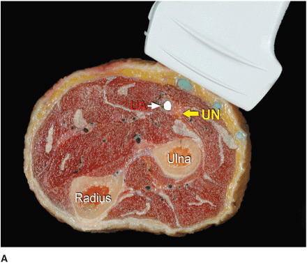
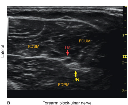
FIGURE 33-5. (A) Anatomy of the ulnar nerve at the midforearm. The ulnar nerve (UN) is closely related to the ulnar artery (UA). (B) Sonoanatomy of the ulnar nerve at the midforearm. UN is shown closely related to the UA, sandwiched between the flexor carpi ulnaris (FCUM) and flexor digitorum profundus muscles (FDPM). FDSM = Flexor Digitorum Superficialis Muscle.
Distribution of Blockade
As is the case with the landmark-based distal blocks, anesthetizing the radial, median, and/or ulnar nerves provides sensory anesthesia and analgesia to the respective territories of the hand and wrist. Note that the lateral cutaneous nerve of the forearm (a branch of the musculocutaneous nerve) supplies the lateral aspect of the forearm, and it may need to be blocked separately by a subcutaneous wheal distal to the elbow if lateral wrist surgery is planned. For a more comprehensive review of the innervation of the hand, see Chapter 1, Essential Regional Anesthesia Anatomy.
Equipment
Equipment needed includes the following:
• Ultrasound machine with linear transducer (8–14 MHz), sterile sleeve, and gel
• Standard nerve block tray
• One 20-mL syringe containing local anesthetic
• A 2-in, 22–25 gauge short-bevel insulated stimulating needle
• Peripheral nerve stimulator (optional)
• Sterile gloves
Related posts:


Stay updated, free articles. Join our Telegram channel

Full access? Get Clinical Tree


