FIGURE 35-1. Transducer position and needle insertion using an in-plane technique to block the femoral nerve at the femoral crease.
General Considerations
The ultrasound-guided technique of femoral nerve blockade differs from nerve stimulator or landmark-based techniques in several important aspects. Ultrasound application allows the practitioner to monitor the spread of local anesthetic and needle placement and make appropriate adjustments, should the initial spread be deemed inadequate. Also, because of the proximity to the relatively large femoral artery, ultrasound may reduce the risk of arterial puncture that often occurs with this block with the use of non-ultrasound techniques. Palpating the femoral pulse as a landmark for the block is not required with ultrasound guidance, a process that can be challenging in obese patients. Although the ability to visualize the needle and the relevant anatomy with ultrasound guidance renders nerve stimulation optional, motor response obtained during nerve stimulation often provides contributory information.
Ultrasound Anatomy
Orientation begins with the identification of the pulsating femoral artery at the level of the inguinal crease. If it is not immediately recognized, sliding the transducer medially and laterally will bring the vessel into view eventually. Immediately lateral to the vessel, and deep to the fascia iliaca is the femoral nerve, which is typically hyperechoic and roughly triangular or oval in shape (Figure 35-2A and B). The nerve is positioned in a sulcus in the iliopsoas muscle underneath the fascia iliaca. Other structures that can be visualized are the femoral vein (medial to the artery) and occasionally the fascia lata (superficial in the subcutaneous layer). The femoral nerve typically is visualized at a depth of 2- to 4-cm.
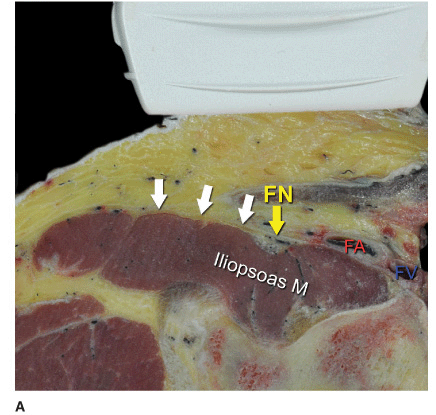
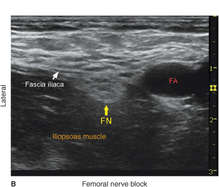
FIGURE 35-2. (A) Cross-sectional anatomy of the femoral nerve (FN) at the level of the femoral crease. FN is seen on the surface of the iliopsoas muscle covered by fascia iliaca. (white arrows). Femoral artery (FA) and femoral vein (FV) are seen enveloped in their own vascular fascial sheath created by one of the layers of fascia lata. (B) Sonoanatomy of the FN at the femoral triangle.
Distribution of Blockade
Femoral nerve block results in anesthesia of the anterior and medial thigh down to the knee (the knee included), as well as a variable strip of skin on the medial leg and foot. It also contributes branches to the articular fibers to both the hip and knee. For a more comprehensive review of the femoral nerve distribution, see Chapter 1, Essential Regional Anesthesia Anatomy.
Equipment
Equipment needed includes the following:
• Ultrasound machine with linear transducer (8–14 MHz), sterile sleeve, and gel
• Standard nerve block tray (described in the equipment section)
• One 20-mL syringe containing local anesthetic
• A 50- to 100-mm, 22-gauge short-bevel insulated stimulating needle
• Peripheral nerve stimulator
• Sterile gloves
Landmarks and Patient Positioning
This block typically is performed with the patient in the supine position, with the bed or table flattened to maximize operator access to the inguinal area. Although palpation of the femoral pulse is a useful landmark, it is not required because the artery should be readily visualized by placing the transducer transversely on the inguinal crease followed by slow movement laterally or medially. If nerve stimulation is used simultaneously, exposure of the thigh and patella are required to monitor the appropriate motor responses (patella twitch).
Exposing the inguinal region in a patient with a large abdominal pannus can be challenging. Using a wide silk tape to retract the abdomen is a useful maneuver prior to skin preparation and scanning. (Figure 35-3).
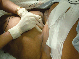
FIGURE 35-3. Obesity is a common problem in patients who present with an indication for femoral nerve block. Taping the adipose tissue away helps optimize the exposure to the femoral crease in patients with morbid obesity.
Technique
With the patient in the supine position, the skin over the femoral crease is disinfected and the transducer is positioned to identify the femoral artery and/or nerve (Figure 35-4). If the nerve is not immediately apparent lateral to the artery, tilting the transducer proximally or distally often helps to image and highlight the nerve from the rest of the iliopsoas muscle and the more superficial adipose tissue. In doing so, an effort should be made to identify the iliopsoas muscle and its fascia as well as the fascia lata because injection underneath a wrong fascial sheath may not result in spread of the local anesthetic in the desired plane. Once the femoral nerve is identified, a skin wheal of local anesthetic is made on the lateral aspect of the thigh 1 cm away from the lateral edge of the transducer. The needle is inserted in-plane in a lateral-to-medial orientation and advanced toward the femoral nerve (Figure 35-5). If nerve stimulation is used (0.5 mA, 0.1 msec), the passage of the needle through the fascia iliaca and contact of the needle tip with the femoral nerve usually is associated with a motor response of the quadriceps muscle group. In addition, a needle passage through the fascia iliaca is often felt as a “pop” sensation. Once the needle tip is witnessed adjacent (either above, below, or lateral) to the nerve (Figure 35-6), and after careful aspiration, 1 to 2 mL of local anesthetic is injected to confirm the proper needle placement (Figure 35-7). When injection of the local anesthetic does not appear to result in a spread close to the femoral nerve, additional needle repositions and injections may be necessary.
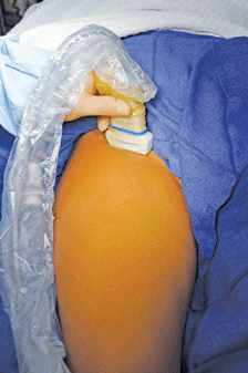
FIGURE 35-4. To image the femoral nerve and/or femoral vessels, the transducer is positioned transversely on the femoral crease as shown on the image.
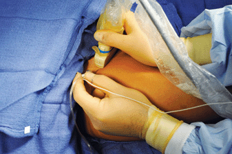
FIGURE 35-5. Transducer position and needle insertion using an in-plane technique to block the femoral nerve at the femoral crease.

FIGURE 35-6. An ultrasound image of the needle path (1,2) to block the femoral nerve. Both needle positions are underneath fascia iliaca, one superficial to the femoral nerve (1) and one deeper to it (2). Either path is acceptable as long as the local anesthetic spreads within the fascia iliaca (white line) to get in contact with the femoral nerve.
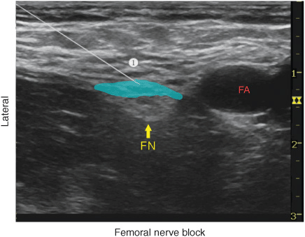
FIGURE 35-7. A simulated needle path (1) and spread of the local anesthetic (blue shaded area) to block the femoral nerve (FN). FA, femoral artery.
Related posts:


Stay updated, free articles. Join our Telegram channel

Full access? Get Clinical Tree


