Chapter 39 The Difficult Airway in Conventional Head and Neck Surgery
I Introduction
Acute airway situations in head and neck surgery should be approached in a systematic manner. The simplest adequate form of control should be selected, and the lowest level (i.e., supraglottis, glottis, subglottis, or trachea) of airway obstruction should be ascertained; control should be established by securing an airway below that level. Acute airway problems often evolve in association with other medical problems. Obvious and potential difficult mask ventilation or difficult intubation should be discussed with the head and neck surgeon, and thoughtful discussion about sequential steps of airway management should take place before anesthetic induction and especially before attempting intubation. Ideally, if time allows, an action plan addressing airway management, including the initial strategy and two backup measures, should be communicated to all team members in the room. Maintaining lines of communication during the intubation procedure can reduce morbidity and mortality associated with difficult airway management.1
The severity or completeness of airway obstruction is categorized as follows:
1. Complete obstruction: no detectable airflow in or out of lungs
2. Partial obstruction: patient with stridor or dyspnea from narrowing of the major airway
3. Potential or impending obstruction: concern that a patient will develop airway compromise because of a known anatomic or physical condition if the respiratory physiology or the consciousness level is altered
II AerodigestIVe Oncologic Surgery
A Preoperative Airway Assessment
1 History
Tobacco and alcohol use are associated with most cases of head and neck cancer and predispose these patients to chronic obstructive pulmonary disease, pneumonia, hypertension, coronary artery disease, and alcohol withdrawal. Information about previous surgical and anesthetic procedures with an emphasis on a history of anesthetic difficulties or difficult intubations, or both, must be obtained and communicated. Previous difficult airway management is considered to be one of the most important predictors of subsequent airway management difficulties.2,3 Patients with a history of obstructive sleep apnea (OSA), especially those without an obvious anatomic abnormality, need careful assessment because their redundant pharyngeal mucosa and soft palate anatomy may hinder BMV and the ability to intubate. Common presenting symptoms of airway obstruction include dyspnea at rest or on exertion, voice changes, dysphagia, stridor, and cough. Physical findings may include hoarseness; agitation; and intercostal, suprasternal, and supraclavicular retraction. Voice changes provide an early suggestion of the anatomic level and severity and progression of the lesion. A muffled voice may indicate supraglottic disease, whereas glottic lesions often result in a coarse, scratchy voice. If there is suspicion of an anterior mediastinal, pharyngeal, or neck mass resulting in partial airway obstruction, initiating anesthesia in the patient in the supine position without first securing the airway may lead to complete airway obstruction and therefore deserves special preoperative evaluation.
2 Physical Examination
A systematic and comprehensive evaluation of the patient’s upper airway is mandatory. The condition of dentition, facial hair (beard), size and mobility of the tongue, thyromental distance, Mallampati score, and limitations in neck flexion and extension must be evaluated. A thyrocervical distance of less than 6 cm in a fully extended adult neck is a good indicator of difficult laryngoscopy and an inability to visualize the vocal cords. The presence and character of stridor should be appreciated, because it may suggest the location of airway narrowing (Table 39-1). Postirradiation changes, neck masses, and previous neck surgery may result in reduced neck mobility, causing difficult mask ventilation and difficult intubation. Findings of morbid obesity, evidence of any oropharyngeal or lip edema, and signs of upper aerodigestive tract bleeding may direct the preferred method of airway management. Lower cranial nerve dysfunction from tumor or previous surgery may also result in airway difficulty related to aspiration or obstruction.
TABLE 39-1 Evaluation of Stridor
| Factor | Features |
|---|---|
| Definition | |
| Typical characteristics | |
| Airway obstruction | |
| Inspiratory versus expiratory | |
| Awake versus asleep |
B Securing the Airway
There are many approaches to securing the airway of patients undergoing head and neck surgery in a safe manner. Determining the optimal approach may depend on the surgery being performed, location of a lesion or infectious mass, or tolerance of the patient. For example, in a patient undergoing maxillomandibular fixation to repair a fractured mandible, nasotracheal intubation is ideal to keep the endotracheal tube (ETT) out of the oral cavity. In a patient with severe supraglottic angioedema, flexible fiberoptic intubation through the nose with the patient upright is preferred to enable identification of the airway with fiberoptic visualization and to avoid pharyngeal collapse in the supine position. Establishing a sequential airway plan with backup options and open communication between the anesthesiology and surgical teams facilitate preparedness and patient safety. An analysis of anesthesia-related cardiopulmonary arrests revealed that up to one third of severe complications result from an inability to establish an optimal airway after the induction of general anesthesia.4
1 Examination of the Airway in the Awake Patient
For a potentially difficult airway, the anesthesiologist may elect to evaluate the airway in an awake patient with the help of judicious intravenous sedation and topical anesthesia. This assessment helps to determine the optimal approach to securing the airway without compromising the patient’s spontaneous breathing. The successful execution of this technique requires constant meaningful contact with the patient and adequate use of topical agents, such as 4% lidocaine spray, with the ultimate goal of not compromising the ability of the patient to breathe spontaneously and to protect the airway. Percutaneous blocks of the superior laryngeal nerves or translaryngeal instillation of lidocaine is best avoided in patients with a head or neck tumor. After the patient is adequately prepared, careful direct laryngoscopy is performed to assess whether to proceed with an awake intubation or to induce general anesthesia for subsequent intubation. A reasonable airway in an awake patient may change to a compromised airway immediately after induction of general anesthesia, with loss of tone of the pharyngeal wall and anterior displacement of the larynx.5 If direct laryngoscopy with sedation is too risky to perform, consideration should be given to awake, flexible fiberoptic laryngoscopy to assess the airway and the possibility of awake, nasotracheal, flexible fiberoptic intubation.
2 Choice of Endotracheal Intubation Technique
a Endotracheal Intubation after Induction of General Anesthesia
In patients with no obvious or expected airway compromise, the ETT can be placed during direct laryngoscopy after induction with a short-acting paralyzing agent, such as succinylcholine. Because it is difficult or impossible to empty the stomach with a nasogastric tube in the case of a large pharyngeal or esophageal tumor, the patient is assumed to have a full stomach.6 Risk of aspiration is reduced by preinduction administration of ranitidine, metoclopramide, and oral sodium citrate–citric acid buffer (Bicitra, Willen Drug Co., Baltimore, MD). The Hollinger anterior commissure laryngoscope is a valuable tool in difficult airway management, and it should be considered when other techniques have failed. This scope may accommodate a 5.0-mm or smaller cuffed ETT, but the insufflation port may become lodged in the barrel of the scope. To avoid this issue, a trial passage of the ETT through the laryngoscope should be attempted before the performance of direct laryngoscopy.7
b Nasotracheal Intubation in the Awake Patient
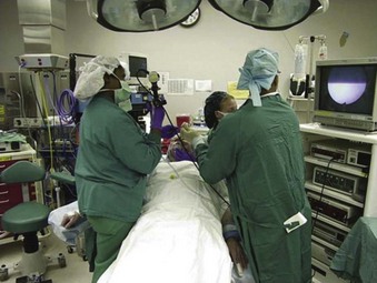
The nasotracheal route is useful in cases of small mouth opening, severe trismus, large tongue, receding lower jaw, large oral cavity tumor, planned maxillomandibular fixation, or tracheal dilatation. Operator and equipment positioning relative to the patient is depicted in Figure 39-1. For nasal intubation, the nose is prepared with a nasal decongestant spray, such as pseudoephedrine (Afrin) and topical 4% cocaine. Cocaine is more advantageous than lidocaine because it is a vasoconstrictor in addition to being a very effective surface anesthetic. The potential for abuse by the personnel is occasionally a deterrent for its routine use. In placing the ETT through the nose, it is necessary to remember that the nasal floor usually runs in a horizontal plane perpendicular to an imaginary vertical line connecting the glabella and the pogonion. The ETT is advanced to approximately the 15-cm mark, and the connector is removed. The flexible fiberoptic bronchoscope (FFB) is then used to visualize the glottic opening and to introduce the tube into the trachea.
d Rigid Bronchoscopy
The rigid bronchoscope, a hollow stainless steel tube through which a rigid telescope is placed, provides excellent access to the airways. The distal end of the rigid bronchoscope is usually beveled to facilitate intubation and lifting of the epiglottis. The updated American Society of Anesthesiologists (ASA) protocol states that in the “cannot intubate, cannot ventilate” (CICV) scenario, “a rigid bronchoscope for difficult airway management reduces airway-related adverse outcomes.”8 This modality is recommended as a technique for cases of difficult mask ventilation. Other indications for rigid bronchoscopy include massive airway hemoptysis, foreign body retrieval, laser or photodynamic therapy, and placement of airway stents.9
e Retrograde Intubation
Though rarely used, retrograde intubation represents another alternative technique for intubation of the difficult airway in head and neck patients. This technique can also be used in patients with trismus, trauma to the cervical spine, temporomandibular joint ankylosis, upper airway masses, or failed intubation. This technique begins with puncture of the cricothyroid membrane (CTM) or cricotracheal membrane with a small needle angled superiorly to pass a guidewire through the larynx, and it is then guided through the nose or mouth. The ETT is then advanced over the guidewire with a Seldinger technique into position in the trachea.10
Retrograde intubation may also be used to facilitate other approaches. For example, a combination of conventional, fiberoptic, and retrograde techniques can be used to intubate successfully.11 An FFB with a suction port can be used as an anterograde guide over a retrograde wire. A direct laryngoscopic view can facilitate placement of the tip of a fiberoptic scope into or near the glottic aperture. After the FFB is past the vocal folds and tracheal rings have been identified, the preloaded ETT may be passed over the FFB into the trachea.
f Tracheostomy with Local Anesthesia
If a patient is in acute respiratory distress because of upper airway obstruction and the patient is considered unable to be safely intubated under general anesthesia after evaluation (including FFB), the best choice is to perform a planned but urgent awake tracheostomy.12 This should be performed under local anesthesia with minimal intravenous sedation to avoid loss of spontaneous breathing. Tracheostomy under local anesthesia in an awake patient is also an excellent method to secure the airway in the following situations: upper airway abscess that may be in the way of or distorting the pathway for endotracheal intubation; bulky, friable supraglottic or glottic mass; and glottic stenosis with presumed bilateral cricoarytenoid joint fixation. In these situations, attempts at direct laryngoscopy and intubation may result in abscess rupture or aspiration of purulent material, blood, or material from a friable tumor, or in the case of cricoarytenoid joint fixation, the fixed, medialized vocal folds do not allow passage of the ETT without severe damage to the membranous vocal folds.
3 Difficult or Failed Intubation
a Adequate Bag-Mask Ventilation
As an alternative, in a patient who is adequately BMV, the ETT may be passed with oral fiberoptic-guided laryngoscopy.13 An OPA bite block is placed in the patient’s mouth, and the regular mask is replaced with an endoscopy port (i.e., Patil-Syracuse endoscopy mask).14 The lubricated FFB is passed through the mask’s diaphragm into the OPA and into the trachea through the glottis. The ETT is then threaded over the FFB into the patient’s trachea. As described earlier, a scope can also be used to pass an ETT through the nose. The nose is decongested with topical Afrin, and topical 4% cocaine is used to anesthetize the nose. The appropriately sized, well-lubricated ETT is then placed along the floor of the nose, and an FFB is passed through the tube. An assistant occludes the other nostril and the mouth, and a triple connector is used so that the scope and O2 delivery can be simultaneously introduced.
Related posts:
 Prehospital Airway Management
 Complications of Managing the Airway
 Ultrasonography in Airway Management
 Nonintubation Management of the Airway: Airway Maneuvers and Mask Ventilation
Stay updated, free articles. Join our Telegram channel

Full access? Get Clinical Tree


