Sonography of Thoracic Paravertebral Space and Considerations for Ultrasound-Guided Thoracic Paravertebral Block
Catherine Vandepitte, Tatjana Stopar Pintaric, and Philippe E. Gautier
Thoracic paravertebral block (PVB) is a well-established technique for perioperative analgesia in patients having thoracic, chest wall, or breast surgery or for pain management with rib fractures. Ultrasound guidance can be used to help identify the paravertebral space (PVS) and needle placement, and to monitor the spread of the local anesthetic. Importantly, interference of the closely related osseous structures with ultrasound imaging and the proximity of the highly vulnerable neuraxial structures make it imperative that all well-described technique precautions are exercised, regardless of the ultrasound imaging. In this chapter, we describe general principles of thoracic PVB, rather than propose a cookbook with specific techniques and step-by-step directions. The reader is advised to use the anatomic information and techniques presented here to devise an approach in line with own clinical experience.
Anatomy and General Considerations
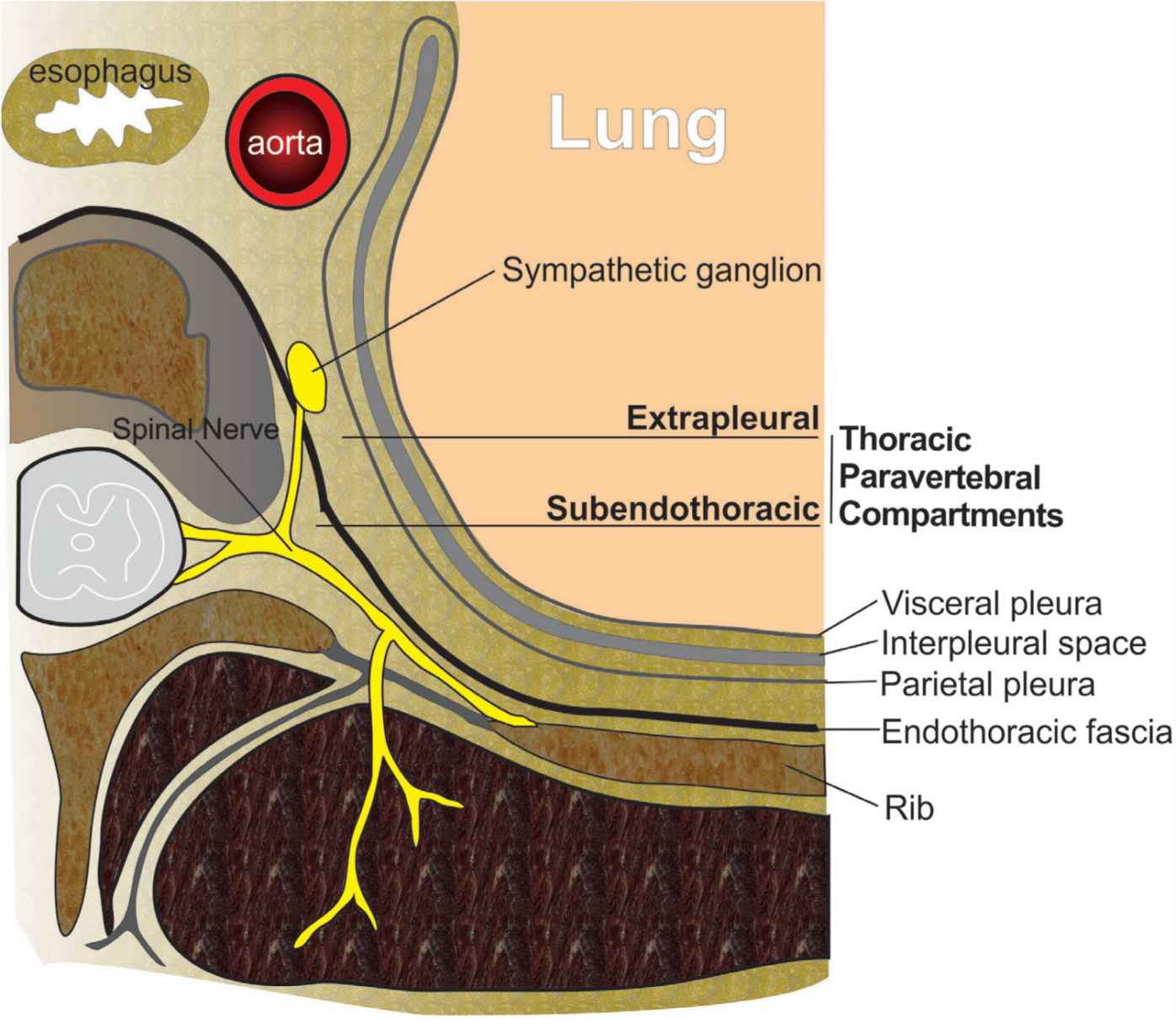
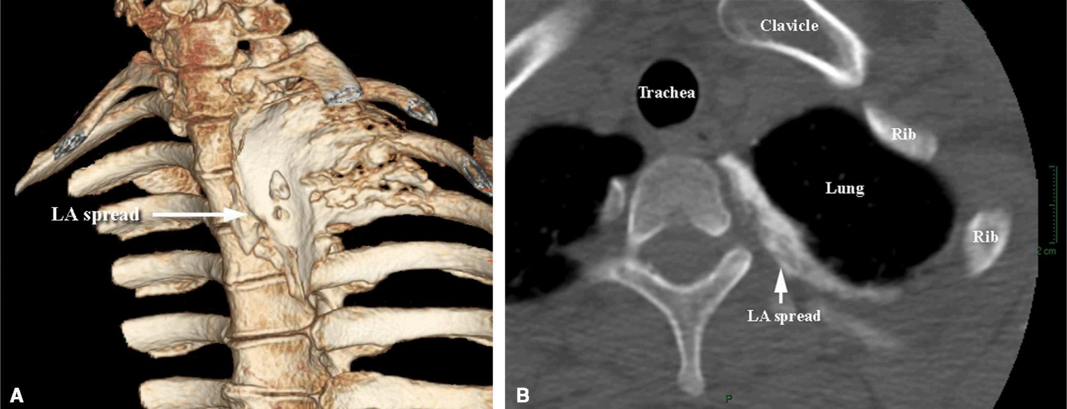
Thoracic PVB is accomplished by an injection of local anaesthetic into the PVS, which contains thoracic spinal nerves with their branches, as well as the sympathetic trunk. Anatomically, the PVS is a wedge-shaped area positioned between the heads and necks of the ribs (Figure 45-1). Its posterior wall is formed by the superior costotransverse ligament, the anterolateral wall is the parietal pleura with the endothoracic fascia, and the medial wall is the lateral surface of the vertebral body and intervertebral disk.1 The PVS medially communicates with the epidural space via the intervertebral foramen inferiorly and superiorly across the head and neck of the ribs.2–5 Consequently, injection of local anesthetic into the PVS space often results in unilateral (or bilateral) epidural anesthesia. The cephalad limit of the PVS is not defined, whereas the caudad limit is at the origin of the psoas muscle at L1.6 Likewise, the PVS space communicates with the intercostal spaces laterally, leading to the spread of the local anesthetic into the intercostals sulcus and resultant intercostal blockade as part of the mechanism of action (Figure 45-2).

FIGURE 45-1. A schematic representation of the thoracic paravertebral space and its structures of relevance to paravertebral block.

FIGURE 45-2. (A) Three-dimensional magnetic resonance reconstruction image of the spread of local anesthetic (5 mL) within the paravertebral space. (B) A computed topography image of the local anesthetic (LA) spread in the thoracic paravertebral space. The contrast is seen spreading in the medial to lateral and anterior to posterior direction underneath the parietal pleura.
Transverse In-line Technique
Similar to techniques not using ultrasound guidance, the patient can be positioned in the sitting or lateral decubitus position with the site of surgical interest uppermost. Either a linear or phased array (curved) transducer can be used however, latter may be used only in slim patients. A high-frequency (10–12 MHz) transducer is used to obtain images in the axial (transverse) plane at the selected level, with the transducer positioned just lateral to the spinous process (Figure 45-3). For most patients, the depth of field is set about 3 cm to start scanning. The transverse processes and ribs are visualized as hyperechoic structures with acoustic shadowing below them (Figure 45-3
Stay updated, free articles. Join our Telegram channel

Full access? Get Clinical Tree


