, Corinna Eleni Psomadakis2 and Bobby Buka3
(1)
Department of Family Medicine, Mount Sinai School of Medicine Attending Mount Sinai Doctors/Beth Israel Medical Group-Williamsburg, Brooklyn, NY, USA
(2)
School of Medicine Imperial College London, London, UK
(3)
Department of Dermatology, Mount Sinai School of Medicine, New York, NY, USA
Keywords
SeborrheaSeborrheic dermatitisSeborrheic eczemaSebopsoriasisDandruffScalpFlakingScalePsoriasisInflammationFungal infectionCradle capCorticosteroidAntifungal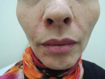
Fig. 6.1
Bilateral scaly erythematous , slightly greasy plaques typically found on the brow and nasolabial folds
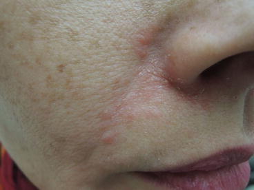
Fig. 6.2
Waxy plaque at the nasolabial fold
Primary Care Visit Report
A 42-year-old female with past medical history of psoriasis presented with a rash on her face. She reported having similar outbreaks in the past, and they would resolve with application of tea tree oil. The present rash started about a week prior to visit, on the left nasal and cheek area. It was itchy, and she treated it by applying apple cider compresses, drinking coconut oil, and taking vitamins. The initial rash on the left side improved, but 3 days prior to visit a similar rash appeared on her right cheek, nasal area, and right chin, and progressively worsened since then. She tried one application of hydrocortisone valerate on the rash about 5 days prior to visit, and it did not help.
Vitals were normal. On exam, the right lateral alar crease had an erythematous scabbing rash, and the right nasolabial fold had a 1 cm × 3 cm erythematous papular rash. Her right lateral chin had a honey-crusted 1 cm × 1 cm erythematous papular rash, and her left alar crease featured a minimal erythematous papular rash .
This was treated as seborrheic dermatitis with overlaying impetigo, and the patient was prescribed hydrocortisone butyrate (Class V steroid) cream 0.1 % twice daily for 10 days for the seborrheic dermatitis and oral cephalexin 500 mg twice daily for 10 days plus mupirocin ointment 2 % three times daily for 10 days for the impetiginization.
Discussion from Dermatology Clinic
Differential Dx
Seborrheic dermatitis
Mild psoriasis
Impetigo
Dermatophyte fungal infection , e.g., tinea capitis, tinea faciei, tinea corporis
Subacute lupus erythematosus
Candidiasis
Favored Dx
Given the distribution and appearance of this rash, the favored diagnosis is seborrheic dermatitis, with one impetiginized area. However, seborrheic dermatitis and mild psoriasis are sometimes clinically indistinguishable when present on the face or scalp , and diagnosis is often times just a matter of how much scale is present, and how thick the plaques are. The term sebopsoriasis is sometimes used in those cases. Both facial seborrheic dermatitis and psoriasis can be successfully controlled with mild topical steroids, but may differ in their long-term management, as described below.
Overview
Seborrheic dermatitis is a common, chronic cutaneous condition involving areas of the body with active sebaceous glands, such as the scalp, face, chest, and back. Its pathogenesis is controversial [1], and it has been associated with presence of yeast of the genus Malassezia . Many factors contribute to its pathogenesis. Malassezia metabolites, such as oleic acid, compromise the permeability of the skin barrier, becoming more susceptible to irritants and triggering an inflammatory response [1]. Seborrheic dermatitis, also sometimes called seborrheic eczema , may be classified as a type of dermatitis, a fungal infection , or an inflammatory process similar to psoriasis .
Related posts:


Stay updated, free articles. Join our Telegram channel

Full access? Get Clinical Tree


