Programmable Intrathecal Pain Pump
There are several ways of delivering pain medication. Typical routes of administration are oral, transdermal, intramuscular, intravenous, and epidural. Programmable intrathecal pain pumps were developed to treat pain on a tertiary level. They deliver medication via a catheter directly to the intrathecal space, the fluid-filled space between the thin layers of tissue that cover the brain and spinal cord. The intrathecal administration of centrally acting agents bypasses the blood–brain barrier, resulting in much higher concentrations of medication in the cerebrospinal fluid (CSF).
Inadequate methods for treatment of cancer pain helped stimulate the development of the programmable intrathecal pump. Some cancer patients with painful forms of the disease require escalating doses of opioids for proper pain control. At a critical point in their cancer-related pain management, the standard administration of escalating pain medications causes systemic side effects with or without pain relief. The equivalent dose of opioids needed to control pain becomes overwhelming, causing sedation, nausea, severe constipation, and emesis. The rationale in the development of the programmable intrathecal pump is that it can deliver a fraction of the systemically needed medication directly to the central nervous system (CNS), achieving the same level of pain control with less drug-induced side effects. The equivalent dose of morphine given intrathecally is only 1/300 the amount given orally in a 24-hour period. Intrathecal administration can allow for much more effective pain control.
The pump delivers a continuous infusion of medication directly to the CNS that can be adjusted according to analgesia and/or side effects (Fig. 30-1). The solution usually infused is morphine but fentanyl or Dilaudid can be used as well, to which an adjuvant medication can be added. The most common adjuvant medication used is the local anesthetic bupivacaine (a longer-acting local anesthetic). Ziconotide and clonidine are used as well. Adding adjuvant drugs to the opioid solution can enhance the treatment regimen, acting synergistically.
When to use
Intrathecal pain pumps are reserved for patients who have failed conservative treatment with medications and primary pain procedures. Intrathecal analgesia is appropriate for approximately 5% to 10% of cancer patients.1
A randomized clinical trial comparing the impact of adding intrathecal analgesia to medical management with that of medical management alone in patients with refractory cancer pain found that intrathecal therapy improved pain scores, reduced the incidence of drug-related toxicity, reduced reliance on systemic analgesia, improved survival rates, and also improved the quality of life of both the patients and their caregivers.2
A randomized clinical trial comparing the impact of adding intrathecal analgesia to medical management with that of medical management alone in patients with refractory cancer pain found that intrathecal therapy improved pain scores, reduced the incidence of drug-related toxicity, reduced reliance on systemic analgesia, improved survival rates, and also improved the quality of life of both the patients and their caregivers.2
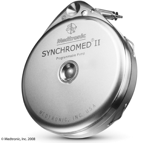 Figure 30-1 Implanted programmable pump for intrathecal administration of medication. |
As with most procedures, proper patient selection helps predict success. For best results, it is essential that patients have chronic pain, defined as lasting longer than 3 to 4 months; be expected to live more than 3 months; have pain that is not relieved by optimal medical management; and have no untreated psychopathology that impedes treatment success. In clinical practice, a number of interventional pain management physicians have stopped using programmable intrathecal pumps for conditions other than cancer. Several complications related to the procedure, device, and its maintenance over time often outweigh the theoretical advantages of programmable intrathecal pain pump use in noncancer-related pain. There is frequently a continued need for oral medications. By the time these complications arise in cancer patients they have succumbed to the disease.
There is one other indication for programmable intrathecal pumps. Investigators have found that these pumps are clinically beneficial in patients with severe spasticity (e.g., cerebral palsy) when oral administration of muscle relaxants has failed. The pumps contain the muscle relaxant baclofen rather than an opioid.
How to Perform the Procedures
The Trial
Before implantation of a permanent system, patients first must undergo a screening trial of intrathecal opioids to see if full implantation is appropriate. The response to the acute administration of intrathecal opioids is thought to predict the treatment’s long-term efficacy. The goal is to see if the patient has pain relief and if relief occurs without side effects that would contraindicate the therapy. There is no proven method of performing these trials, but the following discussion briefly describes two of the most common. One method consists of giving the patient a bolus of medication; after gaining access to the intrathecal space, the practitioner performs a lumbar puncture and injects a bolus dose of drug of interest. Another method, more extensive method which is commonly used, involves placement of an intrathecal catheter under fluoroscopic guidance. The practitioner then connects the catheter to a bag of premixed opioid solution and often an adjuvant medication, typically bupivacaine. The patient is admitted to the hospital, medication infusion occurs over time, with titration until pain relief occurs. The trial is considered positive if the patient has at least 50% pain relief without side effects. This method allows the provider to determine if intrathecal therapy will be successful and what dose of medication is needed to provide pain relief before implanting the full system. There is no standard length for the duration of the screening trial, but the norm is 2 to 4 days.
For the trial the procedure is fully explained to the patient, all questions answered, and informed consent obtained. The patient is given preoperative antibiotics; 2 g of Ancef (cefazolin sodium) or 1 g vancomycin if the patient has a penicillin or cephalosporin allergy. The patient is brought to the operating room and placed on the fluoroscopic table in the prone position. Special care is taken to pad all pressure points. A “time out” is performed prior to the procedure including verbal confirmation of correct patients procedure and procedural site. Noninvasive hemodynamic monitors, pulse oximetry, and a nasal cannulus are placed. The skin of the back is prepped with antiseptic solution; drapes are placed over the area in standard sterile fashion. The fluoroscope is positioned in the anteroposterior (AP) position. The fluoroscope is then tilted if necessary in a cephalad to caudad motion to “square up” the endplates of the vertebral bodies (Fig. 30-2).
Using a metal marker and a felt-tip marking pen, an “X” is placed on the skin overlying the right lamina at L4 for a right paramedian approach. (A left paramedian approach can be used if that is easier for the operator.) The skin and subcutaneous tissue are anesthetized with 2% lidocaine using a 1.5-in, 25-gauge needle. After the skin and subcutaneous tissue are anesthetized, the styleted Tuohy needle that comes in the kit is placed through the “X” using a 45-degree angle, oblique paramedian insertion technique. The Tuohy needle is advanced to the intrathecal space at L2–L3, a level above where the skin was entered (Fig. 30-3). When the needle reaches the ligamentum flavum, the ligamentum flavum will grab the needle. At that point, remove the stylet and slowly advance the needle. When the needle reaches the intrathecal space, CSF will start dripping out, confirming proper needle placement (Fig. 30-4). Needle position is further confirmed on AP and lateral fluoroscopic views. A specially designed radiopaque intrathecal catheter is fed through the Tuohy needle into the intrathecal space. The tip of the catheter is designed so that it can be seen under fluoroscopy. The catheter is most commonly advanced to the level of the T9 vertebral body, under live fluoroscopic guidance. Based on the individual’s anatomy, technically it may be more feasible to place the catheter at a different level. Once proper placement is confirmed, an adapter (included in the kit) is attached to the tip of the catheter. CSF should be returned easily via the catheter when aspirating with a 3-mL syringe. Aspiration should be negative for vascular flow. Proper placement is further confirmed with injection of contrast, allowing visualization of
proper intrathecal spread under fluoroscopy. For the intrathecal trial, the adapter is then removed so that the Tuohy needle can be carefully slipped over the properly situated intrathecal catheter. The catheter is then secured to the skin with a bandage and the adapter is reattached.
proper intrathecal spread under fluoroscopy. For the intrathecal trial, the adapter is then removed so that the Tuohy needle can be carefully slipped over the properly situated intrathecal catheter. The catheter is then secured to the skin with a bandage and the adapter is reattached.
Related posts:


Stay updated, free articles. Join our Telegram channel

Full access? Get Clinical Tree


