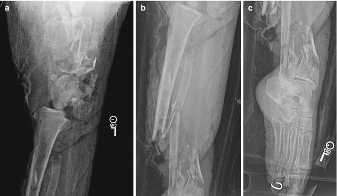
Fig. 22.1
This 28-year-old male was involved in a high-speed motorcycle crash and sustained significant forefoot and midfoot trauma. The heel pad was severely damaged in this case which happens commonly in these injury patterns. This makes subsequent reconstrutive efforts difficult with amputation levels below the midsection of the tibia (i.e., Syme amputations)
Severe upper extremity injuries, which present as complete or near-complete amputations, warrant special consideration and evaluation by a surgeon who is familiar with reconstruction procedures in this area. The decision-making process in the mangled upper extremity can be challenging, especially when limb salvage becomes an option [29]. Primary amputation may not be in the best interest of some patients as it has been suggested that a sensate hand with minimal prehensile function can outperform a prosthesis [30]. Standard principles of wound care should be employed until appropriate consultation can be obtained. When definitive surgical intervention is required, preservation of length is critical and can decrease the energy needed for the patient to suspend their prosthesis (Fig. 22.2). Furthermore, the increased surface area of the limb can help with load distribution, prosthesis propulsion in space, and counterpressure with task performance [26].


Fig. 22.2
This 16-year-old female was involved in a high-speed motor vehicle crash in which the vehicle rolled multiple times. She sustained a traumatic amputation of the forearm including the entire radius and ulna. The proximal soft-tissue involvement was extensive, and she underwent a proximal amputation leaving 14 cm of residual humerus. She was ultimately fit with a myoelectric hand
Absolute indications for primary limb amputation have been suggested in the literature with varying algorithms. Generally, these indications have included a patient presenting with a total or near-total leg amputation or complete tibial or sciatic nerve transection in an adult [14, 31, 32]. Relative indications have included two or more of the following: concurrent severe ipsilateral foot injury, large intercalary soft-tissue or bone loss, warm ischemia time of greater than 6 h, and severe concurrent multiple injuries (Table 22.1) [8, 15, 31, 33–35]. Uniformly, however, these studies indicate that the clinician’s judgment at the time of initial evaluation is critical; amputation decision-making should employ a multitude of factors. We also advise seeking multispecialty input with this difficult decision (i.e., orthopedics, plastic surgery, general surgery). In one study, a combined approach led to 89 % of patients achieving a successful viable limb, and only 11 % went on to secondary amputation [31].
Table 22.1
Primary amputation guidelines
Absolute indications | 1. Presentation with complete or near-complete limb amputation |
2. Complete sciatic or tibial nerve transection in an adult | |
Relative indications | 1. Concurrent ipsilateral severe foot injury |
2. Large intercalary soft-tissue or bone loss | |
3. Warm ischemia time of >6 h | |
4. Severe concurrent multiple injuries |
22.2.1 Outcome of Traumatic Primary Amputations
There is little in the literature reporting the long-term outcome of traumatic amputations. Recently, Dougherty published a study evaluating the outcomes of 123 transtibial amputees from the Vietnam War – 65 % of which were victims of land mines and booby traps. He found that with isolated amputations, these patients led relatively normal lives. However, when concomitant injuries were sustained by these patients, their SF-36 scores lowered, and their incidence of psychological illness increased [36]. Smith et al. [37] published a descriptive study describing outcomes of 20 patients with unilateral transtibial amputations. They found that SF-36 scores were lower than normal age-matched scores in the categories of physical function and role limitations because of physical health problems and pain. Aside from those two sections, scores from the normal population were not significantly different. Lerner et al. [38, 39] evaluated three groups of patients: posttraumatic fracture nonunion, chronic refractory osteomyelitis, and lower-extremity amputation. In their group of 109 patients, they found that the chronic osteomyelitis patients were the most adversely affected among the three groups. Interestingly, 85 % of the amputee patients believed they had been “mentally scarred” by their orthopedic problem, but despite that complaint, they had minimal restriction in lifestyle and activity – a direct contrast to the poorer functioning osteomyelitis group.
In 2004, a study was published which reviewed 161 trauma-related amputation patients that were participants in the LEAP study [27]. This study found no differences in outcomes between the above-the-knee amputees and the below-the-knee amputees. The exception to this finding was with walking speeds in which the below-the-knee group performed better. A key finding in this study was the significantly poorer outcomes of patients that had undergone a through-the-knee amputation. The poorer outcome was associated with worse walking speeds and also less physician-measured satisfaction in terms of clinical, functional, and cosmetic recoveries of their patients. As we noted earlier, we believe the surgeon must critically evaluate the zone of injury prior to proceeding with a through-the-knee amputation. Furthermore, when faced with the decision to proceed with an above-the-knee amputation, surgeons should take whatever steps are necessary to preserve femoral length [40]. It was recently shown that retained length of the femur significantly improves temporospatial and kinematic gait outcomes. Careful attention to the adductors, either with preservation or reconstruction, can benefit this group of patients and improve their mobility.
The outcome of isolated traumatic lower-extremity amputations is mixed but can generally be associated with residual disability and lower outcome scores than the general population. While Dougherty’s [41] study of transtibial amputations demonstrated relatively normal scores with a select population with an isolated lower-extremity injury, other studies indicate substantially poorer outcomes. In another study by Dougherty examining more proximal transfemoral amputations, substantial disability was found in patient follow-up [36]. Smith et al. [37] and the LEAP study [27] also identified significant disability with traumatic amputations in follow-up. These studies indicate that when lower-extremity injuries are among a constellation of traumatic injuries, which they often are, outcomes demonstrate increased disability. An extensive rehabilitation program offered at the treating US Army hospital may have influenced the better outcomes identified in Dougherty’s transtibial amputation study. This finding and those of the LEAP study underscore the need to have high-energy traumatic amputation patients closely followed and managed by a multidisciplinary team including surgeons, rehabilitation physicians, nurses, prosthetists, and therapists. It is also the surgeon’s responsibility to inform patients of expected outcomes and ensure that unrealistic expectations are not confusing patients during their recovery. These discussions can allay patient fears and allow the patient, their families, and support networks to adjust to the trauma and plan ahead for expected changes.
22.3 The Subtotal Amputation Injury: Limb Salvage or Amputation
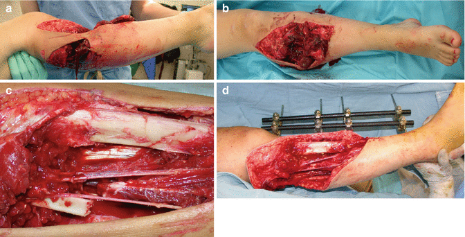
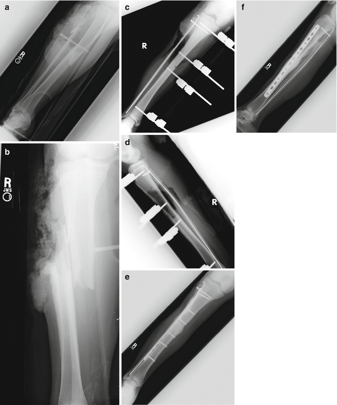
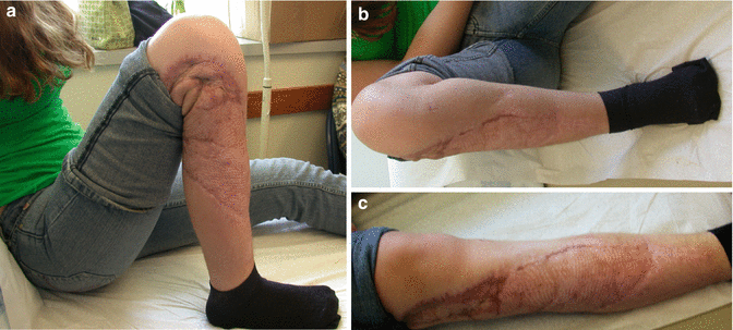
The high-energy trauma patient with a subtotal amputation to an extremity presents immediate challenges to the trauma team. The Lower Extremity Assessment Project (LEAP) was a prospective cohort study of 601 patients who had been admitted to eight Level I trauma centers for the treatment of severe lower-extremity injuries below the distal part of the femur [21]. This study sought to provide evidence for clinicians to use when faced with this dilemma and has recently published 7-year follow-up data [20]. The singular study has produced multiple projects investigating various facets of the lower-extremity injured patient, and many are discussed in the ensuing sections. Inclusion criteria for the LEAP study are listed in Table 22.2 and highlight the severity of trauma evaluated in this study as well as the breadth of injuries included. Please refer to Case 1 in Figs. 22.3a, 22.4, and 22.5c and Case 2 in Figs. 22.6a and 22.7c for limb-salvage and amputation examples.
1. Traumatic amputations below the distal femur |
2. Gustilo Type IIIA fracture with |
(a) Length of hospital stay >4 days |
(b) Two or more surgical limb procedures |
(c) Two or more of the following: (a) severe muscle damage (>50 % loss of one or more major muscle groups or associated compartment syndrome with myonecrosis); (b) associated nerve injury (posterior tibial or peroneal deficit); (c) major bone loss or bone injury (associated fibula fracture; >50 % displacement, comminution, and segmental-type fracture; and >75 % probability of requiring bone graft/transport) |
3. Gustilo Type IIIB tibia fracture |
4. Gustilo Type IIIC tibia fracture |
5. Dysvascular injuries below the distal femur excluding the foot include knee dislocations, closed tibia fractures, and penetrating wounds with vascular injury documented from arteriogram, surgery, or ultrasound |
6. Major soft-tissue injuries below the distal femur excluding the foot include: |
(a) AOa type IC3–IC5 degloving injuries |
(b) Severe soft-tissue crush/avulsion injuries with muscle disruption or compartment syndrome |
(c) Compartment syndrome resulting in myonecrosis and requiring partial or full muscle unit resection |
7. Severe foot injuries including: |
(a) Type IIIB open ankle fractures |
(b) Severe open hindfoot or midfoot injury (i.e., either insensate plantar surfaces, devascularization, major degloving injury, or open soft-tissue injury requiring coverage) |
(c) Open Type III pilon fractures |

Fig. 22.3
This 20-year-old female sustained severe right lower leg trauma after being run over by a personal watercraft. (a–d) Initial surgical evaluation and debridement with subsequent external fixation. (c) Extensive soft-tissue loss and intact neurovascular bundle posterior to the tibia fracture. At this time, we confirmed our decision to salvage the limb. This wound had a vacuum-assisted closure device until the plastic surgery team could evaluate and ultimately place a tissue flap over the wound (Case and photographs courtesy of David P. Barei, MD)

Fig. 22.4
(a, b) Anterior-posterior and lateral radiographic views of the injured lower extremity. Note significant soft-tissue shadow highlighting the extensive damage. This patient was fortunate and did not sustain substantial bone loss. (c, d) Provisional external fixation was employed to restore length, alignment, and rotation to the injured limb. (e, f) One year post-injury radiographs demonstrating complete union of both the tibia and fibula (Case and photographs courtesy of David P. Barei, MD)

Fig. 22.5
(a–c) Clinical follow-up demonstrating good result of limb salvage with this patient. She was able to gain excellent range of motion and had an outstanding support network aiding her in the recovery process (Case and photographs courtesy of David P. Barei, MD)

Fig. 22.6
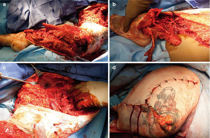
This 40-year-old female was involved in a severe motorcycle crash. In Figures (a–c) profound soft-tissue and osseous damage was sustained. Emergency department evaluation demonstrated the foot be avascular. The patient underwent emergent operative intervention and underwent an acute above-the-knee amputation (d). She returned to the operating suite several times over the ensuing days for further debridement and, ultimately, a disarticulation of the hip joint. Radiographs for this patient are shown in Fig. 22.7
22.3.1 Factors Influencing Initial Salvage Decisions
Initial decisions for the acute trauma patient with a severely injured lower extremity include immediate amputation (i.e., within the first 24 h) or delayed (i.e., secondary procedure with the first hospitalization) [8, 14, 15, 17, 42, 43]. There are a multitude of factors influencing this decision: those related directly to the leg injury itself, the extent and severity of associated injuries, the physiologic reserve of the patient, and their social support network. The training and experience of the attending surgeon may also play a role in the decision-making process [44].
Mackenzie et al. published the results of a survey pertaining to surgeons and their decision to amputate or reconstruct traumatized lower extremities. This study highlighted various factors that different specialties (general surgeons and orthopedic surgeons) deemed most important to consider in the critical decision of amputation versus salvage (Table 22.3). Interesting perspectives representative of specialty-specific training and goals were identified. Namely, the general surgeons tended to emphasize the overall physiologic condition and reserve of the patient as a whole (the injury-severity scale, limb ischemia), whereas the orthopedic surgeon emphasized functional outcome prognosis (nerve integrity, soft-tissue coverage, limb ischemia). The study conclusions suggest that the main factor influencing surgeons on the question of salvageable limbs is apparent soft-tissue damage: muscle injury, absence of sensation, arterial injury, and vein injury. Patient factors were found to play much less of a role, although alcohol consumption and socioeconomic status were noted to be of some influence [44].
Table 22.3
Percent distribution of most important factor typically considered in the decision to amputate vs. reconstruct by specialty
Factor | Total (%) | General surgeons (%) | Orthopedic surgeons (%) |
|---|---|---|---|
Nerve integrity/plantar sensation | 32 | 21 | 38 |
Limb ischemia | 20 | 27 | 15 |
Soft-tissue coverage | 14 | 9 | 17 |
Muscle damage | 7 | 6 | 8 |
Neurovascular damage | 3 | 0 | 6 |
Fracture pattern/bone loss | 4 | 0 | 6 |
High Injury Severity Scale (ISS) | 12 | 31 | 0 |
Patient characteristics | 2 | 0 | 4 |
Others | 6 | 6 | 6 |
22.3.2 Lower-Extremity Injury-Severity Scales and Scores: Tools for Assisting Surgeons with Salvage or Amputation Decisions
Lower-extremity injury-severity scores were developed by clinicians to assist surgical teams in making the often difficult initial decision of whether to attempt limb salvage or amputate a severely traumatized extremity. Surgeons have hypothesized that patients who undergo initial salvage attempts but subsequently require later amputation have worse outcomes than those who have early amputation. This makes intuitive sense and was shown to be correct in the LEAP study [16] and highlights the importance of early and accurate selection on which patients should proceed with a limb amputation during their first hospitalization.
Several studies [31, 33, 45–47] have examined the application of high-energy lower-extremity trauma scoring systems to patients with severe lower-extremity trauma. The LEAP study [21] contained the largest patient cohort of 565 prospectively evaluated high-energy lower-extremity injured patients. Each patient in this study had five well-known injury-severity scoring systems applied to their case in an effort to determine the clinical utility of each system [45]. The five systems evaluated were the Mangled Extremity Severity Score (MESS) [29, 48], the Limb Salvage Index (LSI) [32], the Predictive Salvage Index (PSI) [34], the Nerve Injury, Ischemia, Soft-Tissue Injury, Skeletal, Shock, and Age of Patient Score (NISSSA) [49], and the Hannover Fracture Scale (HFS) [50]. Table 22.4 represents the components of each injury-severity scale with the addition of a newer scale that was developed in India to predict hospital days required, flap requirements, rate of infection, and the number of secondary procedures required. This scale also incorporates patient comorbidities but emphasized primarily the evaluation of type IIIB open tibia fractures [51]. It was not assessed in the LEAP trial but is included for the sake of completeness. See Tables 22.5, 22.6, 22.7, 22.8, and 22.9 for details on each extremity trauma scale.
Table 22.4
Components of lower-extremity injury-severity scoring systems
Severity scale factors | Lower-extremity injury-severity scales | |||||
|---|---|---|---|---|---|---|
MESSa | LSIb | PSIc | NISSSAd | HFSe | GHOISSf | |
Age | X | X | X | |||
Shock | X | X | X | X | ||
Warm ischemia time | X | X | X | X | X | X |
Bone injury | X | X | X | |||
Muscle injury | X | X | X | |||
Skin injury | X | X | X | |||
Nerve injury | X | X | X | X | ||
Deep-vein injury | X | |||||
Skeletal/soft-tissue injury | X | X | ||||
Contamination | X | X | X | |||
Time to treatment | X | |||||
Comorbidities | X | |||||
Score predicting amputation | ≥7 | ≥6 | ≥8 | ≥11 | ≥9 | ≥17 (14–17 gray zone) |
A. Skeletal/soft-tissue injury | |
Low energy (stab; simple fracture; civilian GSW) | 1 |
Medium energy (open or multiple Fxs, dislocation) | 2 |
High energy (close-range shotgun or “military” GSW, crush injury) | 3 |
Very high contamination, soft-tissue avulsion | 4 |
B. Limb ischemia | |
Pulse reduced or absent but perfusion normal | 1a |
Pulseless, paresthesias, diminished capillary refill | 2a |
Cool, paralyzed, insensate limb | 3a |
C. Shock | |
Systolic BP always >90 mmHg | 0 |
Hypotensive transiently | 1 |
Persistent hypotension | 2 |
D. Age (years) | |
<30 | 0 |
30–50 | 1 |
>50 | 2 |
Artery | 0 | Contusion, intimal tear, partial laceration or avulsion (pseudoaneurysm) with no distal thrombosis and palpable pedal pulses; complete occlusion of one of three shank vessels or profunda |
1 | Occlusion of two or more shank vessels, complete laceration, avulsion or thrombosis of femoral or popliteal vessels without palpable pedal pulses | |
2 | Complete occlusion of femoral or popliteal or three of three shank vessels with no distal runoff available | |
Nerve | 0 | Contusion or stretch injury, minimal clean laceration of femoral, peroneal, or tibial nerve |
1 | Parietal transection or avulsion of sciatic nerve; complete transection or partial transection of femoral, peroneal, or tibial nerve | |
2 | Complete transection or avulsion of sciatic nerve; complete transection or avulsion of both peroneal and tibial nerves | |
Bone | 0 | Closed fracture of one or two sites; open fracture without comminution or with minimal displacement; closed dislocation without fracture; open joint without foreign body; fibula fracture |
1 | Closed fracture at three or more sites on the same extremity; open fracture with comminution or moderate to large displacement; segmental fracture; fracture dislocation; open joint with foreign body; bone loss <3 cm | |
2 | Bone loss >3 cm; Type IIIB or IIIC fracture (open fracture with periosteal stripping, gross contamination, extensive soft-tissue injury loss) | |
Skin | 0 | Clean laceration, single or multiple, or small avulsion injuries, all with primary repair; first-degree burns |
1 | Delayed closure due to contamination; large avulsion requiring STSG or flap closure. Second- or third-degree burns | |
Muscle | 0 | Laceration or avulsion involving a single compartment or single tendon |
1 | Laceration or avulsion involving two or more compartments; complete laceration or avulsion of two or more tendons | |
2 | Crush injury | |
Deep vein | 0 | Contusion, partial transection, or avulsion; complete laceration or avulsion if alternate route of venous return is intact; superficial vein injury |
1 | Complete laceration, avulsion, or thrombosis with no alternate route of venous return | |
Warm ischemia time | 0 | <6 h |
1 | 6–9 h | |
2 | 9–12 h | |
3 | 12–15 h | |
4 | >15 h |
Level of arterial injury | |
Suprapopliteal | 1 |
Popliteal | 2 |
Infrapopliteal | 3 |
Degree of bone injury | |
Mild | 1 |
Moderate | 2 |
Severe | 3 |
Degree of muscle injury | |
Mild | 1 |
Moderate | 2 |
Severe | 3 |
Interval from injury to operating room (hr) | |
<6 | 0 |
6–12 | 2 |
>12 | 4 |
Bone loss | Deperiostation | ||
No | 0 | No | 0 |
<2 cm | 1 | Yes | 1 |
>2 cm | 2 | Local circulation | |
Skin injury | Normal pulse | 0 | |
No | 0 | Capillary pulse only | 1 |
<¼ circumference | 1 | Ischemia <4 h | 2 |
¼–½ circumference | 2 | Ischemia 4–8 h | 3 |
½–¾ circumference | 3 | Ischemia >8 h | 4 |
>¾ circumference | 4 | Systemic circulation (syst. BP mm Hg) | |
Muscle injury | Constantly >100 | 0 | |
No | 0 | Until admission <100 | 1 |
<¼ circumference | 1 | Until operation <100 | 2 |
¼–½ circumference | 2 | Constantly <100 | 3 |
½–¾ circumference
Stay updated, free articles. Join our Telegram channel
Full access? Get Clinical Tree
 Get Clinical Tree app for offline access

| |||
