A. Primary surgical procedures in the emergency setting
1. Limb-saving procedures:
Reduction of large joints, such as hip, knee, by close or open means with temporary stabilization by splint or traction
Bony stabilization with urgent vascular surgery for acute damage to vascular supply
Debridement and spanning external fixation for open articular fractures together with appropriate intravenous antibiotics
Fasciotomy and spanning external fixation for articular fractures complicated by compartment syndrome
2. Spanning transarticular external fixation as a damage control procedure
To stabilize floating joint injuries or unstable joint dislocation after reduction in unstable patients
To stabilize peri-articular fractures with unfavourable local soft tissue conditions
B. Secondary surgical procedures that should be done when the general condition of the patient is stabilized or the soft tissue condition has improved
1. Definitive fixation of intra-articular fractures with initially unfavourable soft tissue conditions
2. Definitive fixation of unstable fracture dislocations, e.g. shoulder, acetabulum
3. Definitive fixation of floating joint injuries that are initially treated with spanning external fixation
4. Soft tissue coverage and definitive fixation of open intra-articular fractures
In general, complex fractures around the joints are better managed with a staged strategy [2, 3]. First, the soft tissue condition around the injured joint, especially the knee and the ankle, is usually in an unfavourable condition. There are usually severe oedema and blisters, thus rendering primary fracture fixation very risky with high complication rates. Second, intra-articular fractures are complex injuries. In order to achieve a good outcome, the articular surface should be reconstructed anatomically, the limb axis be restored correctly and a stable fixation connecting the articular block to the metaphysis and diaphysis should be obtained to allow for early joint motion. This often necessitates a good preoperative assessment of the fracture including good quality radiographs, CT scans with reconstruction and in indicated cases, MRI. Good and accurate surgical planning and meticulous surgical skills are crucial in achieving a good fixation. Hence, these difficult definitive reconstructions should not be performed in the setting of emergency surgery in a polytraumatized patient.
Generally speaking, the management of articular fractures in polytrauma patients should include a primary spanning external fixation applied in the emergency setting (Fig. 21.1). The configuration should be simple and allows easy access to the soft tissues during subsequent surgeries. The surgeon applying the external fixation should preferably be the surgeon who fixes the fracture definitively. Definitive fixation should be carried out when both the general condition of the patient and the local soft tissue conditions are optimized. Vacuum-assisted closure (VAC) therapy has been shown to be effective in managing large soft tissue defects and in assisting wound closure [4–6].
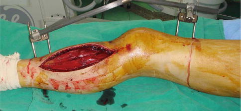
Fig. 21.1
Temporary knee-spanning external fixation in a 34-year-old polytrauma victim with comminuted proximal tibial fracture complicated by compartment syndrome. Emergency fasciotomy was performed. Vacuum-assisted closure was applied and wound closure was performed on day 10 after injury. The definitive fixation was then carried out on day 14
Sometimes in high-energy articular fractures, the stability of the joint is affected, resulting in a fracture-dislocation. In principle, a major joint dislocation that causes significant deformity should be reduced as soon as possible as the distal circulation will be affected. In the case of posterior hip dislocation that commonly occurs with posterior wall fracture of the acetabulum, reduction can usually be done quickly with closed manipulation once the patient is anaesthetized. Fixation of the posterior wall fracture should be done at a later stage after thorough assessment with CT scan. Similarly, fracture dislocations involving the ankle should be reduced urgently to avoid compromises of the soft tissue envelope and the distal circulation.
21.4 Principles of Managing Open Articular Fractures
Open articular fractures are often the result of high-energy trauma and are often associated with severe fracture comminution and bone loss. The common causes include road traffic accidents, industrial accidents or fall from heights. In lower extremity trauma, open injuries are more common especially in the knee and ankle regions [7].
Initial care of open joint injuries includes a good assessment of the patient’s general condition and urgent management of all life threatening events. Repeated debridement should be preformed. The size and the degree of contamination of the open wound are assessed. The wound should be first irrigated with copious amount of normal saline and the debris is removed. Broad-spectrum antibiotics should be started. If wound is grossly contaminated with dirt or soil, antibiotics covering anaerobes should also be started. The devitalized soft tissue including skin, fascia, fat and muscle should be debrided. Exposed cartilage should be covered with viable soft tissue if possible. Important peri-articular structures including tendons, stabilizing ligaments, neurovascular bundle should be debrided with caution. In case of suspected vascular damage associated with open joint injury, vascular surgeon should be brought in to revitalize the distal circulation as soon as possible. The return of blood circulation in the limb is one of the important factors to fight against future complications including infection. Amputation rate can be as high as 86 % if revascularization is delayed [8].
Appropriate imaging studies will always include a CT scan of the injured site that will allow surgeons to formulate an operative plan. One of the challenges in treating open joint injuries is the preservation of free osteochondral fragments. In general, these should be preserved by all means. Small osteochondral fragment can be removed. However, large unstable osteochondral fragment should be stabilized to anatomical position by minimal implants [9]. This can usually be achieved by appropriately sized lag screws or multiple K-wires. The cartilage portion should be cleaned well with copious amount of saline. The bony part should be debrided and contamination removed. Intra-articular drains should be placed to drain all the fluid collection in post-operative period.
Bony fragments which are contaminated or are without any soft tissue attachments should be excised. We use the technique of antibiotic cement spacer placed within the osseous void followed by staged bone grafting [10]. The induced biomembrane formed around the spacer prevents graft resorption, improves vascularity and later corticalization. Unpublished data from our institution between 2009 and 2012 showed four patients with open articular fractures (including two in distal tibia, one in distal femur and one in olecranon) undergoing bone reconstruction by the induced membrane technique. The antibiotic used for cement spacer is either gentamicin or vancomycin. Cancellous bone grafting was done after an average of 44 days (Fig. 21.2). All patients demonstrated radiological consolidation over the defect after treatment.
Fig 21.2
(a) A type IIA open distal femoral fracture in a 25-year-old male sustaining multiple injuries after a motor cycle crash. Debridement and spanning external fixator was applied. (b) Placement of antibiotic cement spacer into the defect after the wound bed had been adequately debrided. (c) The cement spacer was removed and the defect grafted with autograft. Six weeks later, internal fixation was performed. Notice the rapid incorporation of the bone graft into richly vascularized bone (white arrow)
For most of the open articular injuries, joint spanning external fixator is almost a must for the temporary stabilization and immobilization of the damaged joint. The spanning external fixator should be rigid but versatile enough to allow daily observation of the open wound area.
In general, definitive internal fixation of the joint and metaphyseal area is best done as early as possible to minimize joint stiffness and improve cartilage healing. However, this can only be done when the joint has no sign of infection and there is adequate soft tissue coverage. Timing of definitive fixation depends on the general status of the patient as well as local soft tissue and bony conditions. This needs a lot of experience and careful planning. Therefore, open articular fractures are best managed by experienced trauma surgeons in trauma centres where other related experts such as plastic surgeons or vascular surgeons are readily available.
21.5 Floating Joint Injuries
21.5.1 Floating Knee Injury
A floating knee refers to the injury when the ipsilateral femur and tibia are both fractured. A significant force must be needed in order to break these two bones and therefore this injury frequently implies a more substantial mechanism of injury. The patients are commonly haemodynamically unstable and may have significant injuries of other organs and the other extremities. This injury is also associated with complications that carry an increased risk of morbidity and mortality.
Fraser et al. classified floating knee injuries by whether there is joint involvement [11] (Fig. 21.3).
Type I is the injury with extra-articular fractures of both bones.
Type II is subdivided into three groups, as follows:
Type IIa involves femoral shaft and tibial plateau fractures.
Type IIb includes fractures of the distal femur and the shaft of the tibia.
Type IIc indicates fractures of the distal femur and tibial plateau.
This is the commonest classification system for floating knee injury and is of prognostic value since type I fractures have better functional outcome than type II with various extent of intra-articular involvement. This classification was recently modified by Ran et al. to include patellar fractures as a type III fracture [12]. They reported worse outcome with complex articular fractures and type III fractures.
21.5.1.1 Management of Fractures in Floating Knee Injury
Historically, floating knee injuries were totally treated or partially treated non-operatively. However, the results were unsatisfactory [11]. The current recommended treatment of the bony injuries is surgical fixation of both the femoral and the tibial fracture. There is no single ideal method of fixation [13]. The surgeon should take into consideration the extent of soft tissue injury, the location and pattern of the fractures and the associated injuries.
Isolated floating knee injury without significant articular involvement should be treated acutely if the patient is haemodynamically stable. If both fractures occur in the diaphysis, then both the femoral shaft and tibial shaft should be treated with intramedullary nailing. There is still a controversy as to whether antegrade or retrograde femoral nailing should be used. Rethnam suggested that antegrade nailing should be done [14]. Advocates for retrograde femoral nailing suggested that the quickest surgical procedure is to perform a retrograde intramedullary nailing of the femur with an intramedullary nailing of the tibia using a single incision over the knee (Fig. 21.4).
Fig. 21.4
A 48-year-old gentleman was hit by a moving car from front. He sustained a Type I floating knee injury. Distal neurovascular status was normal. He also had a concomitant stable pelvis injury. There was no other major internal organ injury. (a) X-ray of his left femur showed a transverse fracture left mid-shaft of femur. (b) X-ray of his left tibia showed a spiral fracture left mid-shaft of tibia and proximal fibula. Surgical fixation was performed the next day. (c, d) Single medial parapatellar approach was used for the retrograde femur nail and the antegrade tibial nail. The patient was allowed to freely mobilize the hip and knee after surgery with protected-weight-bearing walking
Alternatively, the tibia fracture is temporarily splinted with a cast and an antegrade femoral nailing is done first, followed by the tibial nailing. If either one or both fractures involve the epi-metaphyseal region, then the appropriate peri-articular plate fixation should be performed according to the location (Fig. 21.5). In case of severe soft tissue swelling as in tibial plateau or plafond fractures, the definitive fixation may be delayed until the soft tissue condition improves and there is a lower chance of soft tissue complications. In case of complex articular involvement with significant fracture comminution, such as tibial plateau fracture, then one can also elect to apply an external fixator temporarily and the definitive fixation done at a later stage when the required surgical expertise is available.
Fig. 21.5
(a) A 42-year-old male was injured by a fallen heavy object and sustained a type IIc floating knee injury. (b)Early fixation of both fractures were done after initial stabilization. Good lower limb alignment was obtained. (c, d) Good knee range of motion was obtained at 6 months after injury
On the other hand, in unstable patients or those in extremis, life-threatening injuries such as haemothorax, pneumothorax, intraabdominal haemorrhage, intracranial haematoma must be managed as the first priority. Under these circumstances, a temporary stabilization with a spanning external fixator should be performed, following the principles of damage control orthopaedic surgery. Once the patient’s physiological status is stabilized, conversion to internal fixation and definitive surgery can then be performed.
In the post-operative period, range of motion of the knee joint should be started early. Continuous passive motion can be used until satisfactory knee motion has been achieved. The patient should do partial-weight-bearing walking if both fractures are extra-articular. If one or both fractures involve the knee joint articular surface, then weight bearing should be delayed for 6–8 weeks.
21.5.1.2 Associated Injuries in Floating Knee Injuries
Vascular injuries of the affected limb can occur in a floating knee injury. The reported incidence ranges from 21 to 29 % [15, 16]. Limb ischaemia may occur if the popliteal or posterior tibial arteries are injured. As a result, a thorough vascular assessment is crucial in early detection of this injury. Preoperatively, the peripheral pulses should be assessed with palpation and hand-held Doppler in all floating knee injuries. If arterial injury is suspected, an intra-operative arteriogram should be performed and vascular repair should be performed together with the bony stabilization.
The incidence of open fractures in a floating knee injury can be as high as 50–70 % [16]. The commonest pattern is a closed femoral fracture with an open tibial fracture. Paul et al. reported that 17 of 21 patients had open fractures of one or more bones and 76 % of these were either grade II or grade III [16]. In general, the management of open fractures associated with floating knee injuries should follow the principles of open fracture management. This should include adequate debridement and stabilization of the fractures with either external fixation or intramedullary nailing depending on the grading of the open fractures. It is expected that multiple surgical procedures are usually required and in patients with severe mangled limbs and unstable general conditions, amputation should be considered [16].
Associated ipsilateral knee ligaments injuries are common in the floating knee injury [17]. Anterolateral rotatory instability is the commonest instability pattern. However, there is a diagnostic difficulty as the floating joint cannot be tested for ligamentous injuries. Hence after stabilization of the fractures, stress testing of the knee ligaments must be performed. If a ligamentous injury is suspected, then an acute arthroscopy can be performed and the injured ligaments can be repaired acutely or at a later stage.
21.5.1.3 Complications
The management of the fractures in floating knee injuries is challenging to orthopaedic surgeons. Fraser et al. reported 35 % of patients with floating knee injuries required late surgery for delayed union or non-union, osteomyelitis, refracture and malunion [11]. There are several explanations to this high rate of complications. The first reason is that most of the fracture fixation surgeries are performed in the emergency setting. The level of surgical expertise available is a crucial factor to the success of the surgery since sometimes a good fixation can be difficult for the average surgeon. Moreover, the floating knee segment presents great difficulty in achieving an accurate reduction of either fracture. Hence, floating knee injuries are prone to delayed union or non-union. Rotational mal-alignment can also be difficult to diagnose intra-operatively (Fig. 21.2). The overall leg length should be checked at the end of the surgery and in the early post-operative period. If the patient’s general condition allows, any mal-reduction should be corrected within the first few weeks before hard bone is formed, necessitating an osteotomy surgery.
Fat embolism can occur in a floating knee injury. Karlstrom and Olerud reported 6 out of 31 patients with fat embolism syndrome [18]. Veith et al. reported 13 % incidence of fat embolism syndrome in 54 patients of floating knee injuries [19]. The diagnosis is made if the patient has pyrexia, tachycardia, tachypnoea and altered sensorium within 48 h of admission. To confirm the diagnosis, an arterial blood gas test should be done and will reveal hypoxia. The patient should be managed in an intensive care unit with mechanical ventilation. The fractures should also be provisionally stabilized to minimize further haemorrhage and the chance of the fatty bone marrow entering the circulation. Hence, a spanning external fixator should be applied in the emergency surgery. Definitive fixation of the fractures should be delayed until the patient’s conditions improve, which usually take place after 1 week of supportive care.
21.5.2 Floating Shoulder Injuries
Floating shoulder is an uncommon injury with both clavicle and scapular neck fractured, resulting in gross instability and severe displacement of the shoulder girdle. The term floating shoulder is initially describing the inherent bony instability as described similarly in elbow and knee joints. Later Goss introduced the important concept of superior shoulder suspensory complex [20, 21]. It is a ring of complex soft tissue structures existing between two struts. The middle third of the clavicle acts as the superior strut while the scapular body and spine serves as the inferior strut. The complex maintains a normal relationship between the upper extremity and axial skeleton. The scapula is suspended to the clavicle by ligaments and acromioclavicular joint. It can be further sub-classified into three components [21]:
1.
The clavicle-acromioclavicular joint-acromial strut
2.
The clavicle-coracoclavicular ligamentous-coracoid linkage
3.
The three-process-scapular body junction
A single disruption of the ring is a stable injury. A double disruption will result in unstable injury [21, 22] (Fig. 21.6).
Fig. 21.6
The superior shoulder suspensory complex has three components: 1 – the acromioclavicular joint-aromial strut, 2 – the clavicular-coracoclavicular ligamentous-coracoid linkage, 3 – the three-process-scapular body junction
21.5.2.1 Clinical Presentation and Diagnosis
The clinical presentation of floating shoulder injuries varies with the associated injuries. When there are other serious injuries, the condition is often overlooked. During secondary survey, one can notice that the shoulder is usually grossly swollen and tender. A displaced clavicle fracture or the prominent lateral clavicular end in an acromioclavicular joint dislocation may be visible. Movements in all directions will be severely limited. Ribs fractures are not uncommon. Shortly after the injury, a detailed neurovascular examination around the shoulder may be difficult. Nevertheless, the distal neurovascular status should still be checked as the nearby brachial plexus and axillary vessels may be injured. This is one of the most important prognostic factors with regard to final clinical outcome [23]. Open injuries occur commonly in the area of the clavicle.
Radiological examination includes the anteroposterior view of the scapula and the transcapular lateral view is usually most informative. Important factors to assess include the amount of clavicular displacement, glenoid angulation and medialization, the extent of intra-articular involvement and the extent of communication [24]. If the patient is physiologically stable, further evaluation with CT scan and three-dimensional reconstructions can help to better delineate the fracture pattern.
21.5.2.2 Management
Floating shoulder injuries normally do not require emergency management, unless there is an associated open clavicula fracture, thus requiring urgent debridement. Once diagnosed, the shoulder should be supported with a broad arm sling and additional evaluation along with a CT scan should be performed when the patient’s general condition is stable.
To date there is no consensus regarding the most adequate treatment of floating shoulder injuries because of the small patient number and heterogeneity of all the studies. Based on current literature review, the treatment options are now evenly divided into nonsurgical treatment, as well as open reduction and internal fixation [24]. The degree of displacement of both clavicle and scapular neck fractures plays an important role in deciding the stability of the fractures.
Related posts:
 Psychological Sequelae After Severe Trauma
Outcome After Extremity Injuries
Psychological Sequelae After Severe Trauma
Outcome After Extremity Injuries
 Management of Malunions and Nonunions in Patients with Multiple Injuries
 High-Energy Injuries Caused by Penetrating Trauma
 Posttraumatic Acute and Chronic Osteomyelitis
 Mangled Extremity: Management in Isolated Extremity Injuries and in Polytrauma
Stay updated, free articles. Join our Telegram channel

Full access? Get Clinical Tree


