Fig. 23.1
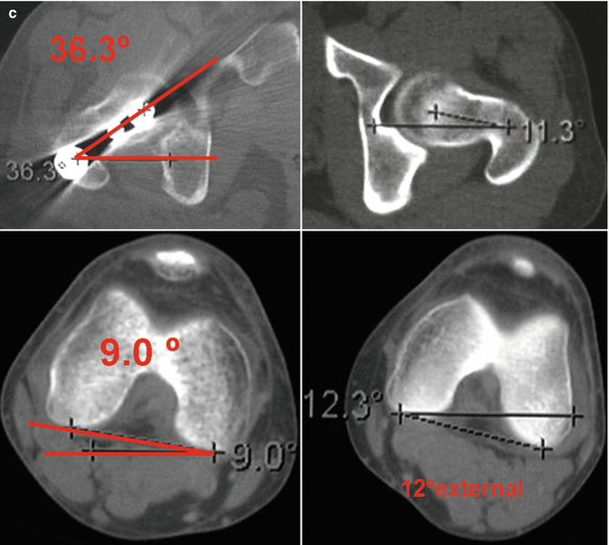
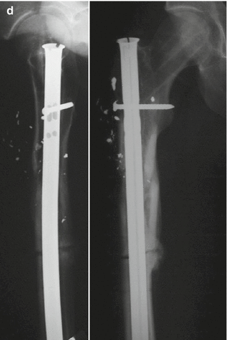
(a) Gunshot fracture to a 25-year-old counter-terrorist fighter, caused by an AK-47. (b) Three months following irrigation, debridement, and fixation with a proximal femoral nail (Synthes, Battlach, Switzerland) with no apparent wound complications. (c) Due to complaints of difficulty running and intoeing, a CT scanogram was performed which revealed almost 40° internal rotation deformity. (d) Fracture revised by a derotation osteotomy and a reamed nail
Bone loss and tissue coverage are a major challenge for tibial fractures especially involving the distal third, when soft tissue availability is scant. Due to these difficulties, our policy in high-energy gunshot wounds is to minimize bone debridement to minimum necessary and preserve as much bone as possible. Many times, these will be osteoconductive and would not result in massive infection. Recent experience in open distal femur fractures demonstrated that more conservative bone debridement does not increase infection rate, but decreases nonunion [33]. Several solutions adopted from the general orthopedic trauma are acceptable. These include rotational flaps, free tissue transfer, and immediate or late bone grafting [34, 35]. Each of these techniques can be used in conjunction with a circular frame for distraction osteogenesis [36], but not exclusively. A case of an IIIB distal tibial gunshot fracture with significant bone loss is depicted in Fig. 23.2.
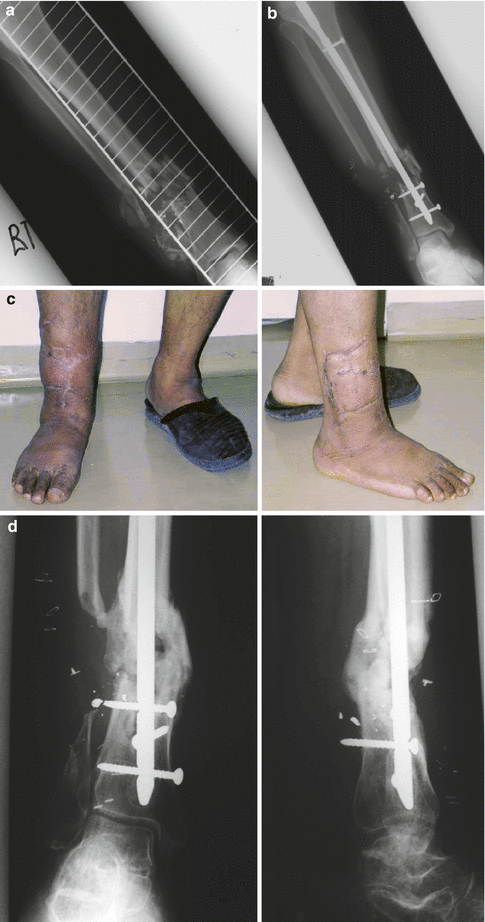
Fig. 23.2
(a) A 50-year-old schizophrenic smoker was injured by an M-16 military assault rifle sustaining a grade III-B open distal tibial fracture. (b) After irrigation and debridement with an attempt to preserve as much bone as possible, and immediate fixation with an unreamed tibial nail. (c, d) A year after a definite treatment that included further wound irrigation, latissimus dorsi free-flap, and iliac crest bone graft. Despite the imperfect ankle alignment, the patient was doing clinically well and returned to function
It should be noted, however, that even with high success rates of these hard-to-treat fractures, one can look at a prolonged recovery period with multiple reconstructive attempts [37]. As with other mangled extremity injuries, the option for primary amputation should be considered. Increased bulk of literature, both civilian and military [38, 39], denounce the use of “scoring systems” such as the MESS score for determination of the fate of a mangled extremity, and prefer to rely on the surgeon’s experience and on the general condition of the gunshot victim as predictors for early amputation.
23.1.4.2 Joints
As with other articular fractures, joint reconstruction and stable fixation should become the primary goal of treatment. However, some unique considerations should be given. First, due to the contaminated nature of gunshot injury, either arthroscopic or open irrigation should be strongly considered even in cases where a joint space violation is suspected [22]. A rare but a relevant example is the case where abdominal penetration occurred concomitant with pelvic or hip involvement. In these cases, contamination and joint sepsis would ultimately result in a catastrophic joint destruction.
Restoring metaphyseal comminution and building the articular block back to the shaft might be challenging in cases of severe comminution (Fig. 23.3), but effort should be made, like with any articular fracture, to restore joint congruency in order to maintain function and early motion [22]. Finally, chronic retention of metallic foreign bodies can result either in a local reaction [40, 41], or in rare cases, in systemic toxicity, such as lead poisoning [42, 43]. These will mandate early removal even in asymptomatic patients.
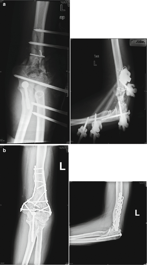
Fig. 23.3
(a) A 40-year-old patient was shot in his left lower arm and treated elsewhere with an external fixator with attempted internal fixation and brought to us for evaluation. (b) A staged protocol was used – removal of external fixator and wound debridement, definite treatment using parallel locked 3.5 reconstruction plates and iliac crest bone graft. Eighteen months after injury, the fracture is solidly healed and the patient has a reasonable range of painless motion
23.2 Blast Injuries
Another penetrating trauma type to be discussed here is related to explosion or blast. These are by far less common injuries than gunshot wounds, but their number is unfortunately growing in both civilian and military setting due to geopolitical reasons [44]. Despite the daunting threat of chemical and biological warfare, conventional terrorism is still the most common form of attacks resulting in high causality events [45]. Examples from recent years include the London attacks of 2005 [46], Madrid train attacks of 2004 [47], and the succession of suicide bombing during the Palestinian uprising between 2000 and 2005 [48, 49]
Blast injuries are different from gunshots mainly because of their multiple mechanisms of injury [16, 50, 51]. They tend to involve more body regions; and generally tend to be of higher severity scores, with increased overall potential for prolonged ICU stay and mortality [16, 52]. Although the surgical management of individual injuries may be similar to that of other types of trauma, the overall management of these patients as individuals as well as in the context of mass casualty event is worthy of consideration.
In this section we will overview the mechanisms of injury, principles of triage, and team approach as well as damage control strategies and definite orthopedic treatment of complex injuries with a special emphasis on the civilian setting.
23.2.1 Mechanisms of Blast Injury
The primary blast effect is related to the rapid pressure wave created during the detonation of an explosive [53]. The scene location and type of explosive used have a direct effect on the severity of injuries. Blast wave energy tends to decrease rapidly in space and dissipate [54]. However, when blast occurs in a closed or confined space, such in a bus or a room, the blast waves are reverberated from the walls instead of dissipating [54–56], thus inflicting more damage on human victims. In a series of suicide bombing in Israel occurring in buses during the years 1995–1996, a threefold increase in primary blast injuries was observed when compared to those of open-space explosions, exemplifying this phenomenon [56]. When the pressure wave created by detonation encounters certain air-fluid interfaces, unique tissue damage may occur. The most common and perhaps the most life-threatening injury involves the lung. Pressure differentials across the alveolar-capillary interface cause disruption, hemorrhage, pulmonary contusion, pneumothorax, hemothorax, pneumomediastinum, and subcutaneous emphysema [57].
The second most common type of primary blast injury is that to hollow viscera. The intestines, most usually the colon, are affected by the detonation wave. Mesenteric ischemia or infarct can cause delayed rupture of the large or the small intestine; these injuries are difficult to detect initially. Rupture, infarction, ischemia, and hemorrhage of solid organs such as the liver, spleen, and kidney are generally associated with very high blast forces or proximity of the patient to the blast center [58].
Tympanic membrane injury was extensively discussed in the literature discussing terror attacks. It is the most common non-lethal injury caused by relatively low-pressure blast waves. Traditionally, its presence was used to predict severe primary blast injuries (such as the lung or bowel), yet it is now referred to as questionable and unreliable [59], as will be further discussed in the triage section.
Traditionally, limb injury due to primary blast was considered a rarity. Hull and Cooper studied primary blast effects on the extremities resulting in traumatic amputations in Northern Ireland [60]. Only 9 out of 52 victims with traumatic amputations due to primary blast survived, demonstrating the high level of energy needed to avulse a limb. However, in the military setting, with the use of body armor becoming a common practice, devastating primary blast amputation has become increasingly common. These can include multiple and higher limb avulsions as well as pelvic and perineal injuries – areas not well protected by armor [4, 61].
Traditionally, the secondary blast effects comprise the core of the orthopedic injuries observed in warfare [62, 63] and also in civilian terror attacks such as the Middle Eastern experience [50, 64, 65]. Secondary blast effects are related to penetrating injuries caused by fragments ejected from the explosives and/or by foreign bodies impregnated within it. The extent of this effect depends on the subject’s distance from the detonation center, the shape and size of the fragments, and the number of foreign bodies implanted or created by the explosive. In contrast to most warfare injuries, the improvised explosives used by terrorists have multiple added fragments including screws, bolts, nails, and other objects that may increase the damage caused by penetrating injuries [66]. Open fractures, severe soft tissue injuries, and multi-organ penetrating injuries are the more common pattern seen in the severely injured victim [66, 67].
Tertiary blast injury refers to the blunt trauma component of the explosion. Flying or falling objects can cause additional traumatic elements to those described above. When structural collapse takes place, a high casualty and mortality event occurs [48]. Our experience in Israel did not demonstrate a significant proportion of additional blunt trauma, but reports from other parts of the world, such as those following the Oklahoma City explosion, state this as the primary mechanism of the injury, as well as the [68] cause of usually devastating results.
The quaternary blast effect is a recently added one, and includes the thermal and chemical damage caused by fire and noxious substances occurring at the vicinity of the explosion. Confined-space explosions significantly increase these types of injuries [56].
23.2.2 Triage and Primary Resuscitation
Perhaps the most significant difference between gunshots and the blast wounded, besides the individual injury pattern, is the “mass-casualty” effect caused by multiple military and civilian attacks. Instead of treating a single patient brought to the treating facility, the surgeon faces a scenario of mass casualty, and is required to simultaneously deal with multiple patients having multiple injuries. Hence, the initial effort should be to establish an orderly triage system, and to allocate medical team and hospital resources even before the first patient arrives to the hospital [69, 70].
In the military setting, front medical teams utilizing “damage control” strategies and performing only emergency surgeries have recently developed, especially in the global war against terrorism [28]. Further procedures are then performed in secondary and tertiary centers after further triage and usually prolonged transportation. However, this is not the case in the civilian setting, which is in the primary focus of this section.
At recent attacks – such in Jerusalem, Madrid, and London and recently the Boston Marathon – evacuation time to a definite care facility ranged between 18 min to 1–2 h [51, 71–73]. Some events, especially those on a large scale, described only a few severely injured patients in a majority of “walking wounded”. The Middle East experience, such as in Jerusalem, paradoxically demonstrated that smaller bombing scenes resulted in overall less causalities but with more critically ill patients (4–8 per event) arriving in very short notice to treating facilities [48].
Logistics of Emergency Department management have a major impact on triage. We recommend evacuation of non-critical non-terror-related patients temporarily to the hospital floors while the seriously ill patients can be treated in designated areas. The trauma bays are thus devoted solely to resuscitative efforts done on critically ill patients, while the rest of the ED serves as an admitting area to the rest of the patients. Each area is staffed with surgeon-in-charge and other members of the treating team (surgical and orthopedic residents, nurses, medical students, etc.). A surgeon-in-charge should be designated beforehand and should serve in critical junctions as suggested by Almogy et al. [71]: Triage at the initial admitting phase and in various treating cycles as well as diagnostics and directions towards the operating room or admitting floors are constantly done, until the general chaos is reduced. Every hospital should explore and identify the logistics mechanism required to provide the best and most efficient setting for disaster management under its capacity, should an actual disaster occur.
The next important principle in managing an event of this nature is to direct the flow of patients in an orderly fashion in a “one way” system. Potential bottlenecks, such as in the CT scanner, ICUs, and limited number of available operating rooms, should be identified and the patients should be directed to their proper destination only after the available resources of the hospital are mapped and identified [48]. Since as many as 50 % of causalities would require a surgical intervention – ICU stay or both [74] – hospital management should be prepared to allocate these facilities in a timely fashion.
23.2.3 Treatment of Specific Injuries
Blast extremity injuries tend to be more varied and less predictable than gunshots [16, 75]. The energy of the penetrating foreign body is extremely variable and greatly depends on the distance from the detonation center [62]. The existence of an extremity fracture, therefore, indicates a high-energy mechanism and has proven many times to implicate a polytrauma situation [76]. This is in contrast to a gunshot patient who can present with an extremity injury as a sole manifestation [16, 77]. In fact, one of the studies performed at our center indicates that fractures caused by blast are highly associated with potentially lethal blast lung injuries [78]. Therefore, an “isolated” fracture caused by blast mechanism should alert the surgeon to aggressively seek and diagnose associated injuries. Although high-energy gunshots wounds may involve a higher rate of vascular injury, compartment syndrome, and higher grade open fractures [16], blast extremity injury can involve more multiple fracture sites than gunshots, as well as a higher ISS and more associated life threatening injuries [16, 79, 80]. In this context, the treatment plans and strategies of each patient should be meticulously defined. As many patients are expedited to the operating room, more orthopedic teams should be available to undertake emergent procedures.
Damage control orthopedic and soft tissue strategies should generally be the rule in these cases since 70 % of bone-injured blast patients have an ISS of above 20 [75, 77] and are highly prone to prolonged ICU stay, respiratory failure, and coagulopathy [52, 79]. An orthopedic surgeon-in-charge should direct the teams in decision making, and the scope of first stage treatment plans should be limited as reconstruction can be planned in subsequent phases.
As in blunt polytrauma situations, tertiary surveys are extremely important in order to identify missed injuries, most of them of musculoskeletal nature [81]. We established a routine of a “morning after” rounds using the records from the ED to allocate the patients in the entire hospital, performing vigilant physical examination and documenting the penetrating injury both in writing and in a graphic form using a burn-unit type chart. A significant number of missed fractures and retained foreign bodies were identified with these surveys.
23.2.3.1 Specific Considerations
Most treatment principles of gunshot injury in regards to bone, soft tissue and vascular management should be applied to blast skeletal injuries. However, specific considerations unique to blast injury in the civilian setting should also be applied. First, as mentioned above, a polytrauma situation dictates decision making in regard to staging of bone and soft tissue treatment that is slightly different than that applied to the typical gunshot wounded victim. An illustrative case is demonstrated on Fig. 23.4.
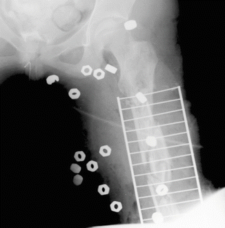
Fig. 23.4
A comminuted femoral fracture caused by secondary blast: note the numerous bolts implanted in the explosive
Also, the metal load and the amount of foreign bodies warrant removal, otherwise unnecessary in gunshot wounds. The mere removal can cause further soft tissue damage, thus mandating minimal invasive techniques. We reported the use of computerized navigation as well as metal detectors to attempt and minimize dissection involved in these removals [40, 82].
Lastly, the fact that more and more suicide bombers are involved in modern terrorism may increase the risk of biological contamination of the victims with tissues originating from the terrorists themselves, such as bone fragments [83]. Concerns of blood borne infections such as Hepatitis B/C and HIV should be taken into consideration when dealing with suicide bombers [84].
Conclusions
At the turn of the twenty-first century, firearm and terror related violence has not shown signs of decline, and it seems the world is still facing a rise in casualties related to these mechanisms. Lots have been learnt during recent years about mechanisms of injury, scenarios of mass casualty events and treatment strategies. Despite this reality, principles of treating an isolated penetrating injury, such as gunshot and multiple penetrating limb injuries, such as blast, are not yet part of the standard medical education and upbringing of the average orthopedic trauma surgeon. Keeping in mind that no part of the world is immune at this point to these devastating injuries, research and investigation of outcome and treatment strategies are in strong need, as well as internalization of the current knowledge and principles among the orthopedic and trauma surgeons community.
References
1.
Bartlett CS. Clinical update: gunshot wound ballistics. Clin Orthop Relat Res. 2003;408:28–57.
2.
3.
Bhatti JA, Mehmood A, Shahid M, Bhatti SA, Akhtar U, Razzak JA. Epidemiological patterns of suicide terrorism in the civilian Pakistani population. Int J Inj Contr Saf Promot. 2011;18:205–11.CrossRefPubMed
Related posts:


Stay updated, free articles. Join our Telegram channel

Full access? Get Clinical Tree


